. 2018 Jan 5;9(1):2.
doi: 10.1186/s13287-017-0750-8.

ERK inhibition promotes neuroectodermal precursor commitment by blocking self-renewal and primitive streak formation of the epiblast

Affiliations

ERK inhibition promotes neuroectodermal precursor commitment by blocking self-renewal and primitive streak formation of the epiblast

Yang Yu et al. Stem Cell Res Ther. .

Abstract

Background: Pluripotent stem cells hold great promise for regenerative medicine. However, before clinical application, reproducible protocols for pluripotent stem cell differentiation should be established. Extracellular signal-regulated protein kinase (ERK) signaling plays a central role for the self-renewal of epiblast stem cells (EpiSCs), but its role for subsequent germ layer differentiation is still ambiguous. We proposed that ERK could modulate differentiation of the epiblast.

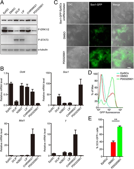
Methods: PD0325901 was used to inhibit ERK activation during the differentiation of embryonic stem cells and EpiSCs. Immunofluorescence, western blot analysis, real-time PCR and flow cytometry were used to detect germ layer markers and pathway activation.

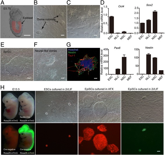
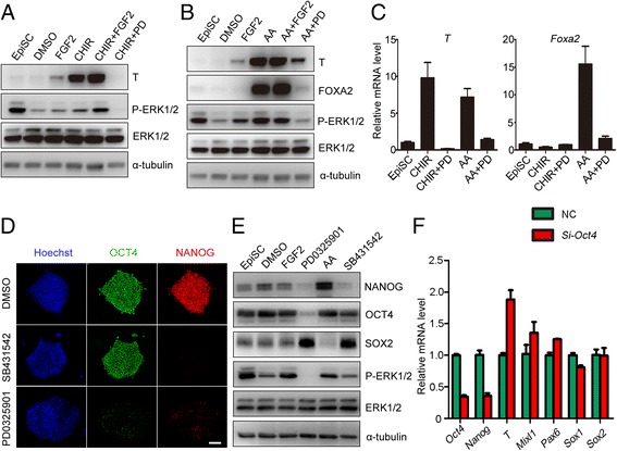
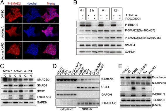
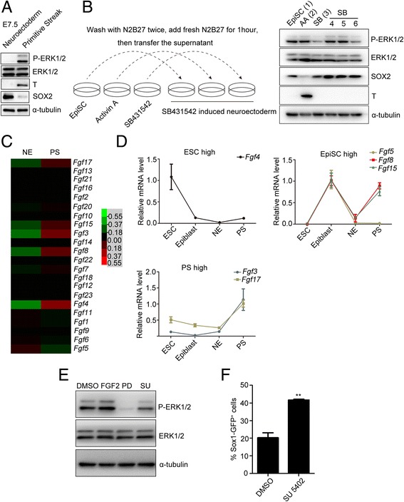
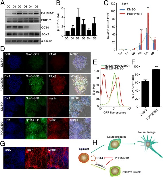
Results: We demonstrate that the ERK phosphorylation level is lower in neuroectoderm of mouse E7.5 embryos than that in the primitive streak. ERK inhibition results in neural lineage commitment of epiblast. Mechanistically, PD0325901 abrogates the expression of primitive streak markers by β-catenin retention in the cytoplasm, and inhibits the expression of OCT4 and NANOG during EpiSC differentiation. Thus, EpiSCs differentiate into neuroectodermal lineage efficiently under PD0325901 treatment. These results suggest that neuroectoderm differentiation does not require extrinsic signals, supporting the default differentiation of neural lineage.

Conclusions: We report that a single ERK inhibitor, PD0325901, can specify epiblasts and EpiSCs into neural-like cells, providing an efficient strategy for neural differentiation.

Keywords: Embryonic stem cells; Epiblast stem cells; Extracellular signal-regulated protein kinase; Neural differentiation; PD0325901.


Conflict of interest statement

Ethics approval and consent to participate

Animal experiments were conducted in accordance with the guidelines of the Institutional Animal Care and Use Committee at Institute of Zoology, Chinese Academy of Sciences.

Consent for publication

All authors have contributed to, read, and approved the final manuscript for submission.

Competing interests

The authors declare that they have no competing interests.

Publisher’s Note

Springer Nature remains neutral with regard to jurisdictional claims in published maps and institutional affiliations.

Figures

Fig. 1
Fig. 1
Epiblast cells were committed to neural lineage in 2i/LIF culture condition. a Epiblasts isolated from mouse E5.75 embryos. b Small domed colonies appeared after culturing epiblast cell clumps on MEF feeder in 2i/LIF medium for 3 days. c All clones exhibited neural-like morphology after two passages in 2i/LIF medium. d Real-time PCR showed the mRNA expression pattern of neural-like clones (NLC) was similar to neural stem cells (NSC) other than ESCs. Pluripotent markers, Oct4 and Sox2; neuroectoderm markers, Sox2 and Pax6; neural stem cell marker, Nestin. RNA collected on day 3 after replating the domed colonies. e Morphology of EpiSCs isolated from mouse E5.75 epiblasts. f NLC emerged by passaging EpiSCs in 2i/LIF medium twice. g NLC expressed the neural stem cell marker NESTIN (green) and the neuron marker TuJ-1 (red). Nuclei counterstained by Hoechst 33342 (blue). h Embryos were obtained from ROSAmT/mG × Nes-cre mouse strains. ROSAmT/mG × Nes-cre E13.5 embryos expressed GFP in the neural system. Only domed ESC clones cultured in 2i/LIF medium and flattened EpiSCs maintained in AFX medium (10 ng/ml Activin A, 5 ng/ml FGF2, and 2 μM XAV939) expressed red tomato; however, domed colonies expressing GFP were observed after EpiSCs were cultured in 2i/LIF for 3 days, indicating CRE activity driven by the Nestin promoter. Bar, 100 μm. E embryonic day, ESC embryonic stem cell, MEF mouse embryonic fibroblasts, EpiSC epiblast stem cell, OCT4 octamer-binding transcription factor 4, Pax6 paired box 6, SOX2 sex determining region Y-box 2
Fig. 2
Fig. 2
PD0325901 in 2i/LIF was responsible for neural differentiation of EpiSCs. a Activities of signal pathways were detected by western blot analysis. β-catenin and STAT3 were activated by 3 μM CHIR99021 and 1000 U/ml LIF, respectively; Phospho-ERK1/2 was inhibited by 1 μM PD0325901. b Real-time PCR results showed that PD0325901 in 2i/LIF induced neuroectoderm marker Sox1, and inhibited pluripotent marker Oct4 and primitive streak markers T and Mixl1. c PD0325901 treatment for 24 hours dramatically increased the proportion of GFP-positive cells, when Sox1-GFP EpiSCs were cultured in N2B27 medium. GFP-positive cells were detected by fluorescence microscopy. Bar, 100 μm. d, e Sox1-GFP EpiSCs were cultured in N2B27 containing 0.01% DMSO or 1 μM PD0325901 for 24 hours. GFP-positive cells were analyzed by FACS. **p < 0.01. DMSO dimethyl sulfoxide, EpiSC epiblast stem cell, ERK extracellular signal-regulated protein kinase, GFP green fluorescent protein, DIC differential interference contrast, LIF leukemia inhibitory factor, Mixl1 mix paired-like homeobox, Oct4 octamer-binding transcription factor 4, T brachyury, Sox1 sex determining region Y-box 1, STAT3 signal transducer and activator of transcription 3
Fig. 3
Fig. 3
PD0325901 prevented formation of the primitive streak and inhibits EpiSC self-renewal. a Western blot analysis showed PD0325901 (PD, 1 μM) prevented expression of PS marker T in the presence of CHIR99021 (CHIR, 3 μM) in EpiSCs cultured in N2B27 for 24 hours. b PD0325901 prevented expression of PS marker T and endoderm marker FOXA2 in the presence of Activin A (10 ng/ml) in EpiSCs cultured in N2B27 for 24 hours. c Real-time PCR showed that PD0325901 inhibited differentiation of PS even in the presence of Activin A or CHIR99021. d Immunostaining showed that PD0325901 inhibited both OCT4 and NANOG expression, whereas SB431542 (2 μM) only inhibited NANOG expression in EpiSCs differentiated in N2B27 for 24 hours. Bar, 100 μm. e PD0325901 inhibited both OCT4 and NANOG expression detected by western blot analysis. f Oct4 knockdown promoted PS differentiation in EpiSCs cultured in N2B27 medium for 24 hours. Pluripotent markers, Oct4 and Nanog; primitive streak markers, T and Mixl1; endoderm marker, Foxa2; neuroectoderm markers, Sox1, Sox2 and Pax6. AA Activin A, NC negative control, DMSO dimethyl sulfoxide, EpiSC epiblast stem cell, FGF fibroblast growth factor, T brachyury, ERK extracellular signal-regulated protein kinase, OCT4 octamer-binding transcription factor 4, FOXA2 forkhead box protein A2, SOX2 sex determining region Y-box 2, Mixl1 mix paired-like homeobox, Pax6 paired box 6
Fig. 4
Fig. 4
PD0325901 blocked formation of the primitive streak by preventing β-catenin accumulation in the nucleus. a Immunostaining showed that PD0325901 could not prevent the translocation of SMAD2 into nucleus in EpiSCs induced by Activin A. Bar, 100 μm. b Inhibition of phospho-ERK1/2 by PD0325901 did not affect phospho-SMAD2 and SMAD4 detected by western blot analysis. c Locations of SMAD2/3/4 were not changed in EpiSCs after PD0325901 treatment for 24 hours. C cytoplasm, N nucleus. d Western blot analysis showed that PD0325901 inhibited translocation of β-catenin into the nucleus. XAV939 (XAV, 2 μM) was used as a positive control that promoted the retention of β-catenin in cytoplasm. e PD0325901 increased expression of E-cadherin protein upon N2B27, CHIR99021, and Activin A treatment. SMAD SMAD family member, DMSO dimethyl sulfoxide, PD PD0325901, ERK extracellular signal-regulated protein kinase, SOX2 sex determining region Y-box 2, CHIR CHIR99021, OCT4 octamer-binding transcription factor 4, T brachyury
Fig. 5
Fig. 5
Low expression of FGF family genes accounted for the reduced ERK activity in the neuroectoderm. a Activity of ERK1/2 in the E7.5 neuroectoderm (NE) and primitive streak (PS) was detected by western blot analysis. b Secreted signals were responsible for regulation of ERK activity. Left panel shows manipulation of cells. Right panel shows western blot analysis results. Lanes 1–3 show EpiSCs, primitive streak (PS-like cells) induced by Activin A (AA) and neuroectoderm (NE-like cells) induced by SB431542 (SB). After 24 hours of differentiation, cells were washed with N2B27 twice, and refilled with fresh N2B27 medium for 1 hour. Supernatants were collected to stimulate NE-like cells. Lanes 4–6 show SB-induced NE-like cells stimulated for 1 hour with the supernatant from EpiSCs (lane 4), PS-like cells (lane 5), and NE-like cells (lane 6). c Heatmap showing relative mRNA levels of FGF family members detected by microarray in the NE and PS. Heatmap was generated by Cluster 3.0. d Expression of main Fgf family members in ESCs, E6.5 epiblasts, NE, and PS examined by real-time PCR. Expression patterns classified into three categories characterized by high expression in ESCs, epiblasts, and PS, respectively. e Phospho-ERK1/2 of EpiSCs detected by western blot analysis after treatment with DMSO, 5 ng/ml FGF2, 1 μM PD0325901, or 2 μM SU-5402 in N2B27 for 6 hours. f FACS showed that FGFR inhibitor SU-5402 treatment on Sox1-GFP EpiSCs increased the proportion of GFP-positive cells. **p < 0.01. AA Activin A, DMSO dimethyl sulfoxide, E embryonic day, EpiSC epiblast stem cell, ESC embryonic stem cell, ERK extracellular signal-regulated protein kinase, SOX2 sex determining region Y-box 2, T brachyury, FGF fibroblast growth factor, GFP green fluorescent protein
Fig. 6
Fig. 6
ERK inhibition at epiblast-like stage promoted differentiation of neuroectodermal precursors from ESCs. a, b ESCs cultured in N2B27 medium for neuroectodermal precursor commitment on gelatin-coated dishes. a Activity of ERK1/2 from D0 to D5 detected by western blot analysis. b Relative intensity of western blot analysis from (a) analyzed by ImageJ software. c PD0325901 treatment on day 3 (48–72 hours) or day 4 (72–96 hours) increased Sox1 mRNA expression. **p < 0.01. d Immunostaining of neuroectoderm marker PAX6 and neural stem cell marker nestin. PD0325901 treatment (72–96 hours) increased expression of these markers. e, f FACS showed that PD0325901 treatment (72–96 hours) increased GFP-positive cells during 46C ESC neural differentiation. **p < 0.01. g GFP-positive cells from PD0325901 treatment (72–96 hours) were further cultured in N2B27 for 3 days and stained with TuJ-1 antibody for neuron detection. Bar, 100 μm. h Working model of PD0325901 on the commitment of epiblast into neuroectoderm precursor cells. PD0325901 inhibits the self-renewal by decreasing the expression of pluripotent genes, and blocks the formation of primitive streak by preventing the accumulation of β-catenin in the nucleus, thus promoting the differentiation of epiblast into neuroectoderm precursor cells. D day, DMSO dimethyl sulfoxide, ERK extracellular signal-regulated protein kinase, OCT4 octamer-binding transcription factor 4, Sox1 sex determining region Y-box 1, SOX2 sex determining region Y-box 2, GFP green fluorescent protein, Pax6 paired box 6

Similar articles

Cited by

References

    1. Evans MJ, Kaufman MH. Establishment in culture of pluripotential cells from mouse embryos. Nature. 1981;292:154–6. doi: 10.1038/292154a0. - DOI - PubMed
    1. Martin GR. Isolation of a pluripotent cell line from early mouse embryos cultured in medium conditioned by teratocarcinoma stem cells. Proc Natl Acad Sci U S A. 1981;78:7634–8. doi: 10.1073/pnas.78.12.7634. - DOI - PMC - PubMed
    1. Brons IG, Smithers LE, Trotter MW, Rugg-Gunn P, Sun B, Chuva de Sousa Lopes SM, et al. Derivation of pluripotent epiblast stem cells from mammalian embryos. Nature. 2007;448:191–5. doi: 10.1038/nature05950. - DOI - PubMed
    1. Tesar PJ, Chenoweth JG, Brook FA, Davies TJ, Evans EP, Mack DL, et al. New cell lines from mouse epiblast share defining features with human embryonic stem cells. Nature. 2007;448:196–9. doi: 10.1038/nature05972. - DOI - PubMed
    1. Bao S, Tang F, Li X, Hayashi K, Gillich A, Lao K, et al. Epigenetic reversion of post-implantation epiblast to pluripotent embryonic stem cells. Nature. 2009;461:1292–5. doi: 10.1038/nature08534. - DOI - PMC - PubMed

Publication types

MeSH terms