Review
. 2018 May;59(5):749-763.
doi: 10.1194/jlr.R082313. Epub 2018 Jan 5.

Is ABCA1 a lipid transfer protein?

Affiliations
Review

Is ABCA1 a lipid transfer protein?

Michael C Phillips. J Lipid Res. 2018 May.

Abstract

ABCA1 functions as a lipid transporter because it mediates the transfer of cellular phospholipid (PL) and free (unesterified) cholesterol (FC) to apoA-I and related proteins present in the extracellular medium. ABCA1 is a membrane PL translocase and its enzymatic activity leads to transfer of PL molecules from the cytoplasmic leaflet to the exofacial leaflet of a cell plasma membrane (PM). The presence of active ABCA1 in the PM promotes binding of apoA-I to the cell surface. About 10% of this bound apoA-I interacts directly with ABCA1 and stabilizes the transporter. Most of the pool of cell surface-associated apoA-I is bound to lipid domains in the PM that are created by the activity of ABCA1. The amphipathic α-helices in apoA-I confer detergent-like properties on the protein enabling it to solubilize PL and FC in these membrane domains to create a heterogeneous population of discoidal nascent HDL particles. This review focuses on current understanding of the structure-function relationships of human ABCA1 and the molecular mechanisms underlying HDL particle production.

Keywords: ATP binding cassette transporter A1; apolipoprotein A-I; cholesterol; high density lipoprotein; phospholipid.


Figures

Fig. 1.
Fig. 1.
Representation of the general molecular structure of ABC transporters. A: Such transporters contain two TMDs and two conserved cytoplasmic ABCs (also designated as NBDs). Any given transporter is either an exporter (e.g., ABCA1) or importer. The two NBDs interact and, at the interface, dashed lines and red half-circles represent the ABC signature motifs and P loops, respectively. Coupling helices transmit conformational changes between the NBDs and the TMDs. B: Schematic of a single NBD showing the locations of conserved and functionally critical motifs. The juxtaposition of conserved motifs in two NBDs creates two ATP-binding sites, each involving the P loop of one NBD and the signature motif of the other. The position of a bound ATP molecule is shown in the diagram. The α- and β-phosphate groups of ATP are bound to the P loop (Walker-A motif) and the switch histidine contacts the γ-phosphate. The purine ring of adenine packs against an aromatic amino acid sidechain in the A loop. The Walker-B motif provides a catalytic glutamate involved in conversion of ATP to ADP and the signature LSGGQ motif holds and orients the ATP molecule during hydrolysis. The D loop is involved in dimerization of the NBDs and plays a role in coupling hydrolysis to transport. A groove in the NBD surface forms the contact interface with the coupling helix of the TMD. Reproduced with permission from Ref. .
Fig. 2.
Fig. 2.
Schematic of the alternating access mechanism of TM movement of substrate by an ABC transporter such as ABCA1. The TMDs form a transport substrate-binding cavity that can open to either the cytoplasmic leaflet or the exofacial leaflet of the cell PM. In the absence of bound nucleotide, the NBDs are far apart (open conformation) and the cavity faces the cytoplasm (left structure in diagram). When transport substrate (red circle) binds in this cavity and ATP binds to the NBDs causing them to move together and dimerize into the closed conformation (the power stroke), the coupled TMDs change conformation and the cavity shifts from inward-facing to outward-facing. This change causes the substrate to be translocated and become available for release on the other side of the membrane (right side of the diagram). Subsequent ATP hydrolysis (usually of both bound ATP molecules) and dissociation of ADP causes the NBDs to separate and adopt the open conformation, and the transporter returns to the starting conformation. Both exporters and importers seem to use the same mechanism, but differ in which state binds the transport substrate with higher affinity. As a PL exporter, ABCA1 binds PL more tightly with the cavity open to the cytoplasmic leaflet of the membrane. For the purpose of 2D illustration, the ATPase sites are shown as above one another, but in reality they are equidistant from the membrane. Reproduced with permission from Ref. .
Fig. 3.
Fig. 3.
Schematic representation of the topology in a membrane of human ABCA1. The six TM α-helices (H) in each of the TMDs are depicted and the segments of the amino acid sequence that form them are listed in Table 1. Two ECDs are formed by amino acids located between H1 and H2 and between H7 and H8. The two cytoplasmic NBDs are close together in 3D space (Figs. 1A, 4).
Fig. 4.
Fig. 4.
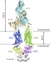
Structure of human ABCA1 determined by single-particle cryoelectron microscopy. The nominal resolution for the overall structure is 4.1 Å and 3.9 Å for the ECD. Individual ABCA1 molecules were visualized by solubilizing them in small digitonin micelles that contain ∼60 molecules of detergent. The various domains in the protein are colored differently and glycosyl groups are shown as black sticks. The picture shows the structure of the transporter in the absence of ATP. The ABCA1 conformation was stable in the digitonin micelles, but the ATPase activity was very low. The presence of two NBD and two TMD domains is characteristic of ABC transporters in general (cf. Fig. 1). The regulatory domain (R domain) contains protein recognition sites (Table 1) from both halves of the transporter (amino acids 1182-1251 and 2155-2220, respectively) and is located near the two NBDs, which are juxtaposed. For the two TMDs, both the topology with respect to the membrane and the helix locations in the amino acid sequence are largely consistent with Fig. 3 and Table 1. Coupling helices that are orientated parallel to the membrane and link the conformations of the NBDs and TMDs (cf. Fig. 1A) are located between residues 3-20, 663-678, 1327-1344, and 1678-1696. The two ECDs characteristic of ABCA transporters are juxtaposed and cofolded in a twisted fashion with ECD1 positioned above TMD2 and ECD2 positioned above TMD1. The overall ECD has a hollow interior because helices in the two domains enclose an ∼60 Å high tunnel that has a predominantly hydrophobic interior. ECD1 and ECD2 contain three and one disulfide bonds, respectively (Table 1). Reproduced with permission from Ref. .
Fig. 5.
Fig. 5.
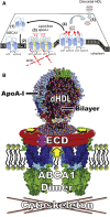
Models of “lipid reservoirs” formed by ABCA1 and their incorporation into nascent HDL particles. A: Schematic drawing of a model of HDL formation involving ABCA1 monomer to dimer conversion, as deduced by single-molecule imaging (31). (1) ABCA1 monomer diffuses freely and translocates PL from the cytoplasmic leaflet to the exofacial leaflet of the PM. (2) ABCA1 forms a reservoir of lipids within its ECDs or nearby and is immobilized as a dimer by being tethered to the actin cytoskeleton. (3) Two molecules of lipid-free apoA-I bind to the ABCA1 dimer. (4) PL and cholesterol are loaded onto apoA-I and the ABCA1 dissociates into monomers. (5) Lipidated apoA-I spontaneously binds to the PM and is in equilibrium between the PM and extracellular medium. Reproduced with permission from Ref. . B: Modification of the mechanism summarized in A showing formation of a lipid reservoir enclosed by the ECDs of ABCA1 (105). PL translocation by ABCA1 (large yellow arrow) is proposed to increase the surface pressure within the exofacial monolayer of the PM bilayer and induce formation of a discoidal PL bilayer pleat that becomes the growing lipid reservoir. apoA-I then binds to the extracellular bilayer pleat to create a discoidal nascent HDL (dHDL) particle. Reproduced with permission from Ref. .
Fig. 6.
Fig. 6.
Summary of the molecular mechanism by which ABCA1 activity in the PM of cells promotes efflux of PL and cholesterol to extracellular apoA-I and formation of nascent HDL particles. As shown at the top of the diagram, direct apoA-I/ABCA1 interaction and apoA-I/membrane lipid interactions occur with the former leading to transporter stabilization and the latter to HDL particle assembly. The PM-activated lipid domain to which apoA-I binds is created as a consequence of the PL translocation induced by ABCA1. As shown in the lower part of the figure, the activated lipid domain is formed by membrane bending and comprises an exovesiculated segment of the PM. Amphipathic α-helices in the apoA-I molecule confer detergent-like properties on the protein, allowing it to solubilize PL by binding to lattice defects in highly curved PL bilayer surfaces, thereby inducing bilayer fragmentation and formation of discoidal nascent HDL particles. These particles comprise small segments of PL/cholesterol bilayer (containing on the order of 100 PL molecules) that are most frequently stabilized by either two or three apoA-I molecules (the smaller and larger nascent HDL particles are formed simultaneously). The membrane solubilization step mediated by apoA-I is rate-limiting for the overall efflux of PL and FC from the cell. The catalytic efficiency (Vmax/Km) of apoA-I is highest for the lipid-free protein so that its efficiency is reduced by prior phospholipidation. See the text for further details. Reproduced with permission from Ref. .
Fig. 7.
Fig. 7.
Demonstration of the size heterogeneity of nascent HDL particles typically created by the incubation of apoA-I with ABCA1-expressing cells. Western blots of 2D native gel electrophoresis of apoA-I-containing nascent HDL particles generated by incubation of J774 macrophages and human skin fibroblasts with human plasma apoA-I are shown. After incubation of cells with human plasma apoA-I (5 μg/ml) for 24 h at 37°C, medium was electrophoresed in the first dimension in a 0.7% agarose gel followed by electrophoresis in the second dimension in a 2–36% concave polyacrylamide gel. The nascent HDL bands from the 2D gel were transferred onto a PVDF membrane and probed with a polyclonal anti-apoA-I antibody. A: J774 macrophage whole medium. B: Human skin fibroblast whole medium. Molecular size markers (hydrodynamic diameter in nanometers) are indicated. Reproduced with permission from Ref. .
Fig. 8.
Fig. 8.
Molecular mechanism responsible for the preferential distribution of cholesterol to larger nascent HDL particles during formation by membrane solubilization. Molecular packing constraints in nanoscale discoidal HDL particles of different sizes modify the cholesterol-solubilizing capacity. The molecular packing of the PL molecules adjacent to the α-helices of the apoA-I molecules wrapped around the circumference of the discs is constrained so that their ability to solvate cholesterol molecules is reduced. The diagram shows a top down view of four HDL discs with their diameters drawn to scale. As explained in Ref. , the areas of each bilayer leaflet occupied by boundary PL and the remaining PL (labeled as PL available for cholesterol) were calculated for 9, 12, 14, and 17 nm HDL discs. The fraction of PL forming the boundary layer varies inversely with diameter for the discoidal HDL particles so that the fractional availability of PL for solvating cholesterol is low in small HDL particles. It is apparent that approximately doubling the particle diameter from 9 to 17 nm leads to a 13-fold increase in the amount of PL into which cholesterol can dissolve (relative available PL area). Reproduced with permission from Ref. .

References

    1. Oram J. F. 2000. Tangier disease and ABCA1. Biochim. Biophys. Acta. 1529: 321–330. - PubMed
    1. Timmins J. M., Lee J. Y., Boudyguina E., Kluckman K. D., Brunham L. R., Mulya A., Gebre A. K., Coutinho J. M., Colvin P. L., Smith T. L., et al. . 2005. Targeted inactivation of hepatic Abca1 causes profound hypoalphalipoproteinemia and kidney hypercatabolism of apoA-I. J. Clin. Invest. 115: 1333–1342. - PMC - PubMed
    1. Brunham L. R., Kruit J. K., Iqbal J., Fievet C., Timmins J. M., Pape T. D., Coburn B. A., Bissada N., Staels B., Groen A. K., et al. . 2006. Intestinal ABCA1 directly contributes to HDL biogenesis in vivo. J. Clin. Invest. 116: 1052–1062. - PMC - PubMed
    1. Oram J. F., and Heinecke J. W.. 2005. ATP-binding cassette transporter A1: a cell cholesterol exporter that protects against cardiovascular disease. Physiol. Rev. 85: 1343–1372. - PubMed
    1. Yokoyama S. 2006. Assembly of high-density lipoprotein. Arterioscler. Thromb. Vasc. Biol. 26: 20–27. - PubMed

MeSH terms

Substances

LinkOut - more resources