. 2018 Jan 23;115(4):E705-E714.
doi: 10.1073/pnas.1709111115. Epub 2018 Jan 5.

Impaired lymphoid extracellular matrix impedes antibacterial immunity in epidermolysis bullosa

Affiliations

Impaired lymphoid extracellular matrix impedes antibacterial immunity in epidermolysis bullosa

Alexander Nyström et al. Proc Natl Acad Sci U S A. .

Abstract

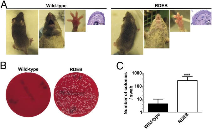
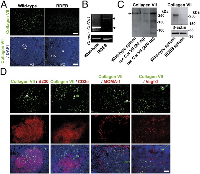
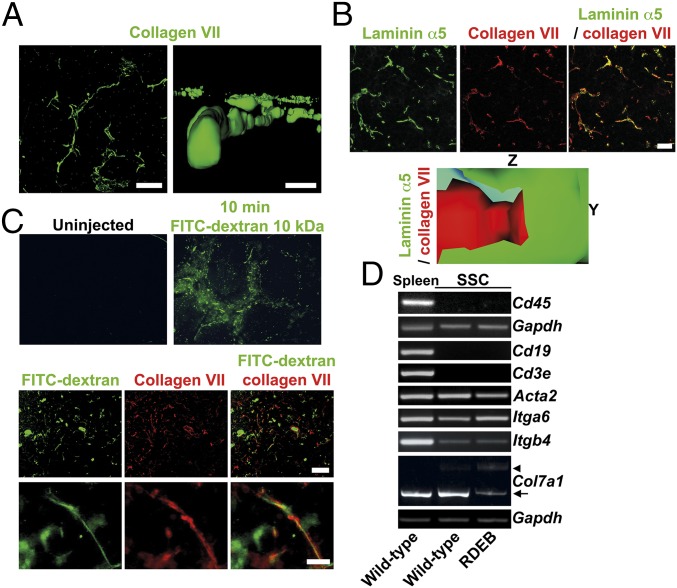
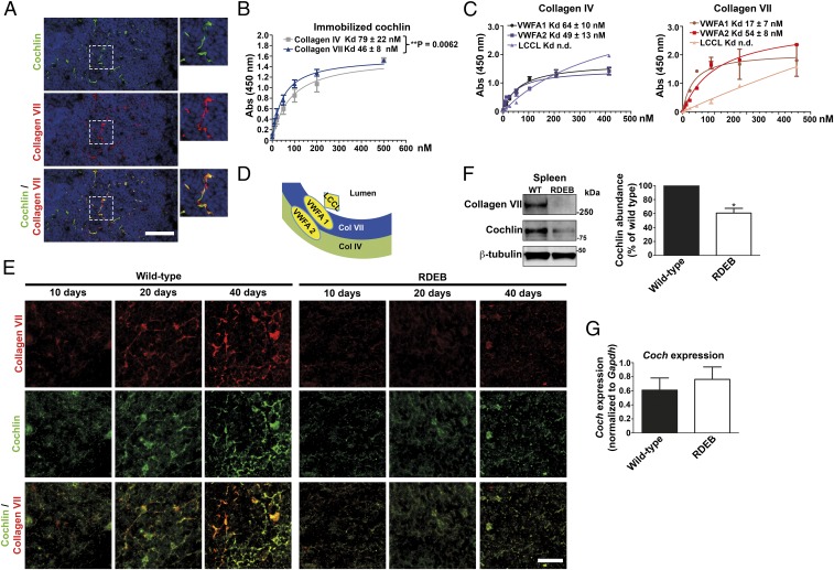
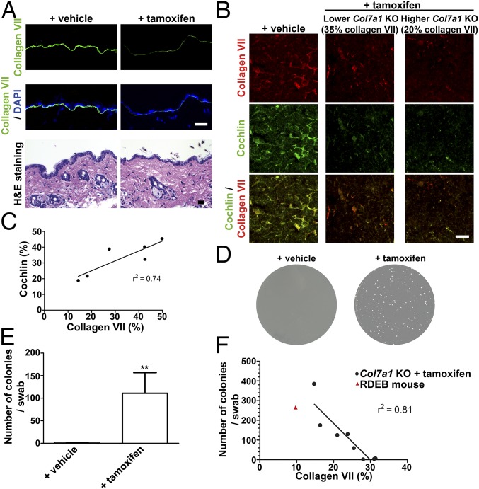
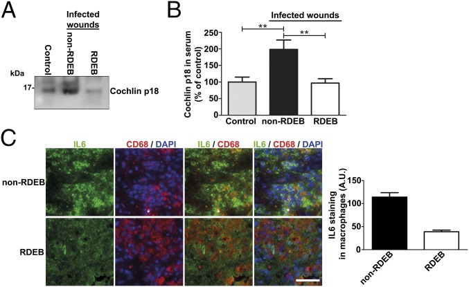
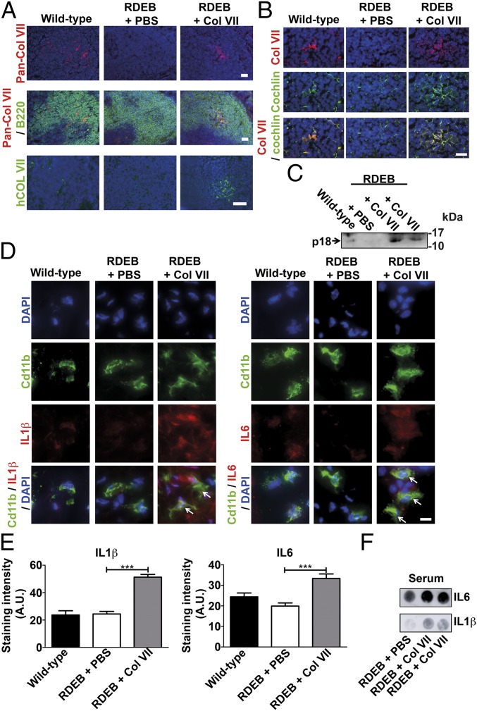
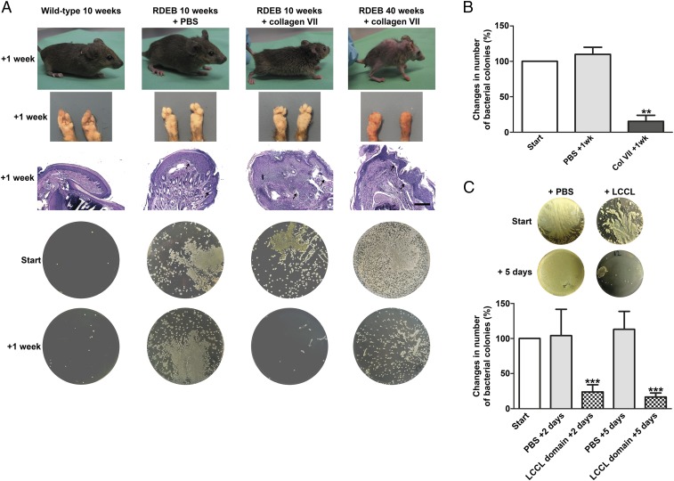
Genetic loss of collagen VII causes recessive dystrophic epidermolysis bullosa (RDEB), a skin fragility disorder that, unexpectedly, manifests also with elevated colonization of commensal bacteria and frequent wound infections. Here, we describe an unprecedented systemic function of collagen VII as a member of a unique innate immune-supporting multiprotein complex in spleen and lymph nodes. In this complex, collagen VII specifically binds and sequesters the innate immune activator cochlin in the lumen of lymphoid conduits. In genetic mouse models, loss of collagen VII increased bacterial colonization by diminishing levels of circulating cochlin LCCL domain. Intraperitoneal injection of collagen VII, which restored cochlin in the spleen, but not in the skin, reactivated peripheral innate immune cells via cochlin and reduced bacterial skin colonization. Systemic administration of the cochlin LCCL domain was alone sufficient to diminish bacterial supercolonization of RDEB mouse skin. Human validation demonstrated that RDEB patients displayed lower levels of systemic cochlin LCCL domain with subsequently impaired macrophage response in infected wounds. This study identifies an intrinsic innate immune dysfunction in RDEB and uncovers a unique role of the lymphoid extracellular matrix in systemic defense against bacteria.

Keywords: bacteria; cochlin; collagen VII; innate immunity; recessive dystrophic epidermolysis bullosa.


Conflict of interest statement

The authors declare no conflict of interest.

Figures

Fig. 1.
Fig. 1.
Adult collagen VII-deficient mice (RDEB mice) display elevated bacterial skin colonization. (A) Photographs of the back, snout, and forepaws of 10-wk-old wild-type and RDEB mouse littermates and corresponding H&E staining of forepaw sections; note the absence of large wounds. (Scale bars, 100 μm.) (B) Blood agar plates of forepaw swab samples from the mice. (C) Quantification of the number of bacterial colonies per swab from wild-type and RDEB mice; n = 8 per group; ***P = 0.0003 (Mann–Whitney U test). Values represent mean ± SEM.
Fig. 2.
Fig. 2.
Collagen VII is expressed in the spleen. (A) Wild-type and RDEB mouse spleen stained for collagen VII (green). [Scale bars: 50 μm (Top); 100 μm (Bottom).] The positions of the central arteriole (CA) and marginal zone (MZ) are indicated. (B) RT-PCR for Col7a1 and Gapdh on RNA extracted from wild-type and RDEB mouse spleens as indicated. Arrow points to wild-type transcript, and arrowhead to aberrantly spliced Col7a1 transcript in RDEB mice (25). (C, Left) Western blot analysis of collagen VII in wild-type mouse spleen lysates; 20 and 200 ng of recombinant human collagen VII were run in parallel lanes. Arrow points to full-length collagen VII. (C, Right) Western blot analysis of wild-type and RDEB mouse spleen lysates for collagen VII and β-actin as loading control. (D) Immunofluorescence analysis of codistribution of collagen VII (green), with B220 (B cells, red), Cd3e (T cells, red), MOMA-1 (red), and Vegf receptor 2 (Vegfr2) (red), respectively. (Scale bar, 100 μm.) Nuclei visualized with DAPI (blue). Asterisks indicate central vessels visible by autofluorescence.
Fig. 3.
Fig. 3.
Collagen VII is part of lymphoid conduits. (A) Confocal analysis of splenic collagen VII structures (green). On the Left side is an XY projection, and the Right side shows a computed YZ 3D rendition. [Scale bars: 10 μm (Left); 1 μm (Right).] (B, Top) Maximum intensity projection of confocal Z stack of splenic follicle stained for laminin α5 (green) and collagen VII (red). (Scale bar, 10 μm.) (B, Bottom) A longitudinal cut, applied through computed 3D model of confocal Z-stack scans, shows collagen VII to be located inside the laminin α5-containing conduit basement membrane. The Y and Z axes are displayed as indicated. (C) Images of spleen sections from a wild-type mouse 10 min after systemic injection of 10-kDa FITC-dextran (green) stained for collagen VII (red). (Scale bars, 5 μm.) (D) RT-PCR analysis of mRNA from wild-type spleen, or cultured SSCs from wild-type or RDEB mice for Cd45, Cd19, Cd3e, Acta2, Itga6, Itgb4, Col7a1, and Gapdh transcripts. Absence of Cd45, Cd19, and Cd3e transcripts confirms cultures to be devoid of leukocytes and lymphocytes. Presence of Itgb4 transcript indicates follicular dendritic cells in the cultures (SI Appendix, Fig. S2) (31). Arrow points to wild-type Col7a1 transcript, and arrowhead to aberrantly spliced Col7a1 transcript (25).
Fig. 4.
Fig. 4.
Collagen VII interacts with cochlin and is indispensable for cochlin deposition in splenic follicular conduits. (A) Immunofluorescence staining of adult wild-type mouse spleen for cochlin (green) and collagen VII (red). (B) Solid-phase binding assay of immobilized human cochlin to human collagen IV (gray squares) and human collagen VII (blue triangles). (C) To test the interaction with the cochlin LCCL, VWFA1, and VWFA2 domains, 100 ng of purified recombinant murine cochlin LCCL, VWFA1, or VWFA2 domain were immobilized on microtiter plates and overlaid with increasing concentrations of human collagen VII or collagen IV. Shown are the binding curves and Kd for collagen IV and VII calculated from four experiments. (D) A schematic model of cochlin interactions and position in lymphoid conduits. (E) Staining of spleen from 10-, 20-, and 40-d-old wild-type and RDEB mice, as indicated. Due to the species used for the collagen VII (red) antibody and the fact that the cochlin LCCL domain may be released, a goat polyclonal antibody detecting the cochlin VWFA domains (green) was used for this and all subsequent double stainings with collagen VII. (F) Western blot on adult wild-type or RDEB mouse spleen lysates, probed for collagen VII, cochlin (rabbit polyclonal antibody), and β-tubulin (Left). Densitometric quantification of three blots; cochlin expression normalized to β-tubulin and expressed as percentage of wild type; *P = 0.0293 (Right), significance tested with paired Student’s t test. (G) qPCR analysis of Coch expression in adult wild-type or RDEB mouse spleen. Values were normalized to Gapdh. Wild-type vs. RDEB mouse, P = 0.5458, significance tested with unpaired Student’s t test. All values represent mean ± SEM.
Fig. 5.
Fig. 5.
Ablation of collagen VII in adult mice leads to loss of cochlin from spleen and increased bacterial skin colonization before affecting skin integrity. (A) Back skin from tamoxifen-inducible Col7a1 knockout mice 12 wk after injection with tamoxifen or corn oil (vehicle). (Top and Middle) Sections stained for collagen VII (green) and nuclei visualized with DAPI (blue). (Bottom) H&E staining showing no apparent dermal–epidermal separation in vehicle- or tamoxifen-treated mice. (Scale bars, 100 μm.) (B) Spleen from tamoxifen-inducible Col7a1 knockout mice and controls 12 wk after knockout induction. Sections stained for collagen VII (red) or cochlin (green, goat polyclonal antibody), as indicated. Shown are a low responder (35% residual collagen VII compared with vehicle-treated controls) and a high responder (20% residual collagen VII compared with vehicle-treated controls), as determined by ImageJ-based quantification of antibody staining intensity for collagen VII on five spleen sections (38). (Scale bars, 100 μm.) (C) Plot of collagen VII abundance vs. cochlin abundance quantified by immunofluorescence staining as described in B. Linear regression analysis reveals a good correlation (r2 = 0.74) of collagen VII vs. cochlin content. (DF) Ablation of collagen VII in adult mice results in increased bacterial colonization. Bacterial swabs from forepaws of mice treated as in A. (D) Photos of representative LB-agar plates. (E) Quantification of number of bacterial colonies per plate; n = 8; **P = 0.0015, significance tested with Mann–Whitney U test. Values represent mean ± SEM. Five tamoxifen-treated mice showed highly and three slightly increased bacterial colonization, virtue of different knockout efficiency. (F) Plot of collagen VII abundance vs. bacterial colonies. Linear regression analysis reveals a good negative correlation (r2 = 0.81) of collagen VII abundance vs. bacterial colonies.
Fig. 6.
Fig. 6.
The innate immunity-regulating collagen VII–cochlin axis is broken in RDEB. (A) Western blot of sera from a healthy donor (control), a donor with a bacterially infected chronic wound (non-RDEB), and a donor with RDEB with infected wounds (RDEB). Five microliters of serum was loaded per lane on 15% tricine gels, and blots probed for cochlin with a monoclonal antibody detecting the released LCCL domain (p18 fragment as indicated) (rat anti-cochlin clone 9A10D2). (B) Densitometric quantification of bands as in A, values expressed as the percentage of the levels in the control group. Values represent mean ± SEM; n = 6; control vs. non-RDEB, **P = 0.0051; RDEB vs. non-RDEB, **P = 0.0032; control vs. RDEB, P = 0.88. Significance tested with unpaired t test with Welch’s correction. (C, Right) Wound granulation tissue from infected non-RDEB and RDEB wounds, stained for IL-6 (green), CD68 (red), and DAPI (blue). Note that, despite the generally increased expression of IL-6 in RDEB wounds (3), its expression in CD68-positive macrophages is significantly lower in RDEB wounds. (Scale bar, 100 μm.) (C, Left) Quantification of the intracellular staining intensity of IL-6 in CD68-positive cells (macrophages).
Fig. 7.
Fig. 7.
Restoration of the collagen VIl–cochlin axis increases circulating levels of cochlin LCCL domain and activates neutrophils and macrophages in skin. (AF) Wild-type and RDEB mice were injected i.p. with either PBS or 20 μg of human recombinant collagen VII 1 wk before sample collection. (A) Spleen stained for pan-collagen VII (red) and B220 (green) or human collagen VII (40, green). (Scale bar, 50 μm.) (B) Spleen stained for cochlin (goat polyclonal, green) and collagen VII (red). (Scale bar, 20 μm.) (C) Western blot of serum (8 μL per lane) probed for cochlin; the arrow shows the released cochlin LCCL domain (p18 fragment, rat anti-cochlin clone 9A10D2 used) (18). (D) High magnification of skin stained for Cd11b (green) and IL-1β (red) or IL-6 (red). Arrows indicate activated IL-1β– or IL-6–positive macrophages. (Scale bar, 5 μm.) (A, B, and D) Nuclei visualized with DAPI (blue). (E) Quantification of the intracellular staining intensity of IL-1β and IL-6 in Cd11b-positive cells; n > 100 cells quantified per group; ***P < 0.0001 (unpaired Student’s t test). (F) Dot blots of 5 μL of serum from one PBS-injected and two collagen VII-injected RDEB mice probed for IL-1β and IL-6. Note the clear increase of IL-1β and IL-6 in the sera from collagen VII-injected RDEB mice, indicating an elevated systemic antibacterial response.
Fig. 8.
Fig. 8.
Restoration of the collagen VIl–cochlin axis reduces skin bacterial load. (A) Photographs of sampled mice, their forepaws, H&E-stained paraffin sections of their forepaws, and corresponding LB-agar plates. Note the epidermal dermal separation in the histological specimens of all PBS and collagen VII-treated RDEB mouse forepaws (arrows), indicating persisting skin fragility. (Scale bar, 100 μm.) The deformities of the paws at 40 wk are due to progressive soft tissue fibrosis in RDEB and develop with advancing course of the disease. Forepaws were swabbed just before and 1 wk after i.p. injection with PBS or 20 μg of collagen VII. (B) The percentage change in colonies in RDEB mice 1 wk after injection with PBS or collagen VII; n > 4; **P = 0.0023 (paired Student’s t test). Values represent mean ± SEM. (C, Top) Representative LB-agar plates from swab samples taken from RDEB mice before and 5 d after the last i.p. injection with PBS or the cochlin LCCL domain. (C, Bottom) The percentage change in colonies in RDEB mice 2 or 5 d after injection with PBS or cochlin LCCL domain. Swab samples collected as in A. Values represent mean ± SEM; n = 6 per group; ***P < 0.0001 (paired Student’s t test).

References

    1. Nyström A, et al. Collagen VII plays a dual role in wound healing. J Clin Invest. 2013;123:3498–3509. - PMC - PubMed
    1. Guerra L, Odorisio T, Zambruno G, Castiglia D. Stromal microenvironment in type VII collagen-deficient skin: The ground for squamous cell carcinoma development. Matrix Biol. 2017;63:1–10. - PubMed
    1. Nyström A, et al. Losartan ameliorates dystrophic epidermolysis bullosa and uncovers new disease mechanisms. EMBO Mol Med. 2015;7:1211–1228. - PMC - PubMed
    1. Fine J-D, Johnson LB, Weiner M, Li K-P, Suchindran C. Epidermolysis bullosa and the risk of life-threatening cancers: The National EB Registry experience, 1986-2006. J Am Acad Dermatol. 2009;60:203–211. - PubMed
    1. Mittapalli VR, et al. Injury-driven stiffening of the dermis expedites skin carcinoma progression. Cancer Res. 2016;76:940–951. - PubMed

Publication types