

Volumetric comparison of hippocampal subfields extracted from 4-minute accelerated vs. 8-minute high-resolution T2-weighted 3T MRI scans
- PMID: 29305751
- PMCID: PMC6033688
- DOI: 10.1007/s11682-017-9819-3
Volumetric comparison of hippocampal subfields extracted from 4-minute accelerated vs. 8-minute high-resolution T2-weighted 3T MRI scans
Abstract
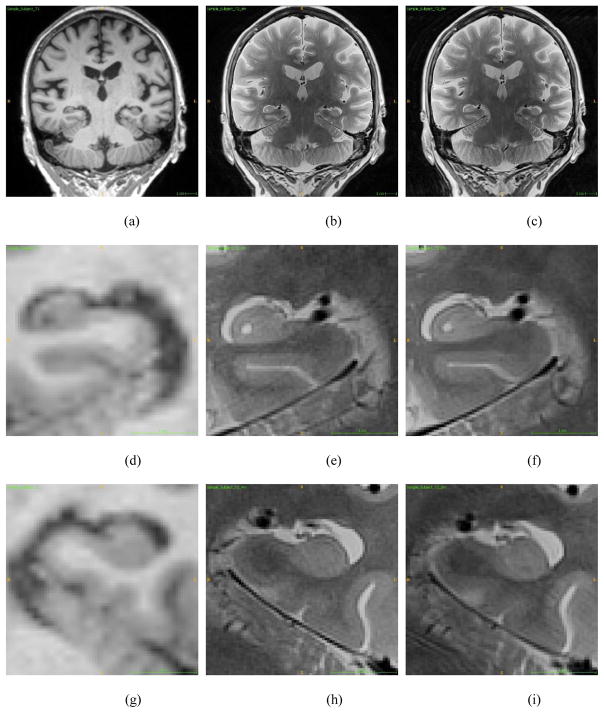
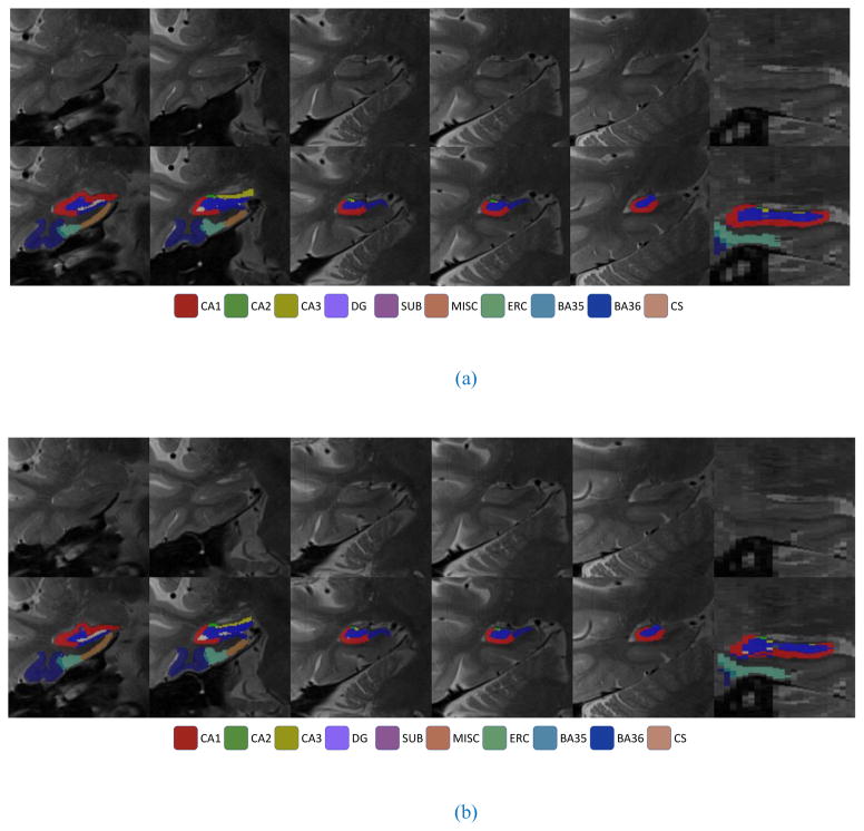
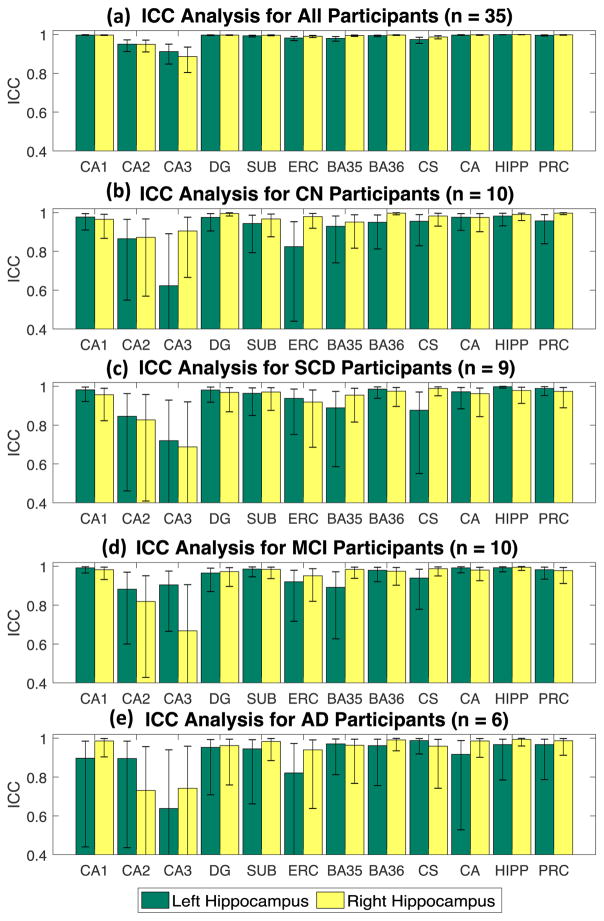
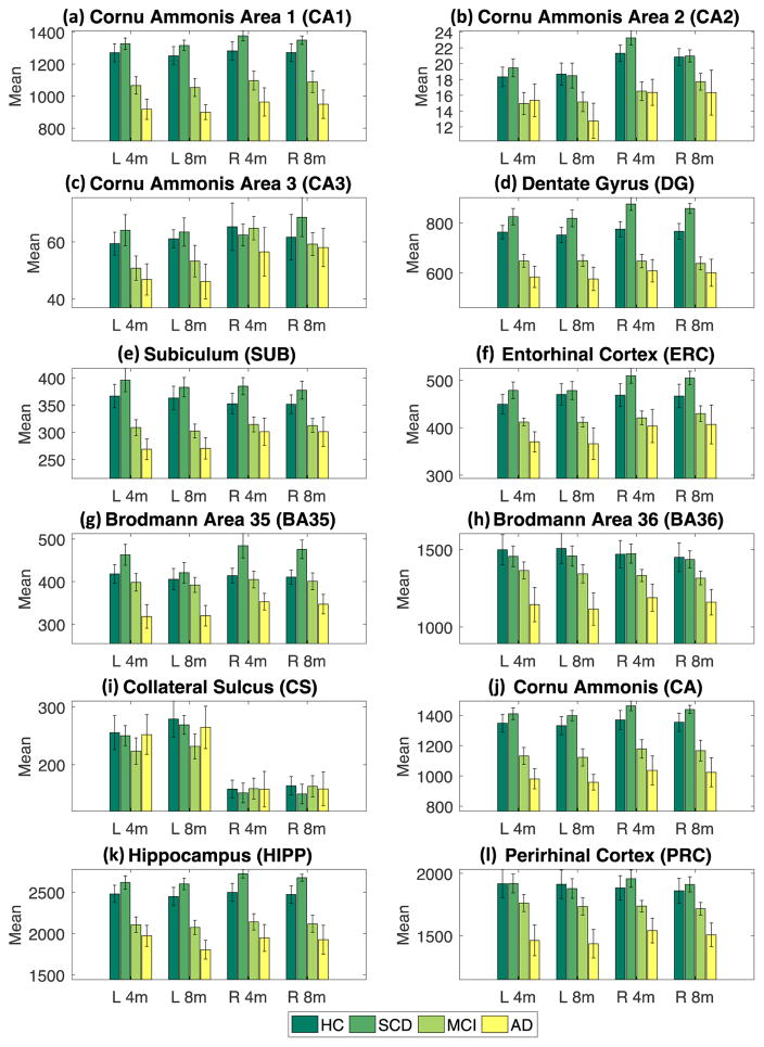
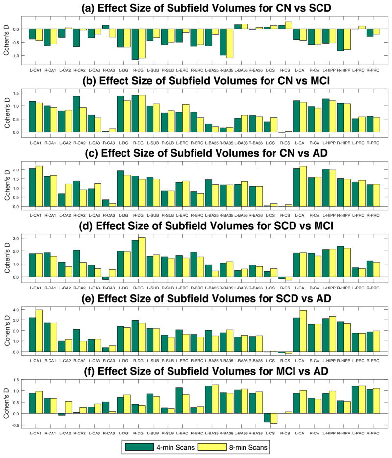
The hippocampus has been widely studied using neuroimaging, as it plays an important role in memory and learning. However, hippocampal subfield information is difficult to capture by standard magnetic resonance imaging (MRI) techniques. To facilitate morphometric study of hippocampal subfields, ADNI introduced a high resolution (0.4 mm in plane) T2-weighted turbo spin-echo sequence that requires 8 min. With acceleration, the protocol can be acquired in 4 min. We performed a comparative study of hippocampal subfield volumes using standard and accelerated protocols on a Siemens Prisma 3T MRI in an independent sample of older adults that included 10 cognitively normal controls, 9 individuals with subjective cognitive decline, 10 with mild cognitive impairment, and 6 with a clinical diagnosis of Alzheimer's disease (AD). The Automatic Segmentation of Hippocampal Subfields (ASHS) software was used to segment 9 primary labeled regions including hippocampal subfields and neighboring cortical regions. Intraclass correlation coefficients were computed for reliability tests between 4 and 8 min scans within and across the four groups. Pairwise group analyses were performed, covaried for age, sex and total intracranial volume, to determine whether the patterns of group differences were similar using 4 vs. 8 min scans. The 4 and 8 min protocols, analyzed by ASHS segmentation, yielded similar volumetric estimates for hippocampal subfields as well as comparable patterns of differences between study groups. The accelerated protocol can provide reliable imaging data for investigation of hippocampal subfields in AD-related MRI studies and the decreased scan time may result in less vulnerability to motion.
Keywords: Alzheimer’s disease; Hippocampal subfields; Magnetic resonance imaging; Segmentation; Volumetric analysis.
Figures
References
-
- Apostolova LG, Dutton RA, Dinov ID, Hayashi KM, Toga AW, Cummings JL, Thompson PM. Conversion of mild cognitive impairment to Alzheimer disease predicted by hippocampal atrophy maps. Arch Neurol. 2006;63:693–699. - PubMed
Publication types
MeSH terms
Grants and funding
- R01 AG040770/AG/NIA NIH HHS/United States
- R01 AG19771/National Institute on Aging
- R01 AG019771/AG/NIA NIH HHS/United States
- P30 AG010133/AG/NIA NIH HHS/United States
- U01 AG024904/AG/NIA NIH HHS/United States
- P30 AG10133/National Institute on Aging
- R01 AG053993/AG/NIA NIH HHS/United States
- R01 EB022574/EB/NIBIB NIH HHS/United States
- U19 AG024904/AG/NIA NIH HHS/United States
- R01 AG040770/National Institute on Aging
- R01 LM011360/U.S. National Library of Medicine (US)
- R01 LM011360/LM/NLM NIH HHS/United States
- K01 AG049050/National Institute on Aging
- R01 EB022574/National Institute of Biomedical Imaging and Bioengineering
- K01 AG049050/AG/NIA NIH HHS/United States
- U01 AG024904/National Institute on Aging
LinkOut - more resources
Full Text Sources
Other Literature Sources
Medical
