. 2018 Apr;102(4):562-569.
doi: 10.1136/bjophthalmol-2017-311150. Epub 2018 Jan 6.

Mechanical stress potentiates the differentiation of periodontal ligament stem cells into keratocytes

Affiliations

Mechanical stress potentiates the differentiation of periodontal ligament stem cells into keratocytes

Jialin Chen et al. Br J Ophthalmol. 2018 Apr.

Abstract

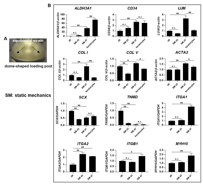
Aims: To explore the role of corneal-shaped static mechanical strain on the differentiation of human periodontal ligament stem cells (PDLSCs) into keratocytes and the possible synergistic effects of mechanics and inducing medium.

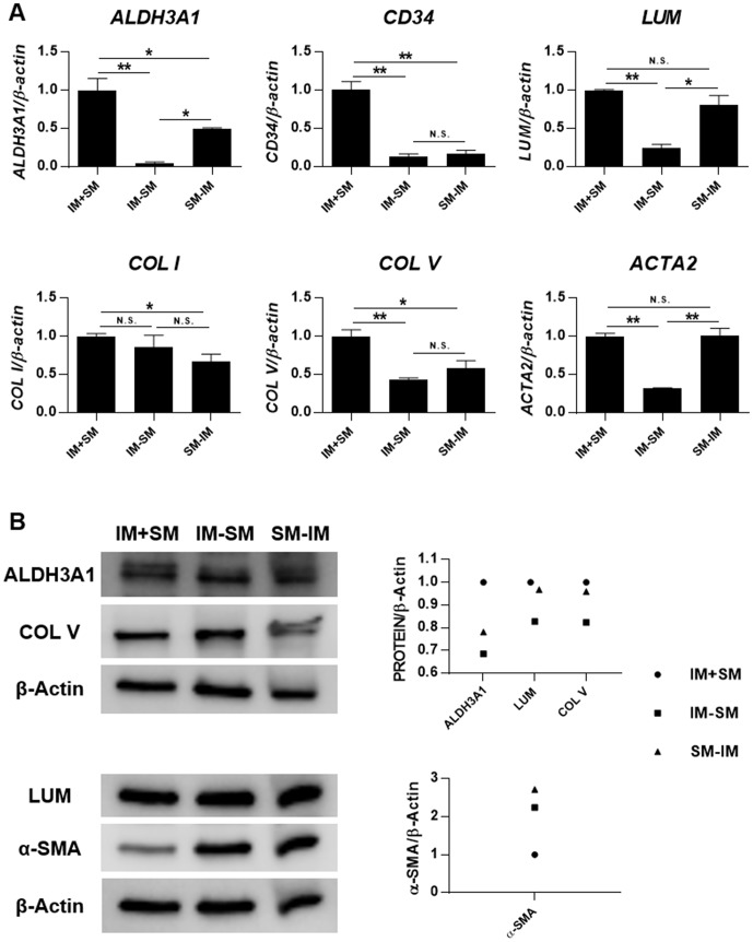
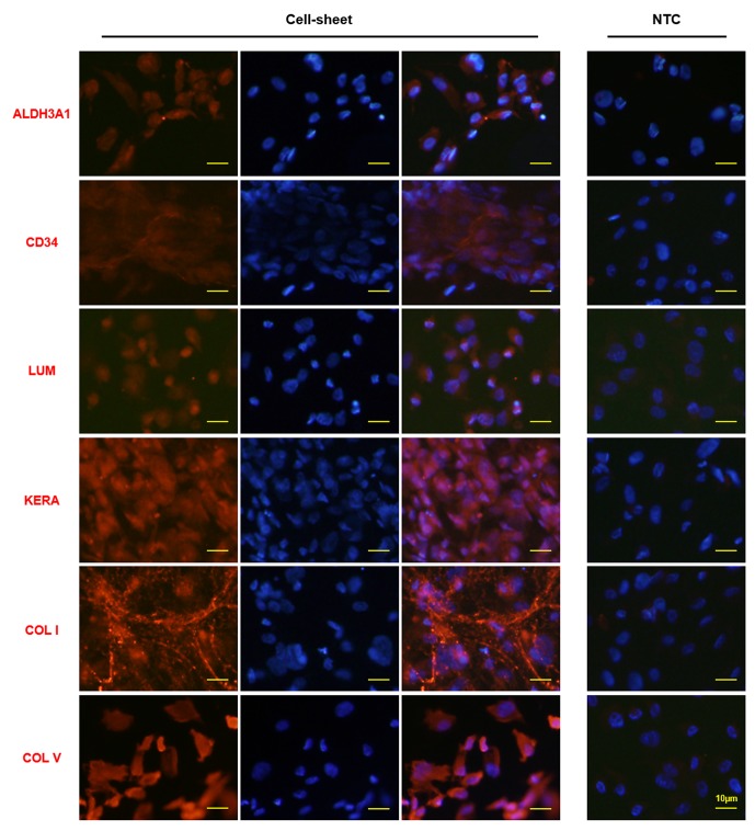
Methods: PDLSCs were exposed to 3% static dome-shaped mechanical strain in a Flexcell Tension System for 3 days and 7 days. Keratocyte phenotype was determined by gene expression of keratocyte markers. Keratocyte differentiation (inducing) medium was introduced in the Flexcell system, either continuously or intermittently combined with mechanical stimulation. The synergistic effects of mechanics and inducing medium on keratocyte differentiation was evaluated by gene and protein expression of keratocyte markers. Finally, a multilamellar cell sheet was assembled by seeding PDLSCs on a collagen membrane and inducing keratocyte differentiation. The transparency of the cell sheet was assessed, and typical markers of native human corneal stroma were evaluated by immunofluorescence staining.

Results: Dome-shaped mechanical stimulation promoted PDLSCs to differentiate into keratocytes, as shown by the upregulation of ALDH3A1, CD34, LUM, COL I and COL V. The expression of integrins were also upregulated after mechanical stimulation, including integrin alpha 1, alpha 2, beta 1 and non-muscle myosin II B. A synergistic effect of mechanics and inducing medium was found on keratocyte differentiation. The cell sheets were assembled under the treatment of mechanics and inducing medium simultaneously. The cell sheets were transparent, multilamellar and expressed typical markers of corneal stroma.

Conclusion: Dome-shaped mechanical stimulation promotes differentiation of PDLSCs into keratocytes and has synergistic effects with inducing medium. Multilamellar cell sheets that resemble native human corneal stroma show potential for future clinical applications.

Keywords: PDLSCs; cell-sheet; corneal stroma; differentiation; inducing medium; mechanics.


Conflict of interest statement

Competing interests: None declared.

Figures

Figure 1
Figure 1
Dome-shaped mechanical stimulation promotes keratocyte differentiation. (A) A dome-shaped loading post was used to apply 3% equibiaxial static strain on PDLSCs in the Flexcell Tension System for 3 days and 7 days. (B) Gene expression was evaluated by quantitative PCR, including the genes for keratocyte makers (ALDH3A1, CD34 and LUM), the main collagens of the corneal stroma (COL I and COL V), myofibroblast maker (ACTA2), tenocyte markers (SCX and TNMD) and integrins (ITGA1, ITGA2, ITGB1 and MYH10). Levels at day 0 (d0, unstimulated PDLSCs) were set as 1. The expression was compared between d0 and day 3 after stimulation of static mechanics (SM d3), d0 and day 7 after stimulation of static mechanics (SM d7), d7 and primary in vitro cultured keratocytes at passage 2. *Significant difference at P<0.05. **Significant difference at P<0.001. N.S., no significant difference (P≥0.05). PDLSCs, periodontal ligament stem cells.
Figure 2
Figure 2
Mechanical stimulation and inducing medium (IM) synergistically promote keratocyte differentiation. PDLSCs were seeded on Bioflex 6-well plates, with treatment of IM, static mechanics (SM) or a combination of both (IM+SM) for 6 days. (A) Gene expression was evaluated by quantitative PCR. Levels at day 0 (unstimulated PDLSCs) were set as 1. The expression was compared between IM and SM, IM and IM+SM, SM and IM+SM, IM+SM and primary in vitro cultured keratocytes at passage 2. *Significant difference at P<0.05. **Significant difference at P<0.001. N.S., no significant difference (P≥0.05). (B) Protein expression was evaluated by Western blot. Densitometry was performed, and the ratio of protein/β-Actin was calculated. Levels at day 0 (d0, unstimulated PDLSCs) were set as 1. PDLSCs, periodontal ligament stem cells.
Figure 3
Figure 3
Mechanical stimulation and inducing medium (IM) exert superior effect on keratocyte differentiation when they are working together at the same time. PDLSCs were seeded on Bioflex 6-well plates, with treatment of IM and static mechanics (SM) together for 6 days (IM+SM), or IM for the first 3 days and SM for the following 3 days (IM-SM), or SM for the first 3 days and IM for the following 3 days (SM-IM). (A) Gene expression was evaluated by quantitative PCR. Levels of IM+SM were set as 1. *Significant difference at P<0.05. **Significant difference at P<0.001. N.S., no significant difference (P≥0.05). (B) Protein expression was evaluated by Western blot. Densitometry was performed, and the ratio of protein/β-Actin was calculated. Levels of IM+SM were set as 1. PDLSCs,  periodontal ligament stem cells.
Figure 4
Figure 4
Mechanical stimulation and inducing medium generate a transparent and multilamellar cell sheet. PDLSCs were seeded on collagen I bonded Bioflex 6-well plates and exposed to static mechanical strain and inducing medium for 12 days to form a cell sheet. The collagen membrane of the Bioflex 6-well plate itself (A) and the collagen membrane with the formed cell sheet (B) were both transparent. (C) The light transmission of the cell sheet was measured in the wavelength from 400 nm to 850 nm. (D) The formed cell sheet was found to be a multilamellar structure, as revealed by DAPI staining; white arrows indicating nuclei that are focused on one layer, and red arrows indicating nuclei that were not focused and thus on another layer. PDLSCs, periodontal ligament stem cells.
Figure 5
Figure 5
Typical markers of corneal stroma are expressed in the cell sheets constructed by PDLSCs exposed to mechanical stimulation and inducing medium. PDLSCs were seeded on Bioflex 6-well plates, with treatment of static mechanical strain and inducing medium for 12 days to form a cell sheet. The expression of typical corneal stroma markers was evaluated by immunofluorescence staining. The right column is the merged picture of left column (corneal stroma markers staining) and middle column (DAPI staining). ‘NTC’ indicates negative control without primary antibody. PDLSCs, periodontal ligament stem cells.

References

    1. Kumar P, Pandit A, Zeugolis DI. Progress in corneal stromal repair: from tissue grafts and biomaterials to modular supramolecular tissue-like assemblies. Adv Mater 2016;28:5381–99. 10.1002/adma.201503986 - DOI - PubMed
    1. Sloniecka M, Backman LJ, Danielson P. Antiapoptotic effect of acetylcholine in fas-induced apoptosis in human keratocytes. Invest Ophthalmol Vis Sci 2016;57:5892–902. 10.1167/iovs.16-19707 - DOI - PubMed
    1. Musselmann K, Alexandrou B, Kane B, et al. . Maintenance of the keratocyte phenotype during cell proliferation stimulated by insulin. J Biol Chem 2005;280:32634–9. 10.1074/jbc.M504724200 - DOI - PubMed
    1. Etheredge L, Kane BP, Valkov N, et al. . Enhanced cell accumulation and collagen processing by keratocytes cultured under agarose and in media containing IGF-I, TGF-β or PDGF. Matrix Biol 2010;29:519–24. 10.1016/j.matbio.2010.05.003 - DOI - PMC - PubMed
    1. Zhang S, Espandar L, Imhof KM, et al. . Differentiation of human adipose-derived stem cells along the keratocyte lineage in vitro. J Clin Exp Ophthalmol 2013;4 10.4172/2155-9570.1000270 - DOI - PMC - PubMed

Publication types