Review
. 2018 Apr;10(2):163-181.
doi: 10.1007/s12551-017-0346-7. Epub 2018 Jan 6.

Unified understanding of folding and binding mechanisms of globular and intrinsically disordered proteins

Affiliations
Review

Unified understanding of folding and binding mechanisms of globular and intrinsically disordered proteins

Munehito Arai. Biophys Rev. 2018 Apr.

Abstract

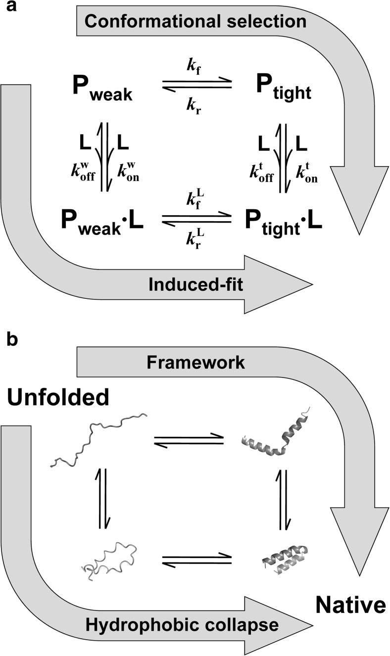
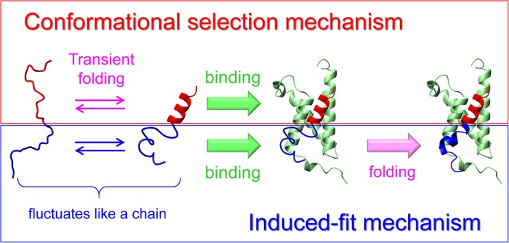
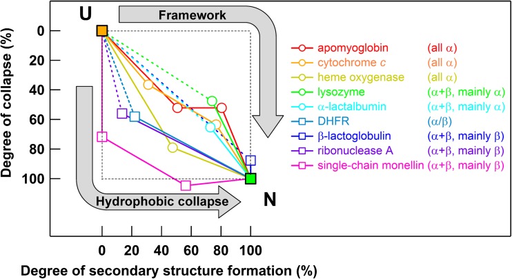
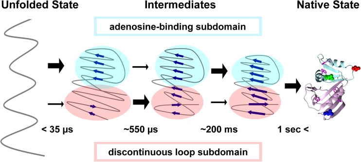
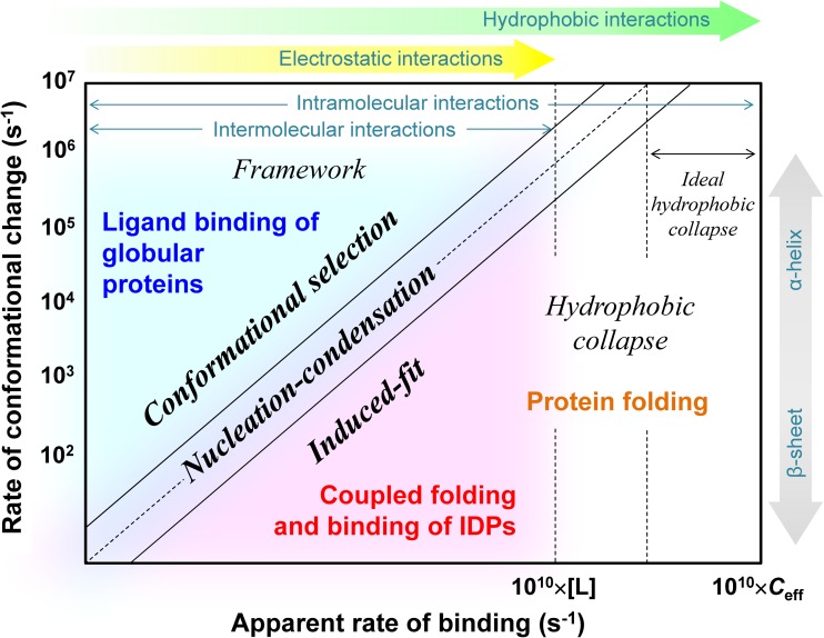
Extensive experimental and theoretical studies have advanced our understanding of the mechanisms of folding and binding of globular proteins, and coupled folding and binding of intrinsically disordered proteins (IDPs). The forces responsible for conformational changes and binding are common in both proteins; however, these mechanisms have been separately discussed. Here, we attempt to integrate the mechanisms of coupled folding and binding of IDPs, folding of small and multi-subdomain proteins, folding of multimeric proteins, and ligand binding of globular proteins in terms of conformational selection and induced-fit mechanisms as well as the nucleation-condensation mechanism that is intermediate between them. Accumulating evidence has shown that both the rate of conformational change and apparent rate of binding between interacting elements can determine reaction mechanisms. Coupled folding and binding of IDPs occurs mainly by induced-fit because of the slow folding in the free form, while ligand binding of globular proteins occurs mainly by conformational selection because of rapid conformational change. Protein folding can be regarded as the binding of intramolecular segments accompanied by secondary structure formation. Multi-subdomain proteins fold mainly by the induced-fit (hydrophobic collapse) mechanism, as the connection of interacting segments enhances the binding (compaction) rate. Fewer hydrophobic residues in small proteins reduce the intramolecular binding rate, resulting in the nucleation-condensation mechanism. Thus, the folding and binding of globular proteins and IDPs obey the same general principle, suggesting that the coarse-grained, statistical mechanical model of protein folding is promising for a unified theoretical description of all mechanisms.

Keywords: Conformational selection; Induced-fit; Intrinsically disordered protein; Ligand binding; Nucleation–condensation; Protein folding.


Conflict of interest statement

Funding

This study was funded by Grants-in-Aids for Scientific Research from the Ministry of Education, Culture, Sports, Science and Technology (MEXT).

Conflict of interest

Munehito Arai declares that he has no conflict of interest.

Ethical approval

This article does not contain any studies with human participants or animals performed by the author.

Figures

Fig. 1
Fig. 1
a Conformational selection and induced-fit mechanisms. Pweak and Ptight denote weakly and tightly binding conformations respectively, and L denotes a ligand. kon and koff denote the second-order binding rate constant and first-order dissociation rate constant respectively. kf and kr denote the forward and reverse rate constants respectively. b Two representative mechanisms of protein folding. The framework model corresponds to the conformational selection mechanism, while the hydrophobic collapse model corresponds to the induced-fit mechanism
Fig. 2
Fig. 2
Mechanisms of the coupled folding and binding reaction of intrinsically disordered c-Myb TAD upon binding to KIX (green). The N-terminal region of c-Myb TAD (red) binds KIX by the conformational selection mechanism (upper), while the C-terminal region of c-Myb TAD (blue) interacts with KIX by the induced-fit mechanism (lower)
Fig. 3
Fig. 3
Folding trajectories of multi-subdomain proteins drawn in the simplified folding landscape. The horizontal and vertical axes show the degree of secondary structure formation, estimated from the change in circular dichroism intensity during folding, and degree of collapse, estimated from the change in the radius of gyration during folding, respectively. The unfolded state (U) and native state (N) are located at the upper left and lower right respectively. Open circles and squares show the location of folding intermediates of α-rich and β-rich proteins respectively. Continuous and dotted lines show the folding trajectories. Folding trajectories for the ideal conformational selection (Framework) mechanism and ideal induced-fit (Hydrophobic collapse) mechanism are indicated with arrows. Adapted with permission from Arai et al. (2007)
Fig. 4
Fig. 4
Subdomain-wise folding of DHFR. Blue arrows in the folding intermediates represent β-strands. Thick and thin black arrows respectively show that large and small conformational changes occur in the indicated subdomain during each phase. Adapted with permission from Arai et al. (2011)
Fig. 5
Fig. 5
Apparent dependence of the folding and binding mechanisms of globular proteins and IDPs on both the rate of conformational change and apparent binding rate of interacting elements. [L] and Ceff denote the free ligand concentration and effective concentration for intramolecular interactions respectively. See text for details

Similar articles

Cited by

References

    1. Akiyama S, Takahashi S, Ishimori K, Morishima I. Stepwise formation of α-helices during cytochrome c folding. Nat Struct Biol. 2000;7:514–520. - PubMed
    1. Ali SA, Iwabuchi N, Matsui T, Hirota K, Kidokoro S, Arai M, Kuwajima K, Schuck P, Arisaka F. Reversible and fast association equilibria of a molecular chaperone, gp57A, of bacteriophage T4. Biophys J. 2003;85:2606–2618. - PMC - PubMed
    1. Arai M, Dyson HJ, Wright PE. Leu628 of the KIX domain of CBP is a key residue for the interaction with the MLL transactivation domain. FEBS Lett. 2010;584:4500–4504. - PMC - PubMed
    1. Arai M, Ferreon JC, Wright PE. Quantitative analysis of multisite protein-ligand interactions by NMR: binding of intrinsically disordered p53 transactivation subdomains with the TAZ2 domain of CBP. J Am Chem Soc. 2012;134:3792–3803. - PMC - PubMed
    1. Arai M, Hamel P, Kanaya E, Inaka K, Miki K, Kikuchi M, Kuwajima K. Effect of an alternative disulfide bond on the structure, stability, and folding of human lysozyme. Biochemistry. 2000;39:3472–3479. - PubMed

LinkOut - more resources