Review
. 2018 Jan;243(2):118-128.
doi: 10.1177/1535370217749494. Epub 2018 Jan 7.

Myogenic regulatory factors: The orchestrators of myogenesis after 30 years of discovery

Affiliations
Review

Myogenic regulatory factors: The orchestrators of myogenesis after 30 years of discovery

Hasan A Asfour et al. Exp Biol Med (Maywood). 2018 Jan.

Abstract

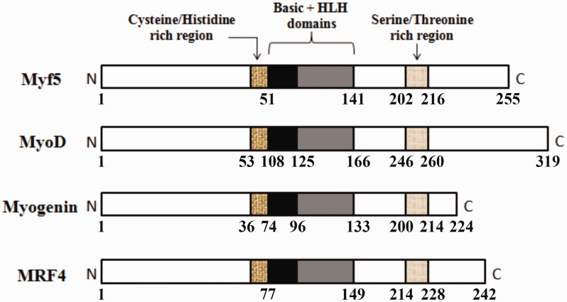
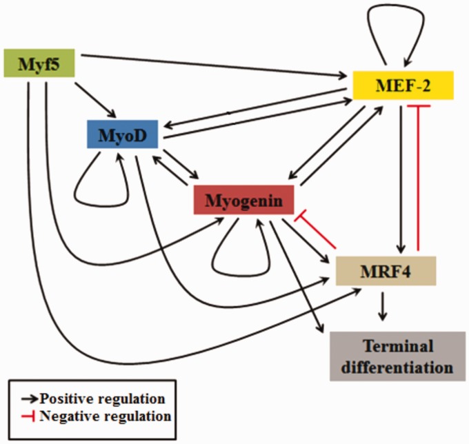
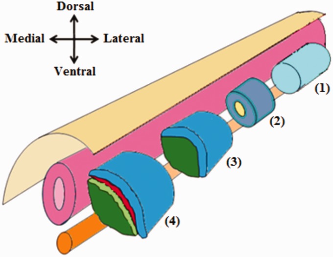
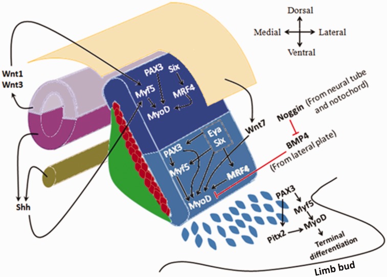
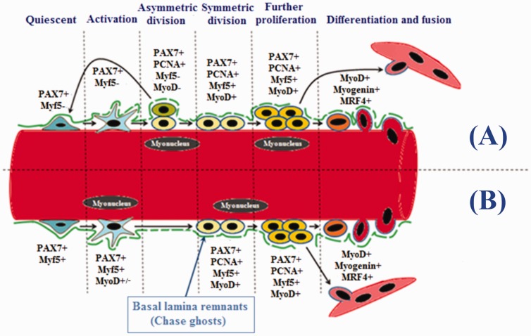
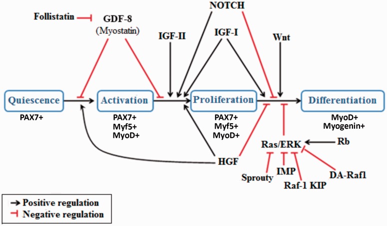
Prenatal and postnatal myogenesis share many cellular and molecular aspects. Myogenic regulatory factors are basic Helix-Loop-Helix transcription factors that indispensably regulate both processes. These factors (Myf5, MyoD, Myogenin, and MRF4) function as an orchestrating cascade, with some overlapped actions. Prenatally, myogenic regulatory factors are restrictedly expressed in somite-derived myogenic progenitor cells and their derived myoblasts. Postnatally, myogenic regulatory factors are important in regulating the myogenesis process via satellite cells. Many positive and negative regulatory mechanisms exist either between myogenic regulatory factors themselves or between myogenic regulatory factors and other proteins. Upstream factors and signals are also involved in the control of myogenic regulatory factors expression within different prenatal and postnatal myogenic cells. Here, the authors have conducted a thorough and an up-to-date review of the myogenic regulatory factors since their discovery 30 years ago. This review discusses the myogenic regulatory factors structure, mechanism of action, and roles and regulations during prenatal and postnatal myogenesis. Impact statement Myogenic regulatory factors (MRFs) are key players in the process of myogenesis. Despite a considerable amount of literature regarding these factors, their exact mechanisms of actions are still incompletely understood with several overlapped functions. Herein, we revised what has hitherto been reported in the literature regarding MRF structures, molecular pathways that regulate their activities, and their roles during pre- and post-natal myogenesis. The work submitted in this review article is considered of great importance for researchers in the field of skeletal muscle formation and regeneration, as it provides a comprehensive summary of all the biological aspects of MRFs and advances a better understanding of the cellular and molecular mechanisms regulating myogenesis. Indeed, attaining a better understanding of MRFs could be utilized in developing novel therapeutic protocols for multiple myopathies.

Keywords: MRF4; Myf5; MyoD; myoblasts; myogenic determination; myogenin; satellite cells.


Figures

Figure 1.
Figure 1.
The primary structures for different MRFs. The three structural homologous domains of different MRFs are shown. These are; a basic domain linked to HLH region, a cysteine/histidine-rich domain that lies on the N-terminal side of the basic domain, and a serine/threonine-rich domain that is located near the C-terminal. The amino acid numbers are represented beneath each structure. (A color version of this figure is available in the online journal.)
Figure 2.
Figure 2.
Signaling cascade of MRFs with their auto and cross-regulatory mechanisms. Myf5 activates all other MRFs (MyoD, Myogenin and MRF4) and MEF-2 proteins. MyoD has an auto-regulatory mechanism and cross-activation mechanism with Myogenin. Myogenin has an auto-regulatory mechanism and can induce terminal differentiation directly or/and indirectly via activation of MRF4. MRF4 directly induces terminal differentiation and can be activated by all other MRFs and MEF-2. MRF4 can inhibit both Myogenin and MEF-2 expressions. MEF-2 has an autoregulatory mechanism and can reciprocally activate both MyoD and Myogenin. (A color version of this figure is available in the online journal.)
Figure 3.
Figure 3.
Differentiation of the paraxial mesoderm. Prior to somite formation, the paraxial mesoderm is composed of a single epithelial tube which flanks the neural tube and notochord (1). Later on, the paraxial mesoderm becomes segmented into somites. Each somite consists of epithelial cells with a mesenchymal core (2). Further differentiation of each somite results in the formation of two distinct layers; the dorsolateral dermomyotome, and the ventromedial sclerotome (3). After that additional two layers formed; the myotome which derived from the dermomyotome and the syndetome which derived from sclerotome (4). (A color version of this figure is available in the online journal.)
Figure 4.
Figure 4.
Different epaxial and hypaxial myogenic factors in dermomyotome and the upstream signals. Wnt1, Wnt3, and Shh signals activate Myf5 in the epaxial dermomyotome which in turn activates MyoD expression. In addition, Six proteins and PAX3 can activate MyoD expression independent of Myf5. However, in the hypaxial dermomyotome, Myf5 expression is dependent on PAX3, Six, and Eya proteins. Never the less, all the aforementioned factors can directly activate MyoD expression, while BMP4 inhibits its expression in the hypaxial dermomyotome. In the limb bud, delaminated (PAX3+/MRFs−) cells do not express MyoD until they reach their destination. MyoD expression in these cells can be induced either canonically through PAX3 and Myf5, or non-canonically by PAX3 and Pitx2. (A color version of this figure is available in the online journal.)
Figure 5.
Figure 5.
Expression of myogenic regulatory factors during different stages of satellite cells. Satellite cells are divided into two groups according to their self-renewal ability; myogenic stem (∼10%, PAX7+/Myf5−) and myogenic precursor (∼90%, PAX7+/Myf5+) cells. (A) Myogenic stem cells can undergo asymmetric divisions where some daughter cells return back to quiescence. (B) Myogenic precursor cells undergo symmetrical divisions where all daughter cells are committed to the myogenic fate. Proliferating satellite cells can be identified by the expression of PCNA. Upregulation of MyoD along with the expression of Myogenin and MRF4 induces these cells into the differentiating stage. Terminally differentiated cells may either fuse into pre-existing myofibers, or into newly formed one. Chase ghost act as a scaffold for myogenic stem and precursor cells during activation, proliferation, and differentiation. (A color version of this figure is available in the online journal.)
Figure 6.
Figure 6.
Illustrating diagram for several growth factors and signaling molecules that regulate different stages of satellite cells. Transition from quiescence into activation and proliferation is stimulated by HGF and inhibited by myostatin (GDF-8). IGF-II induces proliferation. IGF-I can induce both proliferation and differentiation. HGF and NOTCH sustain proliferation and inhibit transition to differentiation. Transition to differentiation is also inhibited by Rb protein through activating Ras/ERK pathway. Transition to differentiation can be induced either directly by the Wnt family of proteins, or indirectly via inhibition of the Ras/ERK pathway by Sprouty, IMP, Raf-1 KIP, and DA-Raf1. (A color version of this figure is available in the online journal.)

References

    1. Yablonka-Reuveni Z. The skeletal muscle satellite cell: still young and fascinating at 50. J Histochem Cytochem 2011; 59:1041–59 - PMC - PubMed
    1. Allouh MZ, Rosser BW. Nandrolone decanoate increases satellite cell numbers in the chicken pectoralis muscle. Histol Histopathol 2010; 25:133–40 - PubMed
    1. Allouh MZ, Jarrar AA, Asfour HA, Said RS, Shaqoura EI. Sustanon induces dose-independent hypertrophy and satellite cell proliferation in slow oxidative fibers of avian skeletal muscle. Histol Histopathol 2017; 19:11871 - PubMed
    1. Waldemer-Streyer RJ, Chen J. Myocyte-derived Tnfsf14 is a survival factor necessary for myoblast differentiation and skeletal muscle regeneration. Cell Death Dis 2015; 6:e2026. - PMC - PubMed
    1. Allouh MZ, Aldirawi MH. Influence of mesterolone on satellite cell distribution and fiber morphology within maturing chicken pectoralis muscle. Anat Rec 2012; 295:792–9 - PubMed

Publication types

MeSH terms

Substances