. 2018 Feb 13;10(2):339-346.
doi: 10.1016/j.stemcr.2017.12.006. Epub 2018 Jan 4.

Cell-Cycle-Specific Function of p53 in Fanconi Anemia Hematopoietic Stem and Progenitor Cell Proliferation

Affiliations

Cell-Cycle-Specific Function of p53 in Fanconi Anemia Hematopoietic Stem and Progenitor Cell Proliferation

Xiaoli Li et al. Stem Cell Reports. .

Abstract

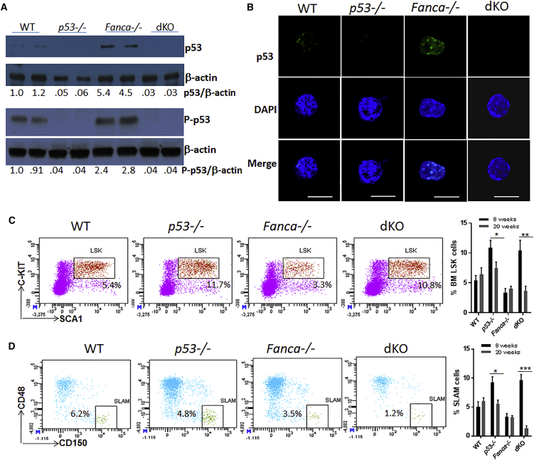
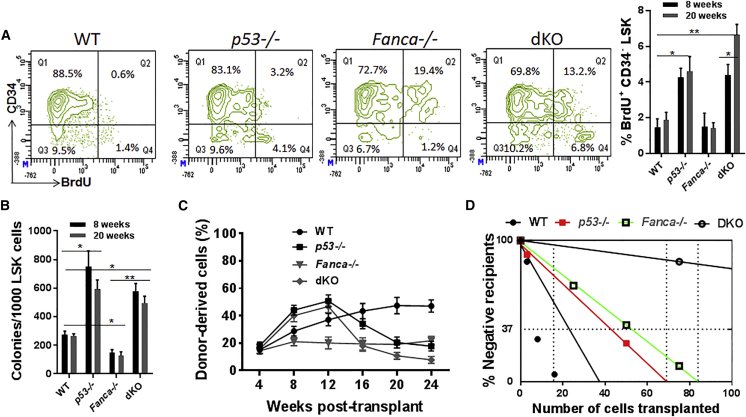
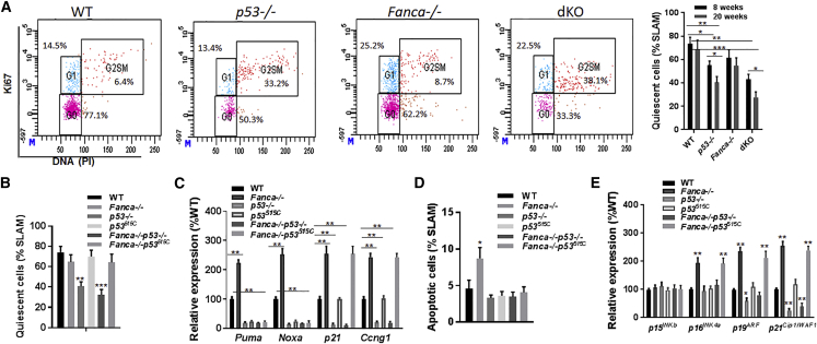
Overactive p53 has been proposed as an important pathophysiological factor for bone marrow failure syndromes, including Fanconi anemia (FA). Here, we report a p53-dependent effect on hematopoietic stem and progenitor cell (HSPC) proliferation in mice deficient for the FA gene Fanca. Deletion of p53 in Fanca-/- mice leads to replicative exhaustion of the hematopoietic stem cell (HSC) in transplant recipients. Using Fanca-/- HSCs expressing the separation-of-function mutant p53515C transgene, which selectively impairs the p53 function in apoptosis but keeps its cell-cycle checkpoint activities intact, we show that the p53 cell-cycle function is specifically required for the regulation of Fanca-/- HSC proliferation. Our results demonstrate that p53 plays a compensatory role in preventing FA HSCs from replicative exhaustion and suggest a cautious approach to manipulating p53 signaling as a therapeutic utility in FA.

Keywords: Fanconi anemia; apoptosis; bone marrow failure; cell cycle; hematopoietic stem and progenitor cells; p53; proliferation.


Figures

Figure 1
Figure 1
Loss of p53 in Fanca−/− Mice Leads to Increased HSPC Pool but Progressive Decline of HSC Reservoir (A) Elevated p53 protein level in Fanca−/− HSPCs. BM LSK (LinSCA-1+C-KIT+) cells were isolated from mice with the indicated genotype, and cell lysates were subjected to immunoblot analysis using antibodies specific for total p53, phosphor-p53 (P-p53), or β-actin. The relative levels of total p53 or of P-p53 to β-actin are indicated below the blot. Each lane contains proteins from ∼30,000 LSK cells. (B) Immunostaining of p53 protein in phenotypic HSCs. Freshly isolated CD34 LSK cells from mice with the indicated genotype were immunostained to detect p53 (green). Nuclei were visualized using DAPI (blue). Scale bars, 10 μm. (C) Progressive decrease of HSPCs in p53-deficient Fanca−/− mice. Whole bone marrow cells (WBMCs) isolated from mice with the indicated genotype were subjected to flow cytometric analysis for LSK staining. Representative plots for 8 weeks (left) and quantification for both 8 and 20 weeks (right) are shown. Results are means ± SD of three independent experiments (n = 9 per group). (D) Progressive decrease of HSCs in p53-deficient Fanca−/− mice. WBMCs isolated from mice with the indicated genotype were subjected to flow cytometric analysis for SLAM (LSK CD150+CD48) staining. Representative plots for 20 weeks (left) and quantification for both 8 and 20 weeks (right) are shown. Results are means ± SD of three independent experiments (n = 9 per group). p < 0.05; ∗∗p < 0.01; ∗∗∗p < 0.001.
Figure 2
Figure 2
p53 Deficiency Leads to Proliferative Exhaustion of Fanca−/− HSCs (A) p53 deficiency decreases Fanca−/− HSC quiescence. BM cells from mice with the indicated genotype at 8 and 20 weeks of age were gated for CD34 LSK population and analyzed for BrdU incorporation. Representative plots for 20 weeks (left) and quantification for both 8 and 20 weeks (right) are shown. Results are means ± SD of two independent experiments (n = 6 per group). (B) Loss of p53 promotes Fanca−/− HSPC proliferation. LSK cells isolated from mice with the indicated genotype at 8 and 20 weeks of age were plated in cytokine-supplemented methylcellulose medium. Colonies were enumerated on day 7 after plating. Results are means ± SD of three independent experiments (n = 9 per group). (C) Loss of p53 causes progressive decline in repopulating potential of Fanca−/− HSCs. 50 SLAM cells from mice with the indicated genotype (CD45.2) at 20 weeks of age, along with 4 × 105 protector BM cells (CD45.1), were transplanted into lethally irradiated Boy J recipients. Donor-derived chimera was detected by flow cytometry at the indicated weeks post-transplant. (D) Competitive repopulating units (CRUs) determined by limiting dilution BM transplantation assay. Graded numbers of test SLAM cells (CD45.2+) from mice with the indicated genotype at 20 weeks of age were mixed with 4 × 105 protector BM cells (CD45.1) and transplanted into irradiated congenic recipients (CD45.1+). Plotted are the percentages of recipient mice containing less than 1% CD45.2+ blood nucleated cells at 16 weeks after transplantation. The frequency of functional HSCs was calculated according to Poisson statistics. p < 0.05; ∗∗p < 0.01.
Figure 3
Figure 3
The Cell-Cycle Regulatory Activity of p53 Is Required for Preventing Fanca−/− HSC Exhaustion (A) Cell-cycle analysis of SLAM cells. BM cells from mice with the indicated genotype at 8 and 20 weeks of age were gated for the SLAM population, and analyzed for cell-cycle phases by flow cytometry. Representative dot plots for 20 weeks (left) and quantification of quiescent (G0) cells within the SLAM population for both 8 and 20 weeks (right) are shown. Results are means ± SD of three independent experiments (n = 9 per group). (B) Quiescence is not compromised in Fanca−/− SLAM cells expressing the p53515C/515C mutation. BM cells from mice with the indicated genotype at 20 weeks of age were gated for the SLAM population, and analyzed for cell-cycle phases by flow cytometry. Quantification of quiescent (G0) cells within the SLAM population is shown. Results are means ± SD of three independent experiments (n = 9 per group). (C) qRT-PCR analysis of the expression of apoptosis and cell-cycle genes. SLAM cells isolated from mice with the indicated genotype at 20 weeks of age were subjected to qPCR analysis using primers for the indicated genes. Levels of the expression in each sample were normalized to the level of GAPDH mRNA, and the expression levels of the WT samples were normalized as 100. (D) The p53515C mutation selectively impairs the p53 function in apoptosis. Cells from mice with the indicated genotype at 20 weeks of age were gated for SLAM population and analyzed for apoptosis by Annexin V and 7AAD. Quantification is shown. Results are means ± SD of three independent experiments (n = 9 per group). (E) qRT-PCR analysis of the expression of negative cell-cycle regulator genes. SLAM cells isolated from mice with the indicated genotype at 20 weeks of age were subjected to qPCR analysis using primers for the indicated genes. Levels of the expression in each sample were normalized to the level of GAPDH mRNA, and the expression levels of the WT samples were normalized as 100. Statistical significance compared with WT. p < 0.05; ∗∗p < 0.01; ∗∗∗p < 0.001.

References

    1. Abbas H.A., Pant V., Lozano G. The ups and downs of p53 regulation in hematopoietic stem cells. Cell Cycle. 2011;10:3257–3262. - PMC - PubMed
    1. Anur P., Yates J., Garbati M.R., Vanderwerf S., Keeble W., Rathbun K. p38 MAPK inhibition suppresses the TLR-hypersensitive phenotype in FANCC- and FANCA-deficient mononuclear phagocytes. Blood. 2012;119:1992–2002. - PMC - PubMed
    1. Bagby G.C., Jr. Genetic basis of Fanconi anemia. Curr. Opin. Hematol. 2003;10:68–76. - PubMed
    1. Bartek J., Lukas J. Mammalian G1- and S-phase checkpoints in response to DNA damage. Curr. Opin. Cell Biol. 2001;13:738–747. - PubMed
    1. Bondar T., Medzhitov R. p53-Mediated hematopoietic stem and progenitor cell competition. Cell Stem Cell. 2010;6:309–322. - PMC - PubMed

Publication types

MeSH terms

Substances