

Decreased glutathione levels cause overt motor neuron degeneration in hSOD1WT over-expressing mice
- PMID: 29307609
- PMCID: PMC5849514
- DOI: 10.1016/j.expneurol.2018.01.004
Decreased glutathione levels cause overt motor neuron degeneration in hSOD1WT over-expressing mice
Abstract
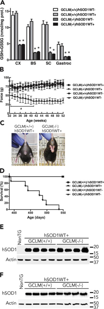
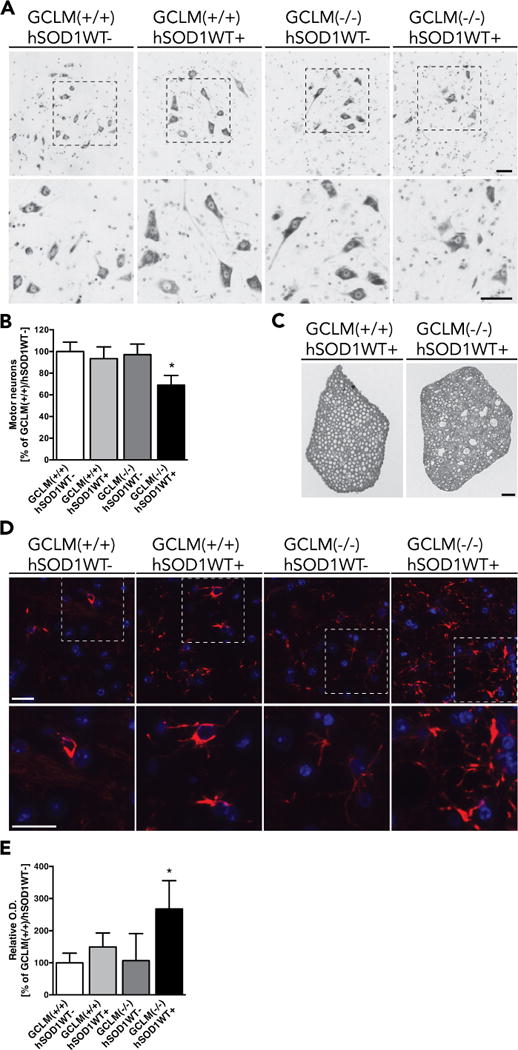
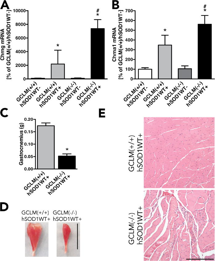
Mutations in Cu/Zn-superoxide dismutase (SOD1) cause familial forms of amyotrophic lateral sclerosis (ALS), a fatal disorder characterized by the progressive loss of motor neurons. Several lines of evidence have shown that SOD1 mutations cause ALS through a gain of a toxic function that remains to be fully characterized. A significant share of our understanding of the mechanisms underlying the neurodegenerative process in ALS comes from the study of rodents over-expressing ALS-linked mutant hSOD1. These mutant hSOD1 models develop an ALS-like phenotype. On the other hand, hemizygous mice over-expressing wild-type hSOD1 at moderate levels (hSOD1WT, originally described as line N1029) do not develop paralysis or shortened life-span. To investigate if a decrease in antioxidant defenses could lead to the development of an ALS-like phenotype in hSOD1WT mice, we used knockout mice for the glutamate-cysteine ligase modifier subunit [GCLM(-/-)]. GCLM(-/-) mice are viable and fertile but display a 70-80% reduction in total glutathione levels. GCLM(-/-)/hSOD1WT mice developed overt motor symptoms (e.g. tremor, loss of extension reflex in hind-limbs, decreased grip strength and paralysis) characteristic of mice models over-expressing ALS-linked mutant hSOD1. In addition, GCLM(-/-)/hSOD1WT animals displayed shortened life span. An accelerated decrease in the number of large neurons in the ventral horn of the spinal cord and degeneration of spinal root axons was observed in symptomatic GCLM(-/-)/hSOD1WT mice when compared to age-matched GCLM(+/+)/hSOD1WT mice. Our results show that under conditions of chronic decrease in glutathione, moderate over-expression of wild-type SOD1 leads to overt motor neuron degeneration, which is similar to that induced by ALS-linked mutant hSOD1 over-expression.
Keywords: Amyotrophic lateral sclerosis; GCLM; Glutathione; SOD1.
Copyright © 2018 Elsevier Inc. All rights reserved.
Figures
Similar articles
-
Decreased glutathione accelerates neurological deficit and mitochondrial pathology in familial ALS-linked hSOD1(G93A) mice model.Neurobiol Dis. 2011 Sep;43(3):543-51. doi: 10.1016/j.nbd.2011.04.025. Epub 2011 May 6. Neurobiol Dis. 2011. PMID: 21600285 Free PMC article.
-
Human Cu/Zn superoxide dismutase (SOD1) overexpression in mice causes mitochondrial vacuolization, axonal degeneration, and premature motoneuron death and accelerates motoneuron disease in mice expressing a familial amyotrophic lateral sclerosis mutant SOD1.Neurobiol Dis. 2000 Dec;7(6 Pt B):623-43. doi: 10.1006/nbdi.2000.0299. Neurobiol Dis. 2000. PMID: 11114261
-
Nuclear localization of human SOD1 and mutant SOD1-specific disruption of survival motor neuron protein complex in transgenic amyotrophic lateral sclerosis mice.J Neuropathol Exp Neurol. 2012 Feb;71(2):162-77. doi: 10.1097/NEN.0b013e318244b635. J Neuropathol Exp Neurol. 2012. PMID: 22249462 Free PMC article.
-
Transgenic mice with human mutant genes causing Parkinson's disease and amyotrophic lateral sclerosis provide common insight into mechanisms of motor neuron selective vulnerability to degeneration.Rev Neurosci. 2007;18(2):115-36. doi: 10.1515/revneuro.2007.18.2.115. Rev Neurosci. 2007. PMID: 17593875 Review.
-
SOD1 in ALS: Taking Stock in Pathogenic Mechanisms and the Role of Glial and Muscle Cells.Antioxidants (Basel). 2022 Mar 23;11(4):614. doi: 10.3390/antiox11040614. Antioxidants (Basel). 2022. PMID: 35453299 Free PMC article. Review.
Cited by
-
Animal Models of Tremor: Relevance to Human Tremor Disorders.Tremor Other Hyperkinet Mov (N Y). 2018 Oct 9;8:587. doi: 10.7916/D89S37MV. eCollection 2018. Tremor Other Hyperkinet Mov (N Y). 2018. PMID: 30402338 Free PMC article. Review.
-
From imbalance to impairment: the central role of reactive oxygen species in oxidative stress-induced disorders and therapeutic exploration.Front Pharmacol. 2023 Oct 18;14:1269581. doi: 10.3389/fphar.2023.1269581. eCollection 2023. Front Pharmacol. 2023. PMID: 37927596 Free PMC article. Review.
-
Emerging Ferroptosis Involvement in Amyotrophic Lateral Sclerosis Pathogenesis: Neuroprotective Activity of Polyphenols.Molecules. 2025 Mar 8;30(6):1211. doi: 10.3390/molecules30061211. Molecules. 2025. PMID: 40141987 Free PMC article. Review.
-
Does wild-type Cu/Zn-superoxide dismutase have pathogenic roles in amyotrophic lateral sclerosis?Transl Neurodegener. 2020 Aug 19;9(1):33. doi: 10.1186/s40035-020-00209-y. Transl Neurodegener. 2020. PMID: 32811540 Free PMC article. Review.
-
Nicotinamide Riboside and Pterostilbene Cooperatively Delay Motor Neuron Failure in ALS SOD1G93A Mice.Mol Neurobiol. 2021 Apr;58(4):1345-1371. doi: 10.1007/s12035-020-02188-7. Epub 2020 Nov 10. Mol Neurobiol. 2021. PMID: 33174130
References
-
- Al-Chalabi A, Jones A, Troakes C, King A, Al-Sarraj S, van den Berg LH. The genetics and neuropathology of amyotrophic lateral sclerosis. Acta neuropathologica. 2012;124:339–352. - PubMed
-
- Andrus PK, Fleck TJ, Gurney ME, Hall ED. Protein oxidative damage in a transgenic mouse model of familial amyotrophic lateral sclerosis. Journal of neurochemistry. 1998;71:2041–2048. - PubMed
-
- Bruijn LI, Beal MF, Becher MW, Schulz JB, Wong PC, Price DL, Cleveland DW. Elevated free nitrotyrosine levels, but not protein-bound nitrotyrosine or hydroxyl radicals, throughout amyotrophic lateral sclerosis (ALS)-like disease implicate tyrosine nitration as an aberrant in vivo property of one familial ALS-linked superoxide dismutase 1 mutant. Proceedings of the National Academy of Sciences of the United States of America. 1997;94:7606–7611. - PMC - PubMed
Publication types
MeSH terms
Substances
Grants and funding
LinkOut - more resources
Full Text Sources
Other Literature Sources
Molecular Biology Databases
Research Materials
Miscellaneous
