

Variant BMP receptor mutations causing fibrodysplasia ossificans progressiva (FOP) in humans show BMP ligand-independent receptor activation in zebrafish
- PMID: 29307777
- PMCID: PMC5866198
- DOI: 10.1016/j.bone.2018.01.002
Variant BMP receptor mutations causing fibrodysplasia ossificans progressiva (FOP) in humans show BMP ligand-independent receptor activation in zebrafish
Abstract
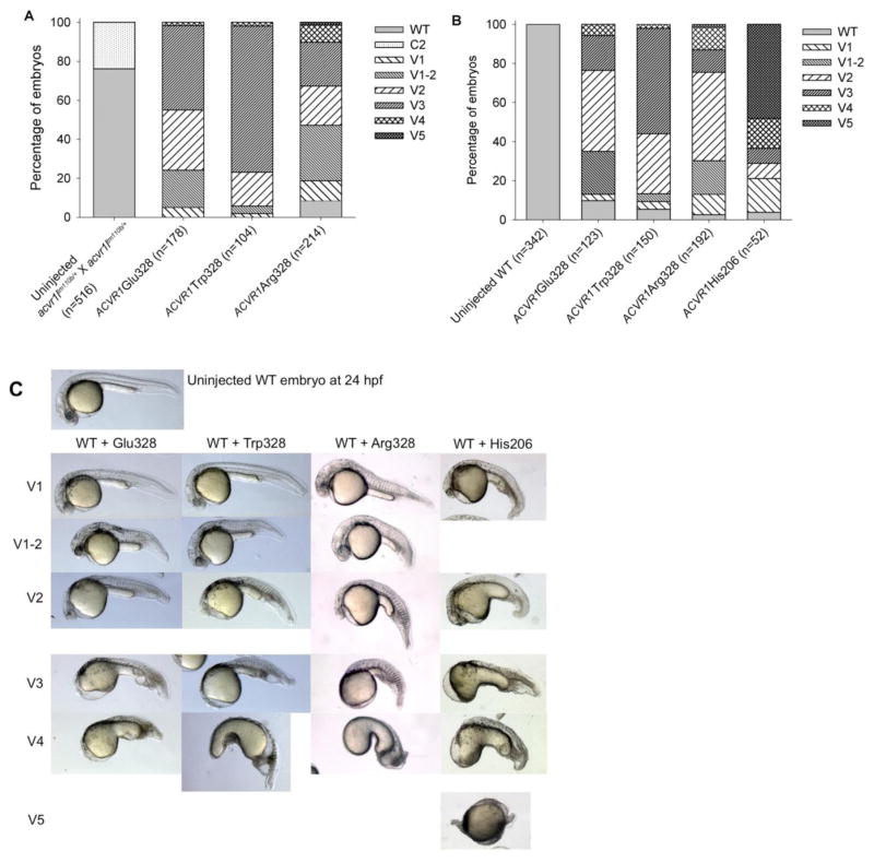
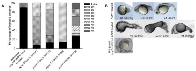
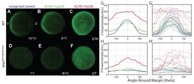
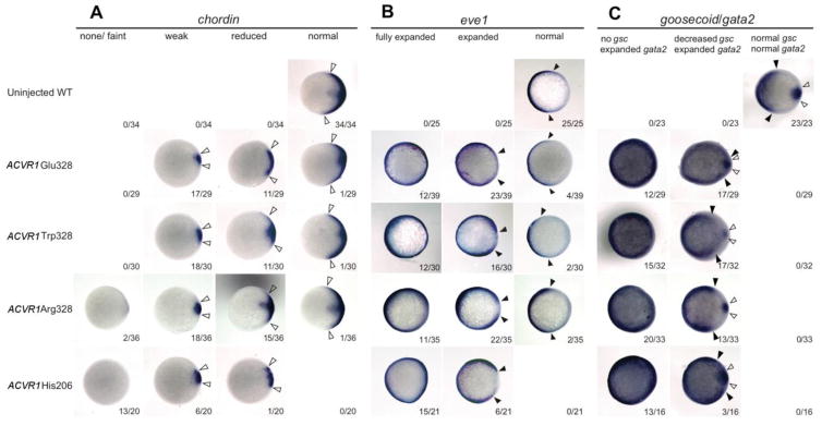
The large majority of cases of the autosomal dominant human disease fibrodysplasia ossificans progressiva (FOP) are caused by gain-of-function Arg206His mutations in the BMP type I receptor ACVR1 (ALK2). The Arg206His mutation is located in the GS domain of the type I receptor. This region is normally phosphorylated by the BMP type II receptor, which activates the type I receptor to phosphorylate its substrate, the signal transducer Smad1/5/8. A small subset of patients with FOP carry variant mutations in ACVR1 altering Gly328 to Trp, Glu or Arg. Since these mutations lie outside the GS domain, the mechanism through which ACVR1 Gly328 mutations cause disease remains unclear. We used a zebrafish embryonic development assay to test the signaling of human ACVR1 Gly328 mutant receptors comparing them to the Arg206His mutant. In this assay increased or decreased BMP pathway activation alters dorsal-ventral axial patterning, providing a sensitive assay for altered BMP signaling levels. We expressed the human ACVR1 Gly328 mutant receptors in zebrafish embryos to investigate their signaling activities. We found that all ACVR1 Gly328 human mutations ventralized wild-type embryos and could partially rescue Bmp7-deficient embryos, indicating that these mutant receptors can activate BMP signaling in a BMP ligand-independent manner. The degree of ventralization or rescue was similar among all three Gly328 mutants. Smad1/5 phosphorylation, a readout of BMP receptor signaling, was mildly increased by ACVR1 Gly328 mutations. Gene expression analyses demonstrate expanded ventral and reciprocal loss of dorsal cell fate markers. This study demonstrates that Gly328 mutants increase receptor activation and BMP ligand-independent signaling through Smad phosphorylation.
Keywords: ACVR1; BMP signaling; Dorsal-ventral embryonic patterning; FOP; Smad1/5; Zebrafish.
Copyright © 2018 Elsevier Inc. All rights reserved.
Conflict of interest statement
COMPETING INTERESTS
The authors declare that they do not have any competing or financial interests.
Figures
Similar articles
-
Reduced GS Domain Serine/Threonine Requirements of Fibrodysplasia Ossificans Progressiva Mutant Type I BMP Receptor ACVR1 in the Zebrafish.J Bone Miner Res. 2023 Sep;38(9):1364-1385. doi: 10.1002/jbmr.4869. Epub 2023 Jul 17. J Bone Miner Res. 2023. PMID: 37329499 Free PMC article.
-
Fibrodysplasia ossificans progressiva mutant ACVR1 signals by multiple modalities in the developing zebrafish.Elife. 2020 Sep 8;9:e53761. doi: 10.7554/eLife.53761. Elife. 2020. PMID: 32897189 Free PMC article.
-
Functional Testing of Bone Morphogenetic Protein (BMP) Pathway Variants Identified on Whole-Exome Sequencing in a Patient with Delayed-Onset Fibrodysplasia Ossificans Progressiva (FOP) Using ACVR1R206H -Specific Human Cellular and Zebrafish Models.J Bone Miner Res. 2022 Nov;37(11):2058-2076. doi: 10.1002/jbmr.4711. Epub 2022 Nov 15. J Bone Miner Res. 2022. PMID: 36153796 Free PMC article.
-
The obligatory role of Activin A in the formation of heterotopic bone in Fibrodysplasia Ossificans Progressiva.Bone. 2018 Apr;109:210-217. doi: 10.1016/j.bone.2017.06.011. Epub 2017 Jun 16. Bone. 2018. PMID: 28629737 Free PMC article. Review.
-
Heterotopic bone induction via BMP signaling: Potential therapeutic targets for fibrodysplasia ossificans progressiva.Bone. 2018 Apr;109:241-250. doi: 10.1016/j.bone.2017.07.024. Epub 2017 Jul 25. Bone. 2018. PMID: 28754575 Review.
Cited by
-
Direct BMP signaling to chordoblasts is required for the initiation of segmented notochord sheath mineralization in zebrafish vertebral column development.Front Endocrinol (Lausanne). 2023 May 8;14:1107339. doi: 10.3389/fendo.2023.1107339. eCollection 2023. Front Endocrinol (Lausanne). 2023. PMID: 37223044 Free PMC article.
-
Zebrafish Models for Human Skeletal Disorders.Front Genet. 2021 Aug 5;12:675331. doi: 10.3389/fgene.2021.675331. eCollection 2021. Front Genet. 2021. PMID: 34490030 Free PMC article. Review.
-
Reduced GS Domain Serine/Threonine Requirements of Fibrodysplasia Ossificans Progressiva Mutant Type I BMP Receptor ACVR1 in the Zebrafish.J Bone Miner Res. 2023 Sep;38(9):1364-1385. doi: 10.1002/jbmr.4869. Epub 2023 Jul 17. J Bone Miner Res. 2023. PMID: 37329499 Free PMC article.
-
BMP2 and BMP7 cooperate with H3.3K27M to promote quiescence and invasiveness in pediatric diffuse midline gliomas.Elife. 2024 Oct 7;12:RP91313. doi: 10.7554/eLife.91313. Elife. 2024. PMID: 39373720 Free PMC article.
-
Pathogenic ACVR1R206H activation by Activin A-induced receptor clustering and autophosphorylation.EMBO J. 2021 Jul 15;40(14):e106317. doi: 10.15252/embj.2020106317. Epub 2021 May 18. EMBO J. 2021. PMID: 34003511 Free PMC article.
References
-
- Bragdon B, Moseychuk O, Saldanha S, King D, Julian J, Nohe A. Bone morphogenetic proteins: a critical review. Cell Signaling. 2011;23(4):609–620. - PubMed
-
- Carvalho DR, Navarro MMM, Martins BJAF, Coelho KEFA, Mello WD, Takata RI, Speck-Martins CE. Mutational screening of ACVR1 gene in Brazilian fibrodysplasia ossificans progressiva patients. Clinical genetics. 2010;77(2):171–176. - PubMed
Publication types
MeSH terms
Substances
Grants and funding
LinkOut - more resources
Full Text Sources
Other Literature Sources
Molecular Biology Databases
