. 2018 Feb 13;57(6):945-962.
doi: 10.1021/acs.biochem.7b01102. Epub 2018 Jan 30.

Structural and Kinetic Studies of Asp632 Mutants and Fully Reduced NADPH-Cytochrome P450 Oxidoreductase Define the Role of Asp632 Loop Dynamics in the Control of NADPH Binding and Hydride Transfer

Affiliations

Structural and Kinetic Studies of Asp632 Mutants and Fully Reduced NADPH-Cytochrome P450 Oxidoreductase Define the Role of Asp632 Loop Dynamics in the Control of NADPH Binding and Hydride Transfer

Chuanwu Xia et al. Biochemistry. .

Abstract

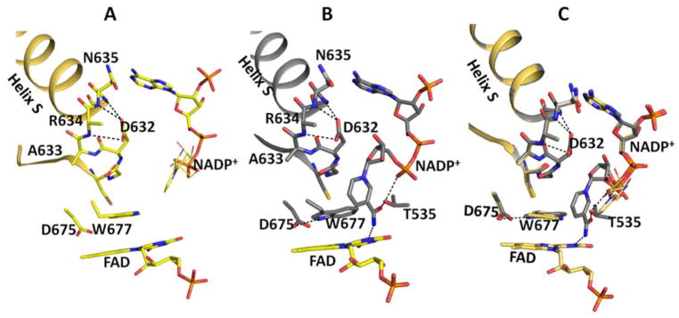
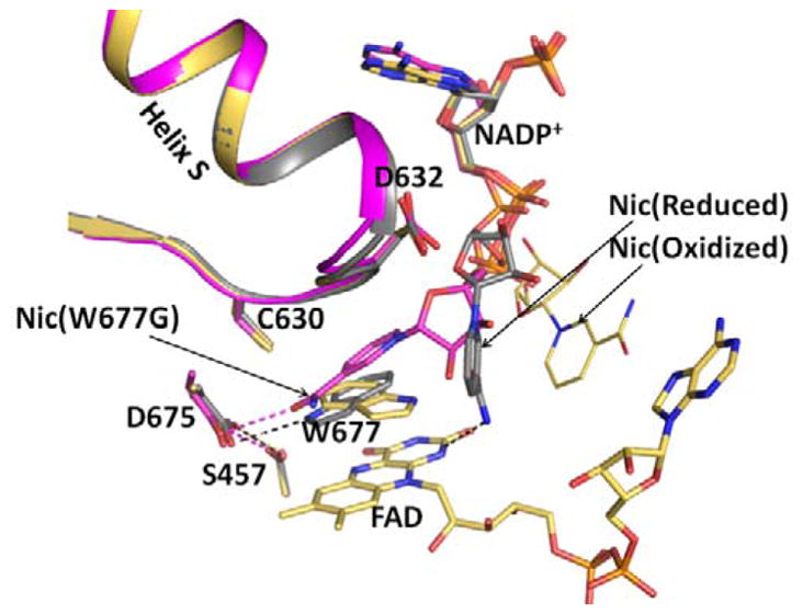
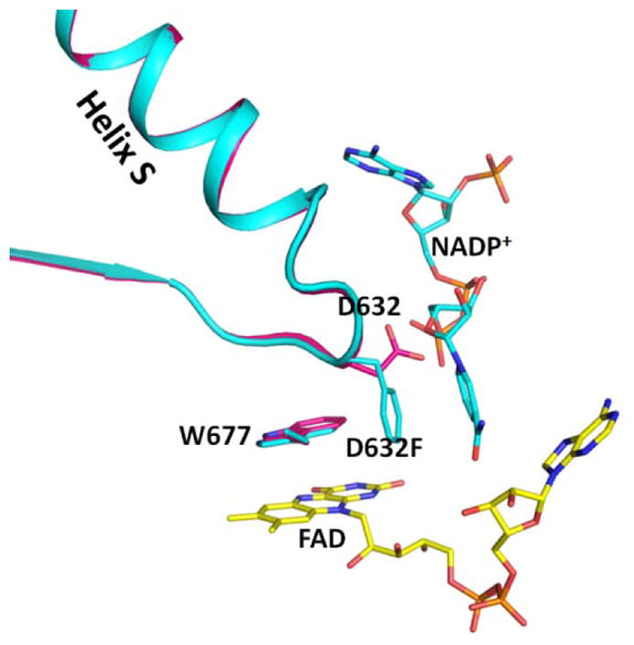
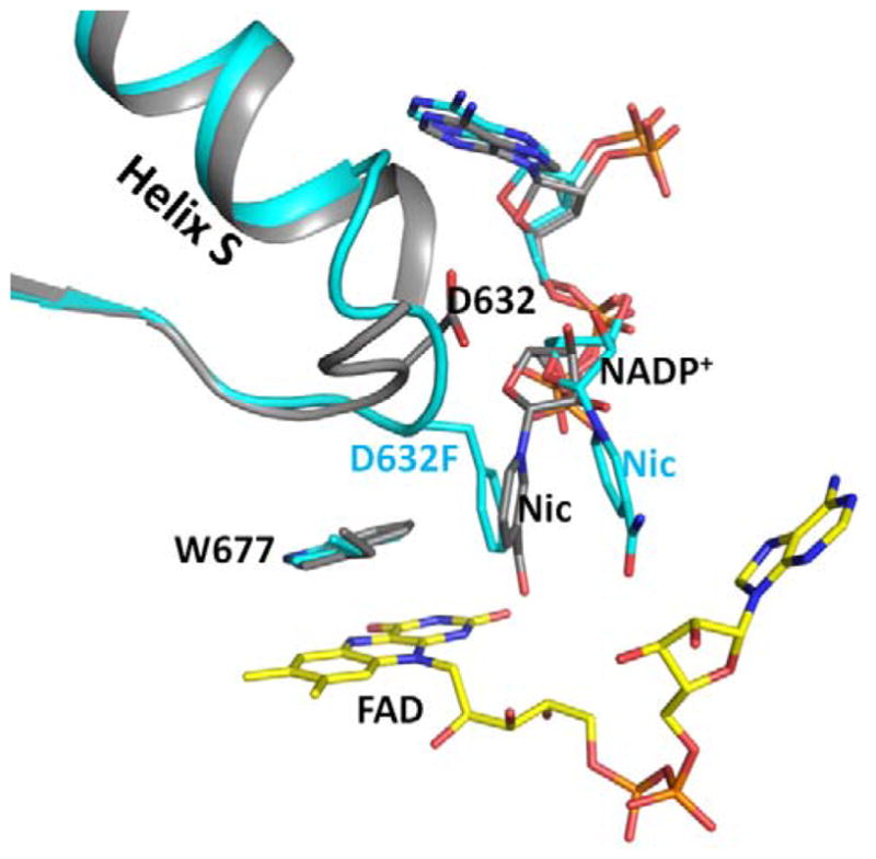
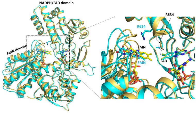
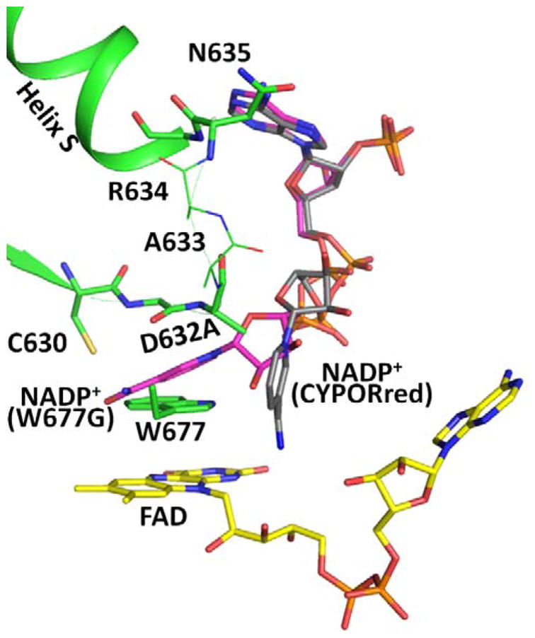
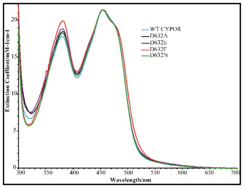
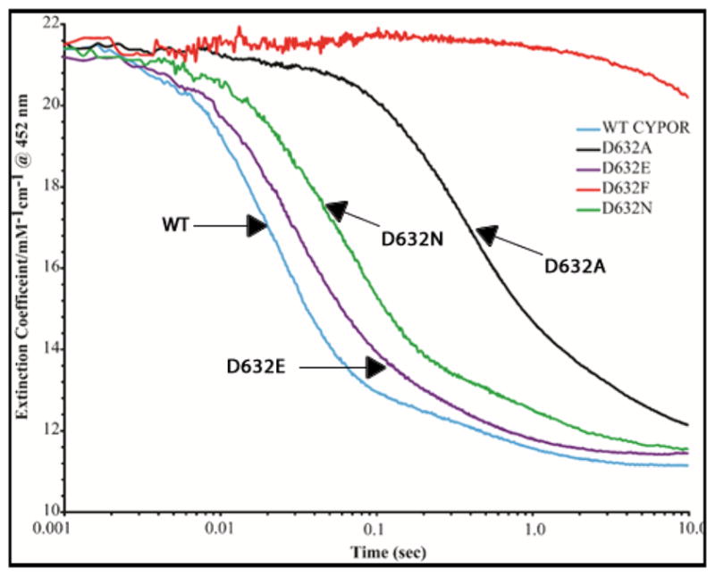
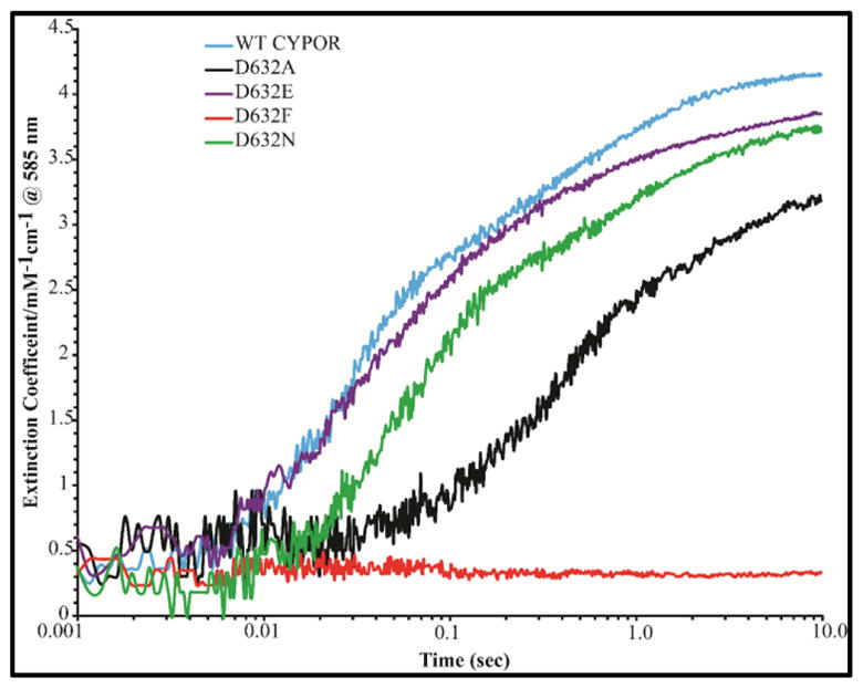
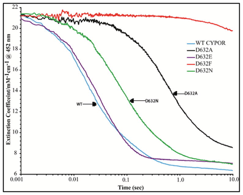
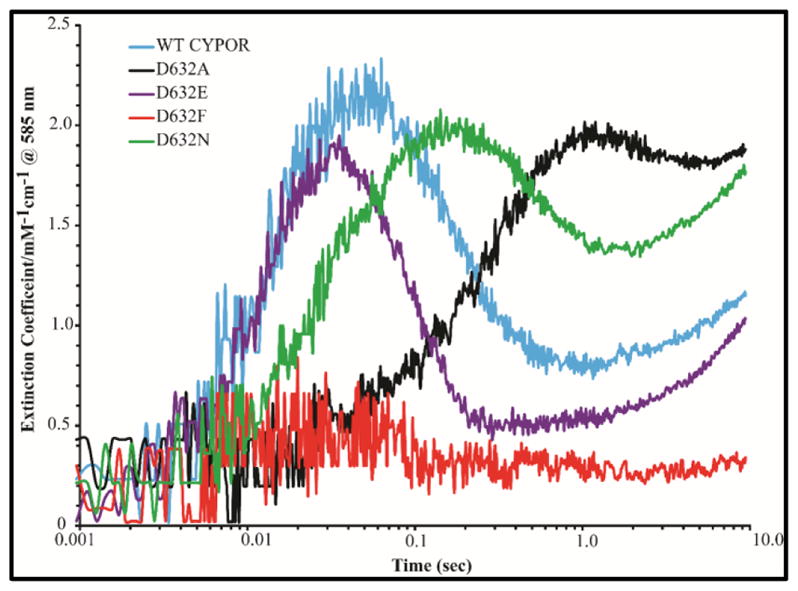
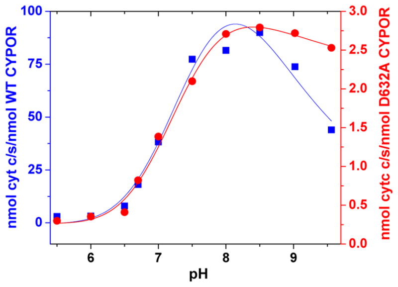
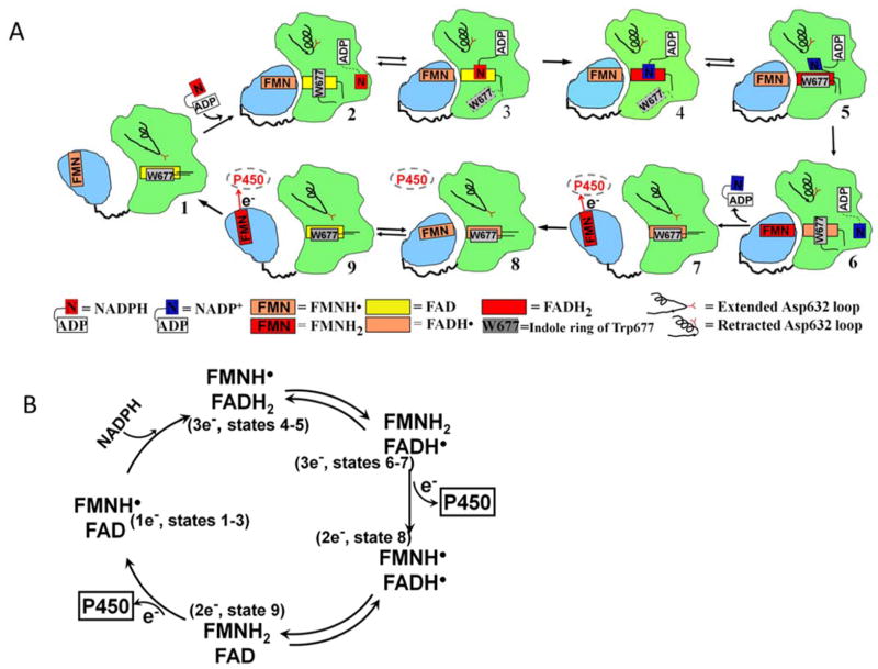
Conformational changes in NADPH-cytochrome P450 oxidoreductase (CYPOR) associated with electron transfer from NADPH to electron acceptors via FAD and FMN have been investigated via structural studies of the four-electron-reduced NADP+-bound enzyme and kinetic and structural studies of mutants that affect the conformation of the mobile Gly631-Asn635 loop (Asp632 loop). The structure of four-electron-reduced, NADP+-bound wild type CYPOR shows the plane of the nicotinamide ring positioned perpendicular to the FAD isoalloxazine with its carboxamide group forming H-bonds with N1 of the flavin ring and the Thr535 hydroxyl group. In the reduced enzyme, the C8-C8 atoms of the two flavin rings are ∼1 Å closer than in the fully oxidized and one-electron-reduced structures, which suggests that flavin reduction facilitates interflavin electron transfer. Structural and kinetic studies of mutants Asp632Ala, Asp632Phe, Asp632Asn, and Asp632Glu demonstrate that the carboxyl group of Asp632 is important for stabilizing the Asp632 loop in a retracted position that is required for the binding of the NADPH ribityl-nicotinamide in a hydride-transfer-competent conformation. Structures of the mutants and reduced wild type CYPOR permit us to identify a possible pathway for NADP(H) binding to and release from CYPOR. Asp632 mutants unable to form stable H-bonds with the backbone amides of Arg634, Asn635, and Met636 exhibit decreased catalytic activity and severely impaired hydride transfer from NADPH to FAD, but leave interflavin electron transfer intact. Intriguingly, the Arg634Ala mutation slightly increases the cytochrome P450 2B4 activity. We propose that Asp632 loop movement, in addition to facilitating NADP(H) binding and release, participates in domain movements modulating interflavin electron transfer.


Conflict of interest statement

Notes

The authors declare no competing financial interests.

Figures

Figure 1
Figure 1
Structures of oxidized and reduced wild type CYPOR in complex with NADP+ in the vicinity of the Asp632 loop and FAD. Oxidized (A, gold), reduced (B, grey), and overlay (C). The notable differences in the FAD domain are the conformations of Trp677 and the nicotinamide moiety of NADP+. In the oxidized structure, the planes of the Trp677 indole ring and the FAD ring are stacking onto each other, but not completely overlapping, and the long axes of their planes are perpendicular. In the reduced structure, the two planes also stack and overlap, and their long axes are parallel. In the oxidized structure, the ribose-nicotinamide moiety is disordered (possible location shown with thin sticks, taken from the structure of the oxidized triple mutant structure, Ser457Ala/Cys630Ala/Asp675Asn (1JA1). In the reduced structure, the nicotinamide is well defined and the carboxamide group of NADP+ forms two H-bonds (dotted lines), the amide group with the negatively charged N1 of the FAD isoalloxazine ring and the carbonyl group with the hydroxyl of the Thr535 side chain, which in turn makes an H-bond with a pyrophosphate oxygen. In the reduced structure, the plane of the nicotinamide ring is perpendicular to both the Trp677 indole ring and the FAD ring, both of which are stacked on each other.
Figure 2
Figure 2
Structures of the oxidized triple mutant (Ser457Ala/Cys630Ala/Asp675Asn) CYPOR, (1JA1) (gold), dithionite-reduced CYPOR (grey), and oxidized Trp677Gly mutant (1J9Z, magenta) were superimposed. They reveal three conformations of the ribityl nicotinamide moiety of NADP+. In the oxidized wild type structure, the indole ring of Trp677 stacks onto the FAD ring and the ribityl nicotinamide (Nic-ox) is flexible (estimated occupancy, ~<50%) with no defined interactions with FAD and Trp677. In the reduced structure, the indole ring also stacks onto the flavin with the long axes of both rings being parallel. There is an H-bond between the Nε1 of Trp677 and Asp675 side chain. In addition, the carboxamide of the ribityl nicotinamide ring (Nic-reduced) forms hydrogen bonds with the negative N1 of the flavin and the Thr535 side chain hydroxyl. The face of the nicotinamide ring interacts with the edge of the Trp677 indole ring. In the Trp677Gly structure, the nicotinamide ring (Nic-W677G) stacks against the re-face of the isoalloxazine ring and is poised for hydride transfer. The three different ribityl-nicotinamide ring conformations may represent the nicotinamide binding pathway into the active site for hydride transfer, after securely anchoring the 2′, 5′-ADP half of NADP+. For clarity, only FAD of the oxidized structure is shown.
Figure 3
Figure 3
Comparison of the structures of the crosslinked NADP+-free structure (3OJW, magenta) and the Asp632Phe mutant structure (cyan) by superimposing the FAD-binding domains. The Trp indole ring conformations in the two structures are the same. The Asp632 loop conformations in the two structures are almost identical, except for the side chains of Asp632 and Phe632. In the Asp632Phe structure, the phenyl side chain is oriented parallel to the nicotinamide ring but both are perpendicular to the indole ring of Trp677 and the flavin ring.
Figure 4
Figure 4
Superimposed structures of the reduced wild type (grey) and Asp632Phe mutant (cyan). The Asp632 loop conformations are different. The loop adopts the tight, retracted conformation in the reduced wild type structure, and a relaxed extended conformation in the Asp632Phe mutant structure. In the Asp632Phe structure, the side chain of Phe632 occupies the position of the nicotinamide ring of the reduced structure, and is almost perpendicular to the Trp677 indole ring in an edge to face conformation. The nicotinamide ring is essentially parallel to the phenyl ring in the Asp632Phe structure.
Figure 5
Figure 5
Demonstration of the 2 Å-greater separation of the FAD and FMN domains of the Asp632Phe structure compared to wild type. Superposition of the structures of oxidized wild type (gold) and oxidized Asp632Phe mutant (cyan). The FAD/NADPH-binding domains (residues Arg243-Ser678) were superimposed. Black labels indicate the wild type and cyan labels indicate the Asp632Phe mutant. Arg634 of the wild type structure is shown as black thin sticks and the position of Arg634 in the Asp632Phe mutant structure is shown as thick cyan sticks for carbon atoms. Right panel: enlarged view of the rectangular dashed area of the left panel. In the Asp632Phe structure the FMN has moved about 2 Å from FAD compared to that observed in the oxidized wild type.
Figure 6
Figure 6
Comparison of the structures of the Asp632Ala mutant with bound NADP+ (green; for clarity, no NADP+ is shown), Trp677Gly (pdb code, 1J9Z, magenta), and reduced wild type CYPOR (grey). For clarity, only the Asp632 loop in the Asp632Ala structure (green) is shown. Nicotinamide rings in the Trp677Gly structure [magenta carbons, NADP+ (W677G)] and in the reduced wild type structure [(grey carbons, NADP+ (CYPORred)] are shown. Ala633 and Arg634 are disordered and their possible positions are shown as thin sticks.
Figure 7
Figure 7
UV visible absorption spectra of oxidized wild type and Asp632 mutants of CYPOR.
Figure 8
Figure 8
Kinetics traces at 452 nm demonstrating the reduction of the FAD of wild type and Asp632 mutants of CYPOR by 1 molar equivalent of NADPH under anaerobic conditions at 25°C.
Figure 9
Figure 9
Kinetic traces at 585 nm demonstrating the rate of formation of the blue semiquinones of wild type and Asp632 mutants of CYPOR by 1 molar equivalent of NADPH under anaerobic conditions at 25°C.
Figure 10
Figure 10
Kinetic traces at 452 nm demonstrating the reduction of the FAD of wild type and the Asp632 mutants of CYPOR by 10 molar equivalents of NADPH under anaerobic conditions at 25°C.
Figure 11
Figure 11
Kinetic traces at 585 nm demonstrating the rate of formation and decay of the blue semiquinones of wild type and the Asp632 mutants of CYPOR by 10 molar equivalents of NADPH under anaerobic conditions at 25°C.
Figure 12
Figure 12
Comparison of the effect of pH on the cytochrome c activity of wild type (blue) and Asp632Ala (red) CYPOR at 30°C.
Figure 13
Figure 13
A) Conformational changes and the cofactor binding during the catalytic cycle of CYPOR - The FMN domain is in light blue and FAD domain in green. The symbols and redox states of the cofactors are shown in the figure, together with the conformations of the Asp632 loop. See text for detailed description of the catalytic cycle involving States 1–9. B) Redox cycling of CYPOR flavins (1:3:2:1 electron cycling model). The numbers in the parentheses indicate the related states shown in Figure 13A.

References

    1. Iyanagi T, Xia C, Kim JJ. NADPH-cytochrome P450 oxidoreductase: prototypic member of the diflavin reductase family. Arch Biochem Biophys. 2012;528(1):72–89. - PMC - PubMed
    1. Kim JJ, Waskell L. Electron Transfer Partners. In: Ortiz de Montellano PR, editor. Cytochrome P450: Structure, Mechanism and Biochemistry. 4. Springer; Switzerland: 2015. pp. 33–68.
    1. Paine MJ, Scrutton NS, Munro AW, Gutierrez A, Roberts GCK, Wolf CR. Electron Transfer Partners of Cytochrome P450. In: Ortiz de Montellano PR, editor. Cytochrome P450: Structure, Mechanism and Biochemistry. 3. Kluwer Academic/Plenum Publishers; New York, Boston, Dordrecht, London, Moscow: 2005. pp. 115–148.
    1. Ortiz de Montellano PR. Hydrocarbon hydroxylation by cytochrome P450 enzymes. Chem Rev. 2010;110(2):932–48. - PMC - PubMed
    1. Guengerich FP. Human Cytochrome P450 Enzymes. In: Ortiz de Montellano PR, editor. Cytochrome P450: Structure, Mechanism and Biochemistry. 3. Vol. 1. Kluwer Academic/Plenum Publishers; New York, NY: 2005. pp. 377–463.

Publication types

MeSH terms

LinkOut - more resources