. 2018 Mar;14(3):270-275.
doi: 10.1038/nchembio.2549. Epub 2018 Jan 8.

Mechanism of intersubunit ketosynthase-dehydratase interaction in polyketide synthases

Affiliations

Mechanism of intersubunit ketosynthase-dehydratase interaction in polyketide synthases

Matthew Jenner et al. Nat Chem Biol. 2018 Mar.

Abstract

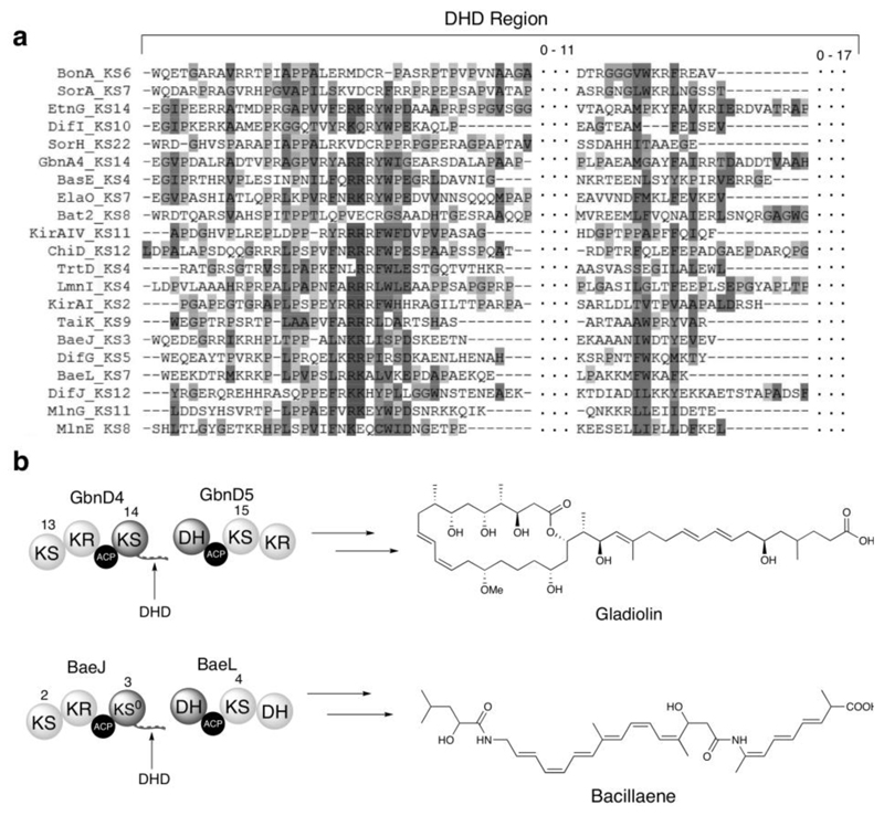
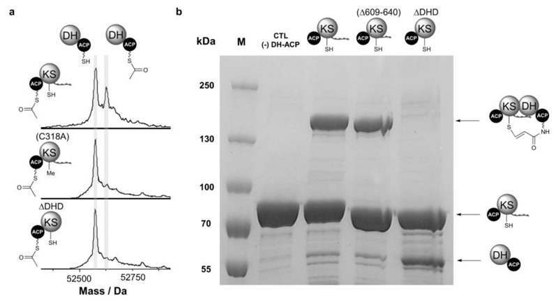
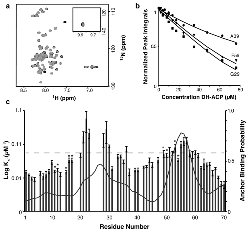
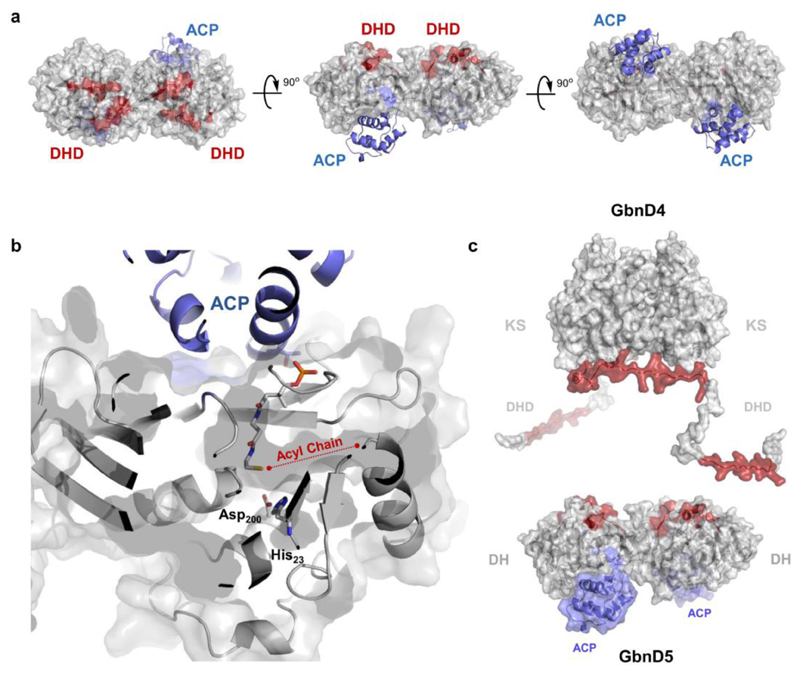
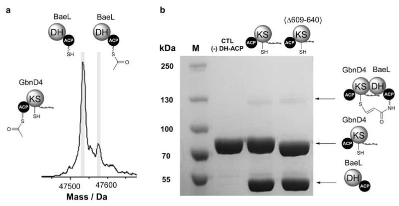
Modular polyketide synthases (PKSs) produce numerous structurally complex natural products that have diverse applications in medicine and agriculture. PKSs typically consist of several multienzyme subunits that utilize structurally defined docking domains (DDs) at their N and C termini to ensure correct assembly into functional multiprotein complexes. Here we report a fundamentally different mechanism for subunit assembly in trans-acyltransferase (trans-AT) modular PKSs at the junction between ketosynthase (KS) and dehydratase (DH) domains. This mechanism involves direct interaction of a largely unstructured docking domain (DD) at the C terminus of the KS with the surface of the downstream DH. Acyl transfer assays and mechanism-based crosslinking established that the DD is required for the KS to communicate with the acyl carrier protein appended to the DH. Two distinct regions for binding of the DD to the DH were identified using NMR spectroscopy, carbene footprinting, and mutagenesis, providing a foundation for future elucidation of the molecular basis for interaction specificity.


Conflict of interest statement

Competeing Financial Interests Statement

The authors declare no competing financial interests.

Figures

Figure 1
Figure 1. Bioinformatics analysis of DHD domains and examples from the gladiolin and bacillaene PKSs.
(a) Multiple sequence alignment of DHD domains identified by manual inspection of KS/DH boundaries in trans-AT PKSs for which the metabolic product is known. Localized conservation is observed in the N-terminal region, but the C-terminal region is poorly conserved. Dotted sections indicate residues between the conserved regions of the DHD domain, and the number of residues located in these regions is indicated above. Sequences from the following pathways were used: Bae, bacillaene (B. amyloliquefaciens); Bas, basiliskamide (B. laterosporus PE36); Bat, batumin/kalimantacin (P. fluorescens); Dfn, difficidin (B. amyloliquefaciens); Etn, etnangien (S. cellulosum So ce56); Ela, elansolid (Chitinophaga pinensis); Gbn, gladiolin (B. gladioli); Kir, kirromycin (S. collinus); Lnm, leinamycin (S. atroolivaceus); Mln, macrolactin (B. amyloliquefaciens); Mmp, mupirocin (Pseudomonas fluorescens); Sor, sorangicin (Sorangium cellulosum); Tai, thailandamide (B. thailandensis E264); Trt, tartrolon (T. turnerae T7901). (b) Examples of DHD domains in the gladiolin (top) and bacillaene (bottom) PKSs and structures of the metabolites produced by these assembly lines.
Figure 2
Figure 2. Acyl transfer and protein crosslinking assays demonstrate that DHD domains play a key role in communication across KS/DH interfaces.
(a) Deconvoluted mass spectra of GbnD5 holo-DH-ACP, following incubation with GbnD4 Ac-ACP-KS-DHD (top), Ac-ACP-KS(C318A)-DHD (middle) and Ac-ACP-KS(ΔDHD) (bottom). Transfer of the acetyl group onto the holo-DH-ACP di-domain results in a +42 Da mass shift. The data show that an acyl group can be transferred across the subunit interface (top), and that the KS active site Cys residue plays an important role in this process (middle). No acetylation of the holo- DH-ACP di-domain is observed when the DHD domain is removed (bottom). (b) SDS-PAGE (6%) analysis of crosslinking reactions between the GbnD5 DH-ACP di-domain loaded with the chloroacrylamide-terminated ppant analogue and the GbnD4 ACP-KS-DHD tri-domain (left), GbnD4 ACP-KS-DHD(Δ609-640) truncated tri-domain (middle) and the ACP-KS(ΔDHD) di-domain (right). Efficient formation of a crosslinked complex (˜130 kDa) is observed for the ACP-KS-DHD tri-domain (left) and GbnD4 ACP-KS-DHD(Δ609-640) truncated tri-domain (middle), but not the ACP-KS(ΔDHD) di-domain (right).
Figure 3
Figure 3. NMR titrations reveal two regions of DHD domains involved in the interaction with DH-ACP di-domains.
(a) Overlaid 2D BEST-TROSY-15N-HSQC spectra of [U-15N]-labeled GbnD4 DHD domain titrated with unlabeled GbnD5 apo-DH-ACP di-domain. The spectrum in black shows the DHD domain alone. The grey spectrum show the DHD domain in a presence of an equivalent amount of the GbnD5 DH-ACP di-domain. As the concentration of the GbnD5 DH-ACP di-domian was increased, several signals were observed to decrease in intensity, suggestive of slow to intermediate exchange with the binding affinity in the lower μM range. (b) Experimental data points and individual fits of normalized signal integrals for selected residues located in the first (Gly29, squares) and second (Phe56, triangles) DH-ACP-interacting regions of the DHD domain, and the region between them (Ala39, circles). (c) LogKa values calculated from signal integral values for individual residues of the DHD domain. Residues with values above the threshold (dotted line – defined in the online methods) are involved in interaction with the DH-ACP di-domain. These data agree well with the binding site probabilities predicted by the ANCHOR web server (solid line). Asterisks indicate residues for which a second set of weaker resonances was observed in the spectra. The error bars correspond to two standard deviations of 1000 iterations of fits using Monte Carlo error analysis.
Figure 4
Figure 4. Mapping DHD and ACP domain interaction sites on the DH domain.
(a) Identification of the interaction sites for the GbnD4 DHD domain and GbnD5 ACP domain on the dimeric GbnD5 DH domain using carbene foot-printing. Regions masked by the DHD and ACP domains are highlighted in red and blue, respectively, on a homology model of GbnD5 DH domain (grey). Residue constraints derived from the foot-printing experiments were used to dock the ACP domain (blue) onto the DH domain. Residues used to constrain the docking calculations are as follows; DH: Leu31, Arg61, Leu298, Val299, Asp300; ACP: Leu394, Ala395, Leu396. The model shows adjacent but distinct binding sites for the DHD and ACP domains. These place the GbnD4 KS domain and the GbnD5 ACP domain in close proximity, facilitating efficient communication across the subunit interface. (b) Predicted location of the ppant arm in the model of the GdnD5 DH-ACP di-domain complex based on the X-ray crystal structure of a crosslinked complex of the ACP and DH from the E. coli fatty acid synthase (PDB accession no: 4KEH). The docking site for the ACP domain is situated near the entrance to the active site of the DH domain. This allows the thiol of the ppant arm to sit adjacent to the catalytic Asp and His residues in the active site of the DH domain. A putative binding pocket for the acyl chain of the substrate sits to the right of the ppant thiol. (c) Overall model for the interaction between the GbnD4 KS-DHD (red) and GbnD5 DH-ACP (grey/blue) di-domains, both of which form homodimers. The red patches on the surface of the DH and DHD domains indicate mutual interaction sites.
Figure 5
Figure 5. DHD domains interact selectively with their cognate DH domain partners.
(a) Deconvoluted mass spectrum of BaeL holo-DH-ACP di-domain following incubation with GbnD4 Ac-ACP-KS-DHD tri-domain. In comparison with the GbnD4 holo-DH-ACP di-domain, only low levels of acetylation can be observed, suggesting that the GbnD4 DHD and BaeL DH domains interact weakly. (b) SDS-PAGE (6%) analysis of the crosslinking reaction between GbnD4 ACP-KS-DHD tri-domain and GbnD4 ACP-KS-DHD(Δ609-640) truncated tri-domain with BaeL DH-ACP di-domain loaded with the β-chloroacrylamido ppant analogue. Only trace amounts of the crosslinked complex (˜130 kDa) can be observed, providing further evidence for a weak interaction between non-cognate DHD and DH domains.

References

    1. Hertweck C. The biosynthetic logic of polyketide diversity. Angew Chem Int Ed Engl. 2009;48:4688–716. - PubMed
    1. Keatinge-Clay AT. The structures of type I polyketide synthases. Nat Prod Rep. 2012;29:1050–1073. - PubMed
    1. Richter CD, Nietlispach D, Broadhurst RW, Weissman KJ. Multienzyme docking in hybrid megasynthetases. Nat Chem Biol. 2008;4:75–81. - PubMed
    1. Weissman KJ, Muller R. Protein-protein interactions in multienzyme megasynthetases. Chembiochem. 2008;9:826–848. - PubMed
    1. Fischbach MA, Walsh CT. Assembly-line enzymology for polyketide and nonribosomal peptide antibiotics: Logic, machinery, and mechanisms. Chem Rev. 2006;106:3468–3496. - PubMed

Publication types

LinkOut - more resources