. 2018 Feb;24(2):239-246.
doi: 10.1038/nm.4470. Epub 2018 Jan 8.

Suppression of luteinizing hormone enhances HSC recovery after hematopoietic injury

Affiliations

Suppression of luteinizing hormone enhances HSC recovery after hematopoietic injury

Enrico Velardi et al. Nat Med. 2018 Feb.

Erratum in

Abstract

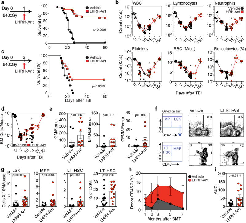
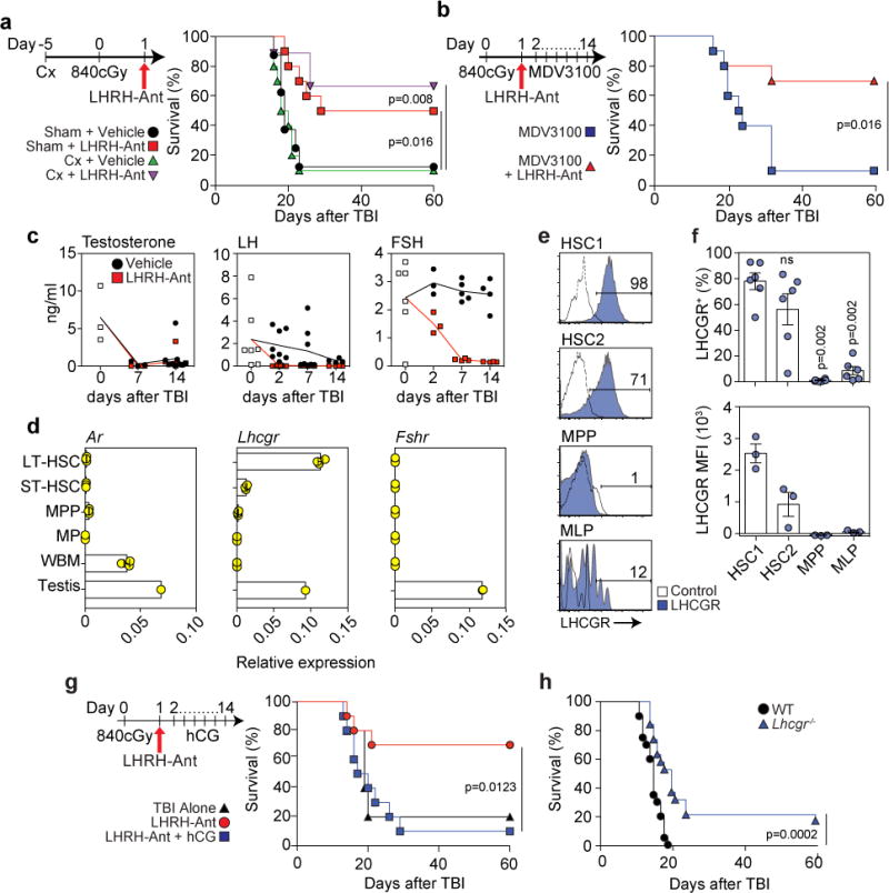
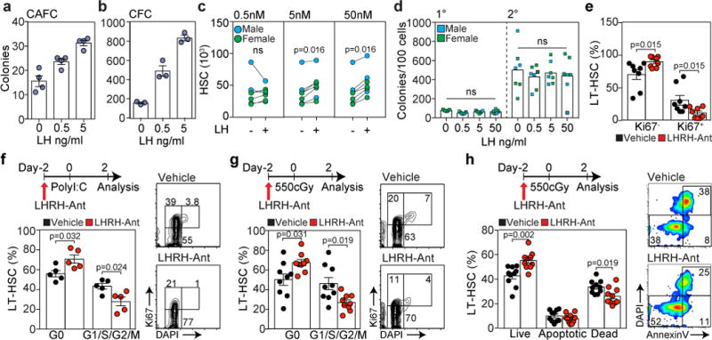
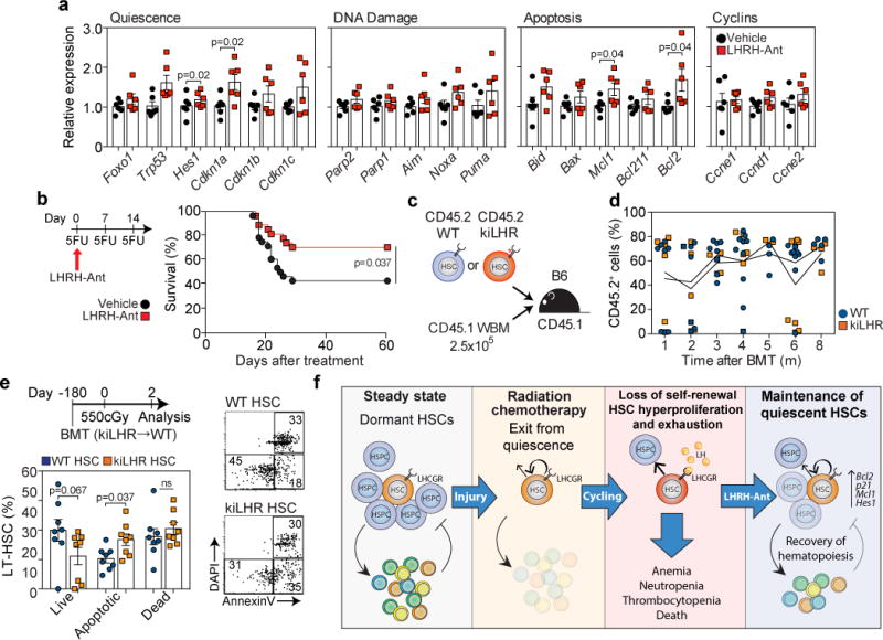
There is a substantial unmet clinical need for new strategies to protect the hematopoietic stem cell (HSC) pool and regenerate hematopoiesis after radiation injury from either cancer therapy or accidental exposure. Increasing evidence suggests that sex hormones, beyond their role in promoting sexual dimorphism, regulate HSC self-renewal, differentiation, and proliferation. We and others have previously reported that sex-steroid ablation promotes bone marrow (BM) lymphopoiesis and HSC recovery in aged and immunodepleted mice. Here we found that a luteinizing hormone (LH)-releasing hormone antagonist (LHRH-Ant), currently in wide clinical use for sex-steroid inhibition, promoted hematopoietic recovery and mouse survival when administered 24 h after an otherwise-lethal dose of total-body irradiation (L-TBI). Unexpectedly, this protective effect was independent of sex steroids and instead relied on suppression of LH levels. Human and mouse long-term self-renewing HSCs (LT-HSCs) expressed high levels of the LH/choriogonadotropin receptor (LHCGR) and expanded ex vivo when stimulated with LH. In contrast, the suppression of LH after L-TBI inhibited entry of HSCs into the cell cycle, thus promoting HSC quiescence and protecting the cells from exhaustion. These findings reveal a role of LH in regulating HSC function and offer a new therapeutic approach for hematopoietic regeneration after hematopoietic injury.


Figures

Figure 1
Figure 1
LHRH-Antagonism improves hematopoietic recovery and mouse survival after L-TBI. (a) Survival of C57BL/6 male mice injected with LHRH-Ant (n=40) or vehicle (n=40) 24h after L-TBI (840cGy) (data combined from five independent experiments). (b) Complete blood counts (CBC) of selected mice in panel “a” at day 0 (n=6), day 7 (n=7 vehicle-treated and 10 LHRH-Ant treated mice), day 10 (n=6 vehicle-treated and 6 LHRH-Ant-treated mice), day 14 (n=11 vehicle-treated and 12 LHRH-Ant-treated mice), day 24 (n=5 LHRH-Ant-treated mice), and day 150 (n=6 LHRH-Ant-treated mice). Data combined from at least two independent experiments. (c) Survival of C57BL/6 male mice treated with LHRH-Ant or vehicle 48h after L-TBI (840cGy) (n=15 mice per group). Data combined from two independent experiments. (d) BM cell counts of the mice in “a” at day 0 (n=11 mice), day 7 (n=11 mice per group), day 10 (n=5 vehicle-treated and 6 LHRH-Ant-treated mice), day 14 (n=8 vehicle-treated and 9LHRH-Ant-treated mice), day 24 (n=5 LHRH-Ant-treated mice), and day 150 (n=7 LHRH-Ant-treated mice), Data combined from at least two independent experiments. (e) CFC colonies of BM cells from vehicle or LHRHAnt treated mice (n=10 mice per group) at day 7 after L-TBI. Data combined from two independent experiments. (f) Concatenated FACS plots and representative gating strategies for the experiment in g (concatenated from three mice). (g) Total numbers of LSK, MPP, LT-HSC and the frequency of LT-HSC in vehicle and LHRH-Ant treated mice 14 days after L-TBI (n=15 vehicle-treated and 14 LHRH-Ant-treated mice). Data combined from four independent experiments. For each mouse, the tibia and femur of both legs were analyzed. (h,i) 14 days after L-TBI, BM and spleen cells from CD45.2 male mice, treated with LHRH-Ant or vehicle, were co-transplanted into lethally irradiated CD45.1 mice with a rescue dose of 2.5×10⁵ CD45.1 whole BM cells. (h) Total peripheral reconstitution measured as the percentage of donor CD45.2 in peripheral blood at 1 month (n=13 vehicle-treated and 12 LHRH-Ant-treated mice), 2 months (n=9 vehicle-treated and 8 LHRH-Ant-treated mice), 3 months (n=7 vehicle-treated and 8 LHRH-Ant-treated mice), 5 months (n=12 mice per group), and 7 months (n=9 vehicle-treated and 8 LHRH-Ant-treated mice). Data combined from at least two independent experiments. (i) Area under the curve for panel “h” was calculated. For panels b, d, e, g, h, and i, data is displayed as the mean ± s.e.m. Statistics for panel a and c were generated using the Mantel– Cox (log-rank) test; panels b, d, e, g, and i were computed using the unpaired nonparametric two-tailed Mann-Whitney U test. In panels b and d *p < 0.05, comparing vehicle and LHRH-Ant treated mice
Figure 2
Figure 2
The regenerative benefit of LHRH-Ant after L-TBI is dependent on suppression of LH not sex steroids. (a) Survival of sham-castrated (sham) and castrated (Cx) mice, treated with LHRH-Ant or vehicle 24 hours after TBI, (n=8 sham-treated; n=10 sham + LHRH-Ant-treated; n=10 castrated; and n=10 castrated + LHRH-Ant-treated mice). Data combined from two of three independent experiments with similar results. (b) Survival of mice receiving daily vehicle or MDV3100 treatment and treated with the LHRH-Ant or vehicle (n=10 mice per group). Data combined from two independent experiments. (c) ELISA of serum levels of LH, FSH and testosterone at timepoints after TBI (Testosterone: day 0, n=3. Vehicle groups: day7, n=3; day14, n=9. LHRH-Ant groups: day 7, n=4; day 14, n=9. LH: untreated group n=7. Vehicle groups: day 2, n=8; day 7, n=8; day 14, n=4. LHRH-Ant groups: day 2, n=8; day 7, n=7; day 14, n=4. FSH: untreated group, n=6. Vehicle groups: day 2, n=3; day 7, n=5; day 14, n=4. LHRHAnt groups: day 2, n=3; day 7, n=4; day 14, n=4 mice). Data combined from at least two independent experiments. (d) Expression of Ar, Lhcgr and Fshr was assessed by qPCR in sorted LT-HSCs, ST-HSCs, MPPs, MPs and whole BM (WBM) from untreated C57BL/6 male mice (n=3 biological replicates; where each replicate was pooled from 2 mice). Data represent one out of two independent experiments with similar findings in both. The gating strategy for each population is shown in Fig. S3a. Expression in one whole testis was assessed as a positive control. Data are plotted as relative quantification normalized to the level of HPRT and beta-actin. (e-f) Expression of LHCGR on HSC1 (Lin-CD34+CD38-CD45RA-CD90+CD49f+), HSC2 (Lin-CD34+CD38-CD45RA-CD90-CD49f+), MPP (Lin-CD34+CD38-CD45RA-CD90- CD49f-) and MLP (Lin-CD34+CD38-CD45RA+CD90-CD10+) cell populations in human UCB. (e) FACS plots showing LHCGR expression for each indicated population. The gating strategy is shown in Fig. S3d and plots are representative of three independent experiments. (f) Top panel, proportion of LHCGR+ cells in human hematopoietic progenitors. Data are combined from three independent experiments using a total of 6 individual UCB units (“ns” = non-statistically significant). Bottom panel, mean fluorescence intensity (MFI) of LHCGR in human UCB samples. Data represent one of three independent experiments with similar findings in each. (g) Survival of mice receiving vehicle (TBI alone) (n=5), LHRH-Ant (n=10) or LHRH-Ant plus daily administration of hCG (n=10 mice). Data combined from two independent experiments. (h) Survival of WT (n=20) or Lhcgr−/− (n=19) mice exposed to 840cGy radiation dose. Data is combined from three independent experiments. For panels d and f data is displayed as the mean ± s.e.m. Statistics for panels a, b, g and h were generated using the Mantel–Cox (log-rank) test; statistics for panel f was computed using the unpaired nonparametric two-tailed Mann-Whitney U test.
Figure 3
Figure 3
LH directly promotes HSC expansion and its suppression after hematopoietic insults decreases HSC entry into cell cycle. (a) LSK cells (2×10³) from a central pool were co-cultured in quadruplicate with MS-5 cells at the indicated concentrations of LH. Total number of CAFC were assessed after 3 weeks. Data represents one out of two independent experiments with similar results. (b) Total number of CFCs generated from LSK cells (3×10³) cultured at the indicated LH concentrations. Data represents one out of two independent experiments performed in triplicate with similar results. (c) CD34+ enriched human UCB cells were cultured in serum and stroma-free cultured conditions in the absence or presence of LH at the indicated concentrations for 7 days, at which time point the number of HSC1 cells (CD34+CD133+CD45RA-CD90+) were quantified by FACS (n=7 individual UCB units plated in triplicate). (d) Primary and secondary hematopoietic colonies generated from HSC1-enriched cells previously cultured with LH as in c (n=7 individual UCB units). (e) Frequency of Ki67+ and Ki67- LT-HSCs in BM of vehicle and LHRH-Ant treated mice 14 days after L-TBI (n=8 mice per group). Data is combined from two independent experiments. (f) Cell cycle analysis of LT-HSCs in vehicle and LHRH-Ant treated mice after poly I:C treatment (n=6 mice per group). Data is combined from two independent experiments. (g) Cell cycle analysis of LT-HSCs in vehicle and LHRH-Ant treated mice after SL-TBI (n=9 mice per group). Data is combined from three independent experiments. (h) Annexin V and DAPI analysis of LT-HSCs using the experimental scheme shown in f (n=10 mice per group). Data is combined from two independent experiments. Concatenated FACS plots of five individual mice are shown on the right. For panels a, b, d, e, f, g and h data is displayed as the mean ± s.e.m. Statistics for panels a, b, d, e, f, g and h were generated using unpaired nonparametric two-tailed Mann-Whitney U test; statistics for panel c were calculated using the non-parametric paired Wilcoxon rank sum test; and statistics for panel d were calculated using the non-parametric paired Friedman test followed by Dunn’s post-hoc test.
Figure 4
Figure 4
LH suppression promotes quiescence and protects HSCs from exhaustion. (a) Expression of the indicated genes, as assessed by qPCR, in LT-HSCs that were sort-purified two days after 300 cGy radiation in surgically-castrated mice treated with vehicle or LHRH-Ant two days prior to radiation. The data are expressed as fold increase relative to the vehicle control group and were normalized to the average of three housekeeping genes (Gapdh, beta-actin, Sdha) (n=6 biological replicates per group, with each replicate comprised of 3 mice). Data is combined from two independent experiments. (b) Survival of vehicle (n=28) and LHRH-Ant (n=27) treated mice following sequential 5-FU treatment. Data is combined from three independent experiments. (c) Schematic representation of the experiment for which the data are shown in d. 500 LT-HSCs from WT or KiLHRD582G mice were sort-purified and transplanted into CD45.1 irradiated hosts together with a rescue dose of 2.5×10⁵ CD45.1 whole BM cells. (d) Total peripheral reconstitution was measured as the percentage of donor CD45.2 in peripheral blood over 8 months (WT: 1 month, n=8; 2 months, n=7; 3 months, n=8; 4 months, n=10; 5 months, n=4; 6 months, n=9; 8 months, n=5; KiLHRD582G mice: 1 month, n=6; 2 months, n=7; 3 months, n=5; 4 months, n=6; 5 months, n=3; 6 months, n=8; 8 months, n=4 mice). Data are combined from at least two independent experiments. (e) The mice in d were exposed to 550cGy and LT-HSCs subjected to Annexin V and DAPI analysis by FACS two days later (n=8 mice per group). Data is combined from two independent experiments. (f) Schematic model of the proposed mechanism by which LHRH-Ant promotes HSC recovery after hematopoietic insult. For panels a, d, and e data is displayed as the mean ± s.e.m. Statistics for panels a and e were generated using the unpaired nonparametric two-tailed Mann-Whitney U test; panel b was calculated using the Mantel–Cox (log-rank) test.

Similar articles

Cited by

References

    1. Dainiak N. Hematologic consequences of exposure to ionizing radiation. Experimental hematology. 2002;30:513–528. - PubMed
    1. Anno GH, Young RW, Bloom RM, Mercier JR. Dose response relationships for acute ionizing-radiation lethality. Health Phys. 2003;84:565–575. - PubMed
    1. Mierzejewska K, et al. Hematopoietic Stem/Progenitor Cells Express Several Functional Sex Hormone Receptors-Novel Evidence for a Potential Developmental Link Between Hematopoiesis and Primordial Germ Cells. Stem cells and development. 2015 - PMC - PubMed
    1. Sanchez-Aguilera A, et al. Estrogen signaling selectively induces apoptosis of hematopoietic progenitors and myeloid neoplasms without harming steady-state hematopoiesis. Cell stem cell. 2014;15:791–804. - PubMed
    1. Nakada D, et al. Oestrogen increases haematopoietic stem-cell self-renewal in females and during pregnancy. Nature. 2014;505:555–558. - PMC - PubMed

Publication types

MeSH terms