. 2018 Jan 8;14(1):e1007146.
doi: 10.1371/journal.pgen.1007146. eCollection 2018 Jan.

An Ichor-dependent apical extracellular matrix regulates seamless tube shape and integrity

Affiliations

An Ichor-dependent apical extracellular matrix regulates seamless tube shape and integrity

Jeffrey B Rosa et al. PLoS Genet. .

Abstract

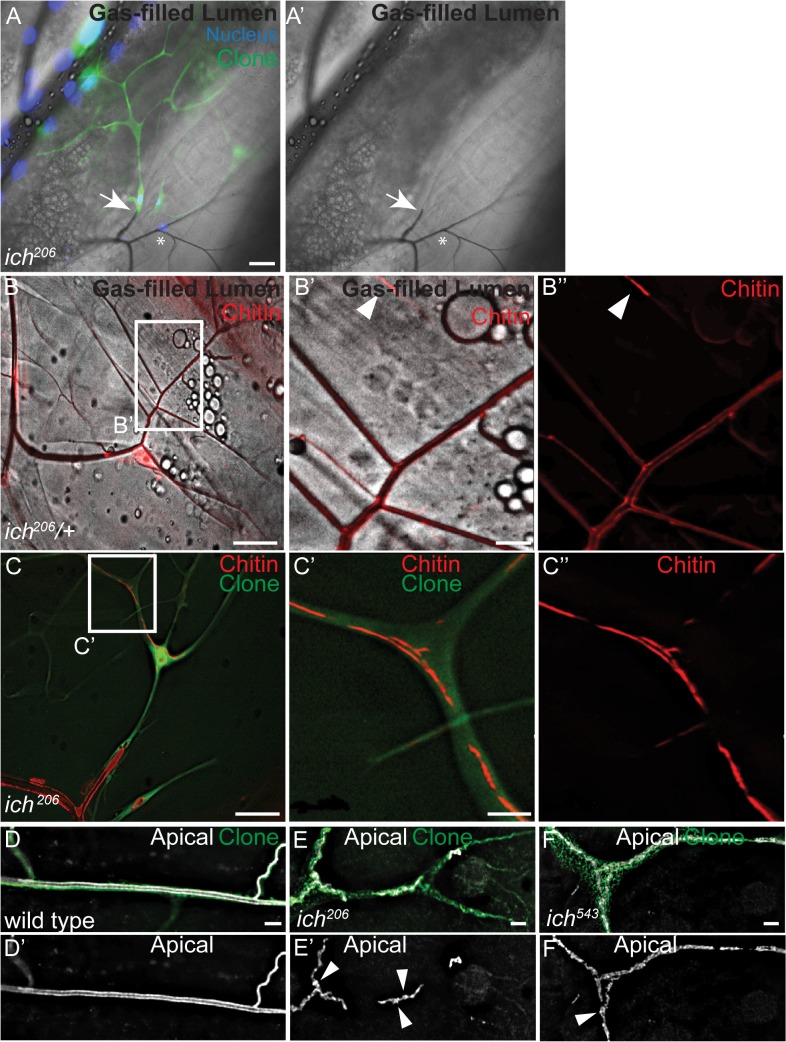
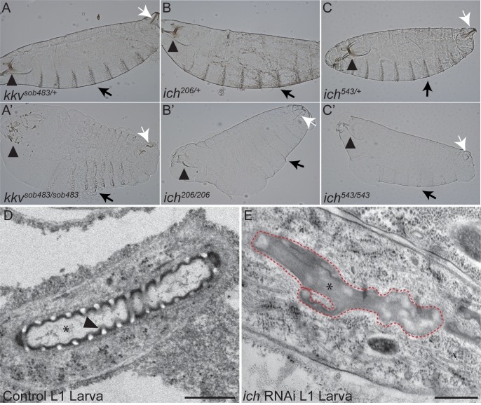
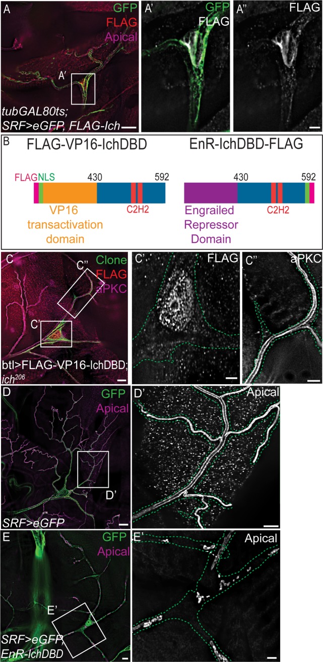
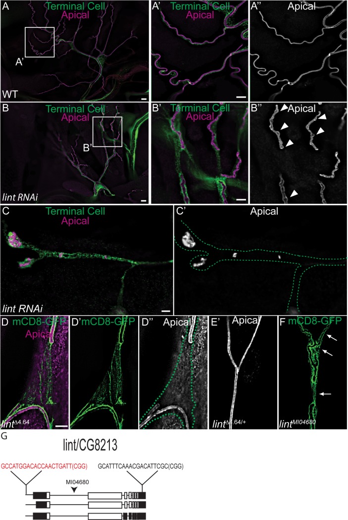
During sprouting angiogenesis in the vertebrate vascular system, and primary branching in the Drosophila tracheal system, specialized tip cells direct branch outgrowth and network formation. When tip cells lumenize, they form subcellular (seamless) tubes. How these seamless tubes are made, shaped and maintained remains poorly understood. Here we characterize a Drosophila mutant called ichor (ich), and show that ich is essential for the integrity and shape of seamless tubes in tracheal terminal cells. We find that Ich regulates seamless tubulogenesis via its role in promoting the formation of a mature apical extracellular matrix (aECM) lining the lumen of the seamless tubes. We determined that ich encodes a zinc finger protein (CG11966) that acts, as a transcriptional activator required for the expression of multiple aECM factors, including a novel membrane-anchored trypsin protease (CG8213). Thus, the integrity and shape of seamless tubes are regulated by the aECM that lines their lumens.


Conflict of interest statement

The authors have declared that no competing interests exist.

Figures

Fig 1
Fig 1. Mutations in ichor (ich) disrupt seamless tube integrity and shape in larval terminal cells.
(A,A’) In a genetic mosaic third instar larva, GFP-labeled ich206/206 terminal cells (green) are visualized next to GFP-negative control terminal cells (ich206/+, asterisk). All tracheal nuclei are labeled (blue). Fluorescent images are superimposed on brightfield. Gas-filled tubes appear as dark lines; the ich206/206 terminal cells lack gas-filled tubes (arrow marks intercellular junction between ich terminal cell and wild type stalk cell). (B-B”) In control terminal cells, ChtVis-TdTomato (red) labels cuticle-lined gas-filled tracheal tubes (B’, B”) as well as fluid-filled lumens found at the tips of terminal branches (arrowheads in B’, B”). In ich206/206 terminal cells (C), ChtVis-TdTomato accumulates in discontinuous, fluid-filled tubes (C’C”). (D-F) The apical membranes in wild-type (D,D’) and ich (E-F’) seamless tubes is visualized by an antiserum raised against a Whacked peptide [69]. In contrast to the continuous apical membranes in control (D,D’), ich terminal cell tubes display numerous apical membrane discontinuities and regions of cystic apical membrane (arrowheads in E’, F’). (Scale Bars: A, A’, B, and C 50 μm; B’-B”, C’-C” 10 μm; D-F’ 5 μm).
Fig 2
Fig 2. ichor encodes a zinc finger protein.
(A) Domain map of Ichor polypeptide and position of EMS-induced lesions. (B-B’) In contrast to internal control (Df(3R)osk/+) seamless tubes (arrowheads in B’), GFP-labeled Df(3R)osk terminal cell clones exhibit ich-like apical membrane discontinuities. Apical membrane in B-D is detected using anti-Wkdpep sera. Outline of Df(3R)osk/Df(3R)osk terminal cell shown as green dotted line. (C-D’) SRF>eGFP wild-type control terminal cells exhibit a patent seamless tube (C-C’), whereas SRF>eGFP, CG11966 RNAi terminal cells (D-D’) contain discontinuous blind-ended tube fragments. Terminal cell outline shown as green dotted line. (E-F) Restoring expression of full-length CG11966 cDNA in ich terminal cells rescues the ich206 discontinuous tube defect, as observed by visualizing apical membranes with aPKC staining (E, E’). (F) Quantification of rescue experiments using Ich transgenes (P = 8.82 x 10⁻¹⁷ for UAS-ich, P = 1.83 x 10⁻¹² for UAS-flag-ich, P = 2.01 x 10⁻¹⁸, using one-sided Fisher’s exact probability test). (Scale Bars: B,C,D,E 20μm; B’, C’,D’, E’ 5 μm).
Fig 3
Fig 3. Ichor is required for assembly of the mature aECM.
Cuticle preparations of heterozygous control (A-C) and homozygous mutant kkvsob483 (A’), ich206 (B’), and ich543 (C’) St.17 embryos or first instar larvae. Chitin-deficient embryos (A’) exhibited a bloated cuticle and degenerated head skeleton (arrowhead in A’). The ich embryos exhibited a mild bloated phenotype as well as cuticle defects distinct from chitin-deficient embryos. The head skeleton (arrowheads in B’, C’) appeared intact but poorly pigmented. Other cuticular structures, such as the epidermal denticles (arrows in B’, C’) and spiracular chambers (white arrows in B’, C’), were also poorly pigmented in ich mutants. By contrast, chitin synthesis is not absolutely required for pigmentation of the head skeleton remnants (arrowhead in A’), denticle belts (black arrow in A’), or spiracular chambers (white arrow in A’). (D, E) Longitudinal sections of control (btl>GFP, D) and ich RNAi (SRF>eGFP, ich RNAi; E) first instar (L1) terminal cell tubes visualized by transmission electron microscopy (TEM). Control terminal cell branches contain tubes (*) of locally uniform dimensions lined by an electron-dense aECM (cuticle). The arrowhead in D points to a clearly defined taenidium with an electron-luscent chitinous core. By contrast, the lumens (*) of ich RNAi terminal branches adopt a highly irregular morphology (red dashed line). These lumens are devoid of taenidiae and instead are occluded with disorganized electron-dense material. (Scale Bars: D, E = 500 nm).
Fig 4
Fig 4. Ichor functions as a transcriptional activator in terminal cells.
(A-A”) tubGAL80ts; SRF>eGFP, FLAG-Ich larvae were upshifted to the restrictive temperature around the start of the third larval instar to induce GAL4-dependent FLAG-Ich expression. FLAG-Ich accumulates at steady-state in the nucleus of terminal cells (A’, A”). (B) Domain organization of FLAG-VP16AD-IchDBD and EnR-IchDBD-FLAG chimeras is schematized. VP16AD (orange) is a potent transcriptional transactivating domain from a viral protein, while EnR (purple) is the transcriptional repressor domain of the Engrailed transcription factor. IchDBD (blue) is the C-terminal portion of the Ich protein that includes the zinc finger domains (red) presumed to confer DNA binding. (C-C”) ich206 MARCM terminal cell clones expressing UAS-FLAG-VP16AD-IchDBD transgene. By virtue of an exogenous nuclear targeting sequence (see B, green), FLAG-VP16AD-IchDBD chimera accumulates in the nucleus (C, C’), restoring seamless tube integrity (C”), as observed by staining for the apical marker aPKC. (D-E’) Wild-type control SRF>eGFP (D-D’) and SRF>eGFP, EnR-IchDBD-FLAG (E, E’) terminal cells. The EnR-IchDBD-FLAG chimera behaves like an Ich dominant negative, inducing the ich loss of function phenotype. In contrast to the fully patent lumens of control terminal cells (D’), SRF>eGFP, EnR-IchDBD-FLAG terminal cells exhibited blind-ended, discontinuous lumens similar to ich mutant terminal cells. (Scale Bars: A, C, D, E 20 μm; A’ and A”, C’ and C”, D’, E’ 5 μm).
Fig 5
Fig 5. The aECM component chitin is essential for seamless tube shape and integrity.
(A) Schematic of the kkv gene and the encoded Chitin Synthase protein structure with the position of the sob mutations indicated. (B-B”) kkvsob404 null terminal cell clone (green) exhibited apical membranes with cystic, irregular morphology. For the areas marqueed in white, the apical membrane is shown enlarged in (B’, B”) as indicated. Terminal cell outlines in B’, B”, C‘, D’ are shown by green dots. In (C) and (D), kkvsob483 and kkvsob761 clones, respectively, are shown. In distal terminal branches (B”, D ‘), discrete apical membrane cysts (arrowheads) are interspersed with tube of apparently normal morphology. (C-D’) limited regions of discontinuous apical membrane (C’,D’) were also observed with all kkv alleles. (E) Penetrance of tube phenotypes for the kkvsob404 null allele and kkvsob483, a putative recessive antimorphic allele (P<0.0001, Chi-Square test). (Scale Bars: B, C, D 20 μm; B’,B”, C’, D’ 5 μm).
Fig 6
Fig 6. Ichor is required for the pan-tracheal expression of osi18 and osi19.
Expression of osi18 mRNA (A-F) was examined in sibling (WT; A-C) and ich206/Df(3R)osk embryos (D-F) by in situ hybridization. Expression of osi19 mRNA (G-N) was examined in siblings (G-I) and ich206/Df(3R)osk embryos (J-L) by in situ hybridization. In contrast to the wild type control heterozygous siblings (A-C and G-I), ich206 hemizygotes (D-F, J-L) lacked osi18 and osi19 expression throughout the trachea. However, by late Stage 16 (F, L) osi18 and osi19 mRNA became detectable in the multicellular dorsal trunks branches of the tracheal system. Stage 16 control (2Xbtl>GFP) embryos (M) detected the expected pattern of osi19 mRNA expression in contrast to embryos (N) expressing a transcriptional repressor domain (EnR) fused to the Ich DNA binding domain (IchDBD). The embryos expressing the Ich chimera (2Xbtl>GFP, EnR-IchDBD) exhibited pan-tracheal reduction of osi19 mRNA.
Fig 7
Fig 7
Ichor is required for CG8213 expression in the trachea (A-D) Wild-type w1118 embryos hybridized with DIG-labeled antisense (A-C’) and sense (D) CG8213 RNA probes. CG8213 expression is first detected at St. 13 in the posterior spiracles (arrowhead in A, A’) and foregut primordium (arrow in B). By St. 15, CG8213 is expressed specifically in cuticle-secreting epithelia, including the trachea (black arrowheads in C), foregut (arrow in C’), hindgut (white arrowhead in C’), and epidermis (see asterisks in F’, I). This signal is specific since a sense probe (D) shows no such pattern. (E) Transcripts and polypeptide encoded by CG8213 locus. Black bar denotes position of RNA probes, designed to detect all spliceoforms. (F-G’) ich206/ich543 mutant (G, G’) and control siblings (F,F’) hybridized with antisense CG8213 RNA probe. Whereas control siblings show a WT CG8213 expression pattern, ich mutant embryos exhibited a significant reduction or loss of CG8213 expression in the trachea. Only a subset of cells in the posterior spiracles (black arrowhead in G) retain CG8213 expression. Hindgut expression (white arrowhead in G’) is also reduced. (H-I) 2Xbtl>GFP (H) and 2Xbtl>GFP, EnR-IchDBD embryos hybridized with antisense CG8213 probe. Whereas control embryos (H) expressed CG8213 prominently in the major dorsal trunk (black arrowhead in H) of the trachea, tracheal-autonomous expression of the dominant-negative Ich transgene caused a loss of tracheal expression; only cells in the posterior spiracles retain CG8213 message, similar to ich loss-of-function phenotype. CG8213 expression in the epidermis (asterisk in I), foregut (arrow in I), and hindgut (white arrowhead in I) is unaffected, indicating a tracheal-autonomous loss of expression.
Fig 8
Fig 8. lumens interrupted (lint) is required for seamless tube integrity in terminal cells.
Wild type (WT, SRF>eGFP) control (A-A”) and short hairpin RNA against CG8213 expressing terminal cells (B-B”) were examined for apical membrane integrity. In contrast to control terminal branch seamless tubes (A’, A”), seamless tubes in lint-depleted terminal cells exhibit cystic dilations (arrowheads in B”), and discontinuities of the apical membrane with 100% penetrance (n = 33 terminal cells) (B’.B”). The most severely affected SRF>eGFP,CG8213 RNAi terminal cells are severely pruned with terminal branches largely devoid of luminal membrane, except for isolated inclusions. Terminal cell outlines are shown by green dots (C,C’). (D-D”) Details of lintΔ4.64 seamless tube defect from a mCD8-GFP-expressing terminal cell clone. The outline of the terminal cell is indicated by green dots. The seamless tubes of a neighboring heterozygous terminal cell (mCD8-GFP negative) are shown in (E’). The gRNA sequences used to target lumens interrupted (lint) using CRISPR/Cas9 are indicated in (G) on a schematic depicting lint gene organization with CDS in white and UTR sequences in black. The gRNA site indicated in red appears to be the site targeted by Cas9. (F) Detail of a lintMI04680 terminal cell clone expressing membrane-anchored GFP, which accumulates preferentially at the apical membrane. The arrows indicate discontinuities in the apical membrane. The position of the lethal transgene insertion is indicated in (G). (Scale Bars: A, B 20 μm; A”, B”, C and C’, D-F 5 μm).

References

    1. Fedeles SV, Tian X, Gallagher A-R, Mitobe M, Nishio S, Lee SH, et al. A genetic interaction network of five genes for human polycystic kidney and liver diseases defines polycystin-1 as the central determinant of cyst formation. Nature Genetics. 2011. June 19;43(7):639–47. doi: 10.1038/ng.860 - DOI - PMC - PubMed
    1. Tual-Chalot S, Oh SP, Arthur HM. Mouse models of hereditary hemorrhagic telangiectasia: recent advances and future challenges. Frontiers in Genetics [Internet]. 2015. February 18; 6 Available from: http://journal.frontiersin.org/Article/10.3389/fgene.2015.00025/abstract. - DOI - PMC - PubMed
    1. Lubarsky B, Krasnow MA. Tube Morphogenesis: making and shaping biological tubes. Cell. 2003. January;112(1):19–28. - PubMed
    1. Buechner M. Tubes and the single C. elegans excretory cell. Trends in Cell Biology. 2002. October 1;12(10):479–84. - PubMed
    1. Berry KL. A C. elegans CLIC-like Protein Required for Intracellular Tube Formation and Maintenance. Science. 2003. December 19;302(5653):2134–7. doi: 10.1126/science.1087667 - DOI - PubMed

Publication types

MeSH terms