. 2018 Jan-Feb:99:51-61.
doi: 10.1016/j.diff.2017.12.003. Epub 2017 Dec 15.

The ubiquitin ligase ITCH coordinates small intestinal epithelial homeostasis by modulating cell proliferation, differentiation, and migration

Affiliations

The ubiquitin ligase ITCH coordinates small intestinal epithelial homeostasis by modulating cell proliferation, differentiation, and migration

Heather L Mentrup et al. Differentiation. 2018 Jan-Feb.

Abstract

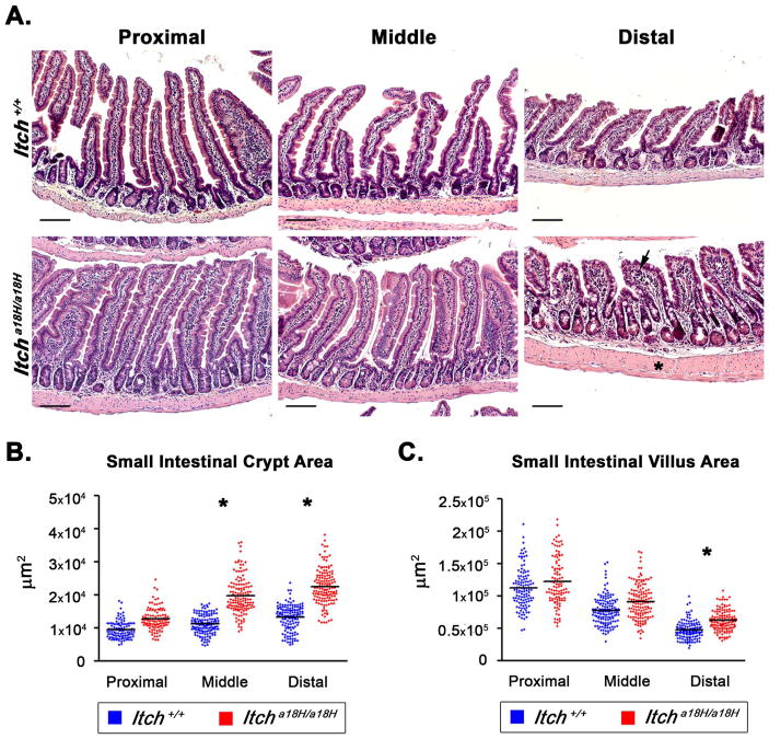
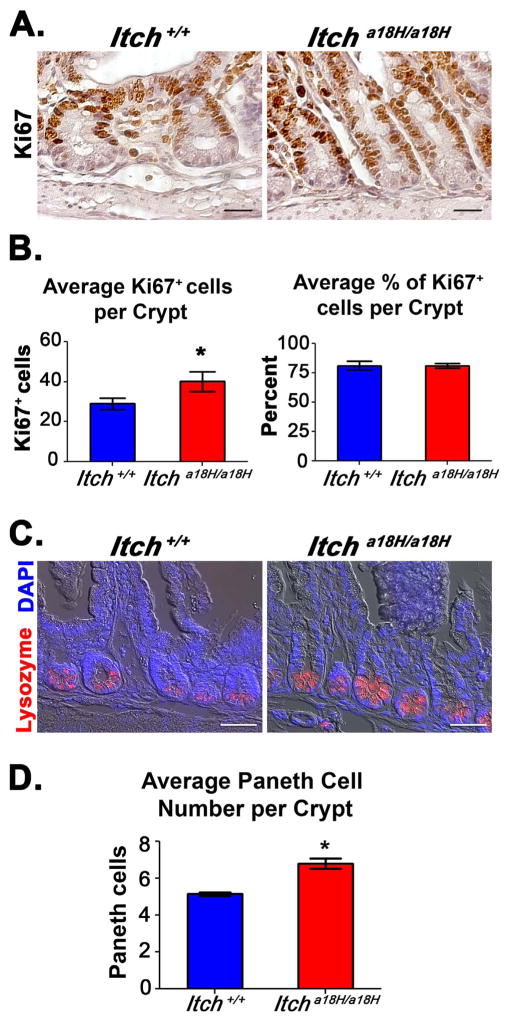
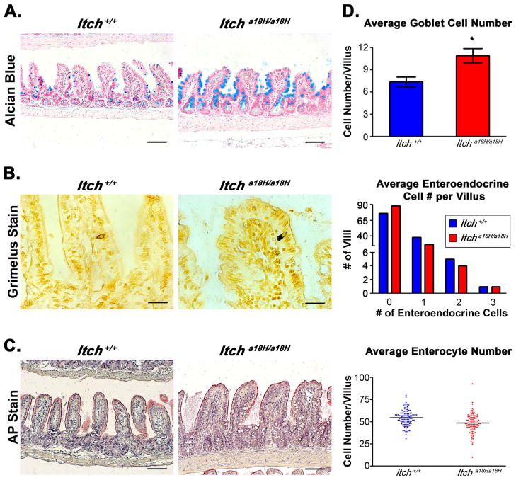
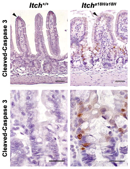
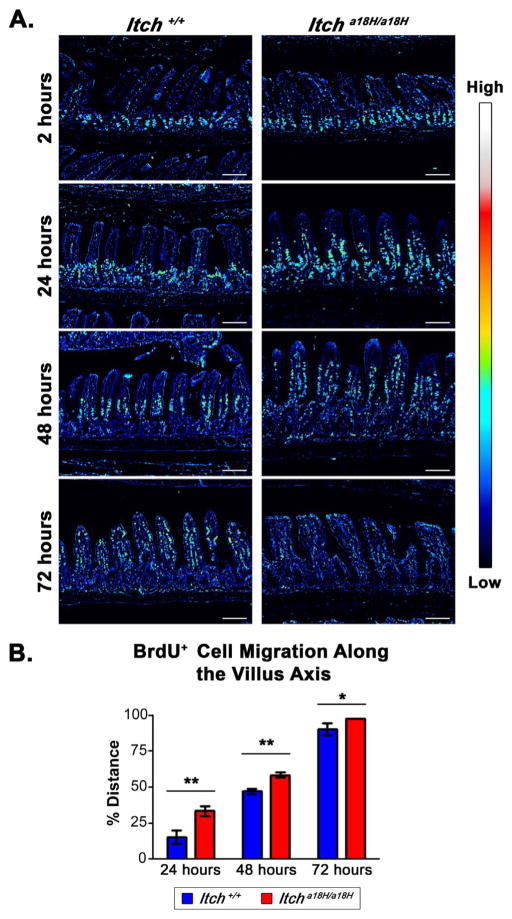
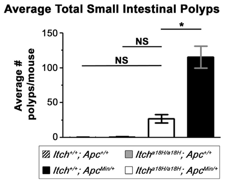
Maintenance of the intestinal mucosa is driven by local signals that coordinate epithelial proliferation, differentiation, and turnover in order to separate antigenic luminal contents from the host's immune system. Breaches in this barrier promote gastrointestinal pathologies ranging from inflammatory bowel disease to cancer. The ubiquitin ligase ITCH is known to regulate immune responses, and loss of function of ITCH has been associated with gastrointestinal inflammatory disorders, particularly in the colon. However, the small intestine appears to be spared from this pathology. Here we explored the physiological mechanism that underlies the preservation of mucosal homeostasis in the small intestine in mice lacking ITCH (Itcha18H/a18H). Histological analysis of the small intestines from young adult mice revealed architectural changes in animals deficient for ITCH, including villus blunting with cell crowding, crypt expansion, and thickening of the muscularis propria relative to age-matched mice sufficient for ITCH. These differences were more prominent in the distal part of the small intestine and were not dependent upon lymphoid cells. Underlying the observed changes in the epithelium were expansion of the Ki67+ proliferating transit amplifying progenitor population and increased numbers of terminally differentiated mucus-secreting goblet and anti-microbial producing Paneth cells, which are both important in controlling local inflammation in the small intestine and are known to be dysregulated in inflammatory bowel disease. Homeostasis in the small intestine of Itcha18H/a18H animals was maintained by increased cell turnover, including accelerated migration of epithelial cells along the crypt-villus axis and increased apoptosis of epithelial cells at the crypt-villus junction. Consistent with this enhanced turnover, Itcha18H/a18H mice carrying the Min mutation (Itcha18H/a18H; ApcMin/+) displayed a 76% reduction in tumor burden as compared to ApcMin/+ littermates with normal levels of ITCH. These findings highlight the role of ITCH as an important modulator of intestinal epithelial homeostasis.

Keywords: Cell migration; Colorectal cancer; E3 ubiquitin ligase; Epithelial differentiation; ITCH; Intestinal homeostasis.


Figures

Fig. 1
Fig. 1
Small intestinal architecture is altered in Itcha18H/a18H animals. (A) Representative H & E stained paraffin-embedded small intestinal sections derived from the proximal (left most column), middle (center column), and distal (right hand column) segments of young adult Itch+/+ and Itcha18H/a18H animals accentuate an enlargement of the intestinal crypt, villus blunting with crowding of epithelial cells, and thickening of the muscularis propria (asterisk) in Itcha18H/a18H animals, becoming more pronounced distally. Mild inflammation was also occasionally noted in the lamina propria (arrow). Scale bars = 200 μm. (B) Small intestinal crypt and villus areas from young adult Itch+/+ (n = 4 or 5) and Itcha18H/a18H (n = 5) were measured in μm2 using ImageJ. A minimum of 25 well-orientated crypts or villi from at least seven different 10x fields were measured per animal. Average crypt area was significantly increased (*p < 0.005) in the middle and distal small intestine of Itcha18H/a18H compared to Itch+/+. (C) Average villus area was significantly increased (*p < 0.05) distally in the small intestine of Itcha18H/a18H animals compared to Itch+/+.
Fig. 2
Fig. 2
Crypt expansion in Itcha18H/a18H animals is accompanied by an increase in proliferating TA cells and differentiated Paneth cells. (A) Pictured are representative micrographs of IHC performed on paraffin-embedded distal intestinal sections derived from young adult Itch+/+ and Itcha18H/a18H animals (n = 5 for each genotype) using an anti-Ki67 antibody and subsequently counterstained with hematoxylin. Animals lacking ITCH appear to have increased cellular proliferation in the crypt. Scale bars = 50 μm. (B) Average number and average percentage of Ki67+ cells in the crypts from adult Itch+/+ (n = 5) and Itcha18H/a18H (n = 5) animals. Ki67+ cells and total cell numbers were counted from 25 well-oriented crypts in a minimum of nine different 20x fields per animal. Itcha18H/a18H animals had, on average, 13 more Ki67+ cells per crypt which was statistically significant (*p < 0.01). No difference was observed in the percentage of proliferating cells within the crypt. Error bars represent SD. (C) Representative phase contrast-coupled immunofluorescent images of paraffin-embedded sections of distal intestine from five adult Itch+/+ and five age-matched Itcha18H/a18H animals stained with anti-lysozyme (red) and counterstained with DAPI (blue) to detect nuclei. Scale bars = 100 μm. (D) Average number of Paneth cells per crypt from adult Itch+/+ (n = 5) and Itcha18H/a18H (n = 5) animals. Paneth cell numbers from 30 crypts in a minimum of 12 different 20x fields were averaged to determine that Itcha18H/a18H animals have significantly more Paneth cells (*p < 0.05) than Itch+/+ animals. Error bars represent SEM (For interpretation of the references to color in this figure legend, the reader is referred to the web version of this article.).
Fig. 3
Fig. 3
Loss of ITCH promotes goblet cell differentiation. (A) Paraffin-embedded intestinal sections stained with alcian blue, pH 2.5 and counterstained with nuclear fast red distinguish goblet cells from adult Itch+/+ and Itcha18H/a18H animals. Scale bars = 200 μm. (B) Pictured are representative Itch+/+ and Itcha18H/a18H derived paraffin-embedded sections stained with 1% silver nitrate to identify enteroendocrine cells in the small intestine. Scale bars = 200 μm. (C) Paraffin-embedded sections stained with AP highlights the brush boarder of enterocytes on the villi of Itch+/+ and Itcha18H/a18H animals. Scale bars = 200 μm. (D) Average number of goblet cells, enteroendocrine cells, and enterocytes located on the villus of Itch+/+ (n = 4) and Itcha18H/a18H (n = 4) animals. Indicated type of cells were counted on 30 villus structures from a minimum of eight different 10x fields per animal. Enteroendocrine cell counts are presented as a histogram of the total number of villus structures bearing no, one, two or three cells in Itch+/+ (n = 4) and Itcha18H/a18H (n = 4) animals. Itcha18H/a18H animals have a statistically significant increase (*p < 0.05) in goblet cells. There was no statistically significant difference in enteroendocrine or enterocyte cell number. Error bars represent SEM for goblet cell counts.
Fig. 4
Fig. 4
Loss of ITCH promotes epithelial apoptosis in the small intestine. Paraffin embedded sections from the distal small intestine of young adult Itch+/+ and Itcha18H/a18H animals (n = 4 for each genotype) were immunostained for cleaved-caspase 3 and counterstained with hematoxylin. Representative photomicrographs are shown. While rare apoptotic cells could be found at the tips of villi in animals of both genotypes (arrowheads, top row of micrographs), increased apoptosis was seen at the crypt-villus junction in only those animals lacking ITCH (bottom row, 63x). Scale bars = 100 μm (top row) or 50 μm (bottom row).
Fig. 5
Fig. 5
Epithelial turnover is increased in animals that lack ITCH. (A) IHC with an anti-BrdU antibody was conducted on paraffin-embedded sections obtained from young adult Itch+/+ and Itcha18H/a18H animals intraperitoneally injected with 100 mg/kg of BrdU. Brightfield images were taken 2, 24, 48, and 72 h post injection and representative examples from each genotype and time point were converted to spectral images using the royal look-up table available in ImageJ. An intensity scale for this spectrum is pictured to the right. These images demonstrate enhanced migration of epithelial cells in Itcha18H/a18H animals with complete sloughing by 72 h following the BrdU pulse. Scale bars = 200 μm. (B) Average cell migration of BrdU-labeled cells along the villus axis in Itch+/+ (n = 3) and Itcha18H/a18H (n = 3) animals at 24, 48, and 72 h after BrdU incorporation was calculated by dividing the measured distance of the furthest migrated labeled cell from the base of the villus by the entire length of the villus from 25 villi in a minimum of five different 10x fields per animal. Since all cells were localized to the crypt in both genotypes at the 2 h timepoint, this data point is not shown. A statistically significant increase in cell migration at was detected in Itcha18H/a18H derived tissues at 24 (**p < 0.005), 48 (**p < 0.005), and 72 h (*p < 0.05) versus tissues derived from Itch+/+ animals. Error bars represent SD.
Fig. 6
Fig. 6
Itcha18H/a18H animals have altered cell-cell junctions. Representative transmission electron micrographs of cell-cell junctions (tight junctions marked by asterisks, adherens junctions by arrows, and desmosomes by arrow head) from the proximal small intestine of nine week-old Itch+/+ and Itcha18H/a18H animals are depicted here. In total samples from four animals of each genotype were examined. Itcha18H/a18H animals lack identifiable desmosomes and adherens junctions appeared more disorganized than the ITCH sufficient control samples. Scale bar = 200 nm.
Fig. 7
Fig. 7
Loss of ITCH is protective in ApcMin/+-induced tumorigenesis. Polyps from the small intestine of 15 week-old Itch+/+; Apc+/+ (n = 11), Itcha18H/a18H; Apc+/+ (n = 11), Itch+/+; ApcMin/+ (n = 11), and Itcha18H/a18H; ApcMin/+ (n = 11) animals were visualized by staining the flushed and fixed small intestine with 0.1% methylene blue. Polyps from the entire length of the intestine were counted under a dissecting microscope. The graph shown here depicts the average number of small intestinal polyps per animal. Itcha18H/a18H; ApcMin/+ animals were observed to have a significant reduction in tumor burden compared to Itch+/+; ApcMin/+ littermates (*p < 0.001, NS = not significant). Error bars represent SD.

References

    1. Agace WW, McCoy KD. Regionalized development and maintenance of the intestinal adaptive immune landscape. Immunity. 2017;46:532–548. - PubMed
    1. Barker N. Adult intestinal stem cells: critical drivers of epithelial homeostasis and regeneration. Nat Rev Mol Cell Biol. 2014;15:19–33. - PubMed
    1. Barker N, van Es JH, Kuipers J, Kujala P, van den Born M, Cozijnsen M, Haegebarth A, Korving J, Begthel H, Peters PJ, Clevers H. Identification of stem cells in small intestine and colon by marker gene Lgr5. Nature. 2007;449:1003–1007. - PubMed
    1. Basak O, van de Born M, Korving J, Beumer J, van der Elst S, van Es JH, Clevers H. Mapping early fate determination in Lgr5+ crypt stem cells using a novel Ki67-RFP allele. Embo J. 2014;33:2057–2068. - PMC - PubMed
    1. Basheer WA, Harris BS, Mentrup HL, Abreha M, Thames EL, Lea JB, Swing DA, Copeland NG, Jenkins NA, Price RL, Matesic LE. Cardiomyocyte-specific overexpression of the ubiquitin ligase Wwp1 contributes to reduction in Connexin 43 and arrhythmogenesis. J Mol Cell Cardiol. 2015;88:1–13. - PMC - PubMed

Publication types

LinkOut - more resources