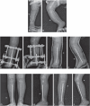
Wrapping grafting for congenital pseudarthrosis of the tibia: A preliminary report
- PMID: 29310362
- PMCID: PMC5728763
- DOI: 10.1097/MD.0000000000008835
Wrapping grafting for congenital pseudarthrosis of the tibia: A preliminary report
Abstract
Objective: Treatment of congenital pseudarthrosis of the tibia (CPT) remains a challenge. The autogenic iliac bone graft is important consistent of treatment for CPT. The purpose of this study was to investigate the role of wrapping autogenic iliac bone graft in improvement of the curing opportunities of CPT.
Methods: We combined Ilizarov fixator with intramedullary rodding of the tibia and wrapping autogenic iliac bone graft for treatment 51 cases of CPT between 2007 and 2010. The mean age is 3.2 years at index operation, of which 31 patients (61%) were below 3 years old. According to Crawford classification, 5 tibia had type-II morphology; 3, type-III; 43, type-IV.
Results: In the postoperative follow-up of 3.5 months (range from 3 to 4.5 months), all cases were found that the bone graft sites of pseudarthrosis of the tibia showed a significant augmentation and spindle-shaped expansion as obvious change. All cases of this series have been followed-up, average followed-up time were 1.6 years (range from 7 to 3.1 years), of which 19 cases were more than 2 years. The average time of removed the Ilizarov ring fixator was 3.5 months (range from 3 to 4.5 months). According to Johnston Clinical evaluation system, 26 cases had grade I, 21 cases, grade II, 4 cases, grade III. Following the Ohnishi X-ray evaluation criteria, union of pseudarthrosis of the tibia were 42 cases, delayed union 5 cases, nonunion 4 cases.
Conclusion: Autogenic iliac bone graft is able to offer the activity of osteoblasts and osteogenesis induced by bone morphogenetic protein (BMP) and glycoprotein, meanwhile enclosing bone graft could help keep cancellous bone fragments in close contact around pseudarthrosis of the tibia, allowing the formation of high concentration of glycoprotein and BMP induced by chemical factors because of established the sealing environment in location, all of which could enhance the healing of pseudarthrosis of the tibia.
Conflict of interest statement
The authors have no conflicts of interest to disclose.
Figures


References
-
- Erni D, De Kerviler S, Hertel R, et al. Vascularised fibula grafts for early tibia reconstruction in infants with congenital pseudarthrosis. J Plast Reconstr Aesthet Surg 2010;63:1699–704. - PubMed
-
- Johnston CE., II Congenital pseudarthrosis of the tibia: results of technical variations in the Charnley-Williams procedure. J Bone Joint Surg Am 2002;84-A:1799–810. - PubMed
-
- Ohnishi I, Sato W, Matsuyama J, et al. Treatment of congenital pseudarthrosis of the tibia: a multicenter study in Japan. J Pediatr Orthop 2005;25:219–24. - PubMed
-
- Granchi D, Devescovi V, Baglio SR, et al. A regenerative approach for bone repair in congenital pseudarthrosis of the tibia associated or not associated with type 1 neurofibromatosis: correlation between laboratory findings and clinical outcome. Cytotherapy 2012;14:306–14. - PubMed
MeSH terms
Supplementary concepts
LinkOut - more resources
Full Text Sources
Other Literature Sources
Medical

