

Cavernous hemangiomas of the temporalis muscle with prominent formation of phleboliths: Case report and review of the literature
- PMID: 29310392
- PMCID: PMC5728793
- DOI: 10.1097/MD.0000000000008948
Cavernous hemangiomas of the temporalis muscle with prominent formation of phleboliths: Case report and review of the literature
Abstract
Rationale: Hemangiomas are benign tumors characterized by an abnormal proliferation of blood vessels, most often occur in the skin and subcutaneous tissue, intramuscular hemangioma, a distinctive type of hemangioma within the skeletal muscle, account for <1% of all hemangiomas, temporalis muscle is a very uncommon site, cavernous hemangioma of the temporalis muscle with prominent formation of phleboliths is rare reported.
Patient concerns: A 62-year-old man presented with a slowly increased mass in his right temporal fossa.
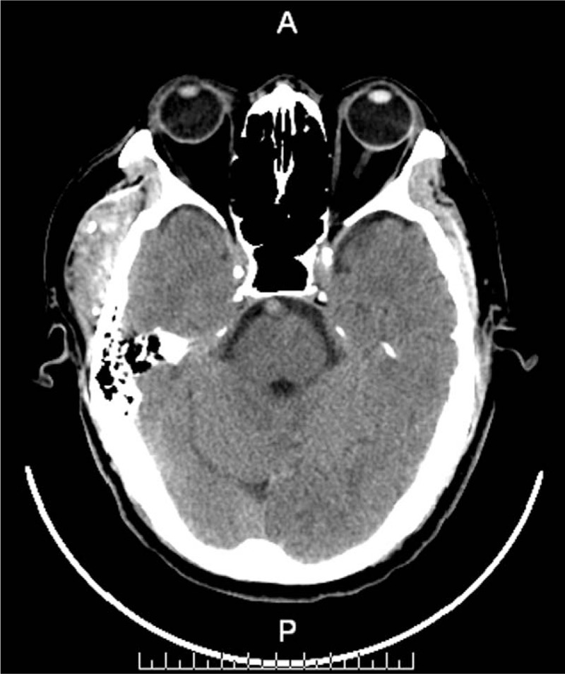
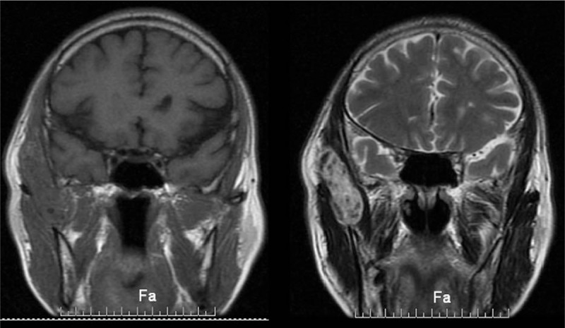
Diagnoses: Computed tomography (CT) scan showed the lesion across the zygomatic arch, with many calcified nodules differ in sizes and no erosion to the bone, magnetic resonance imaging (MRI) showed an oval lesion with hypointense and isointense on T2-weighted imaging within the temporal muscle, and preoperation diagnosis was hemangioma.
Interventions: The tumor was resected under general anesthesia.
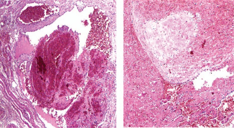
Outcomes: The mass was excised completely, and the histopathology examination confirmed the diagnosis of cavernous hemangioma with prominent formation of phleboliths. The patient recovered very well without dysfunctions.
Lessons: Cavernous hemangioma should be suspected when mass occurs in this region. CT and MRI are important for the early diagnosis of tumor, and resection the tumor completely is recommended.
Conflict of interest statement
The authors have no funding and conflicts of interest to disclose.
Figures
References
-
- Enzinger FM, Weiss SW. Soft tissue tumors. 1988;St Louis, MO: CV Mosby, 512–514.
-
- Fergusson ILC. Hemangiomata of skeletal muscle. Br J Surg 1972;59:634–7. - PubMed
-
- Scott JES. Hemangiomata in skeletal muscle. Br J Surg 1957;44:496–501. - PubMed
-
- Morris SJ, Adams H. Case report: paediatric intramuscular haemangiomata—don’t overlook the phlebolith!. Br J Radiol 1995;68:208–11. - PubMed
-
- Odabasi AO, Metin KK, Mutlu C, et al. Intramuscular hemangioma of the masseter muscle. Eur Arch Otorhinolaryngol 1999;256:366–9. - PubMed
Publication types
MeSH terms
LinkOut - more resources
Full Text Sources
Other Literature Sources
