. 2018 Feb 23;62(3):e01966-17.
doi: 10.1128/AAC.01966-17. Print 2018 Mar.

Crystal Structure of NisI in a Lipid-Free Form, the Nisin Immunity Protein, from Lactococcus lactis

Affiliations

Crystal Structure of NisI in a Lipid-Free Form, the Nisin Immunity Protein, from Lactococcus lactis

Jin Hee Jeong et al. Antimicrob Agents Chemother. .

Abstract

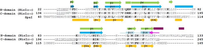
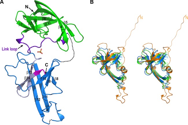
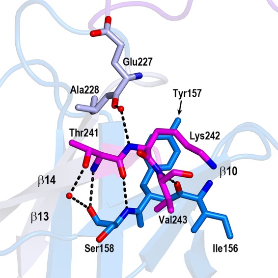
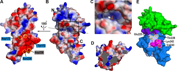
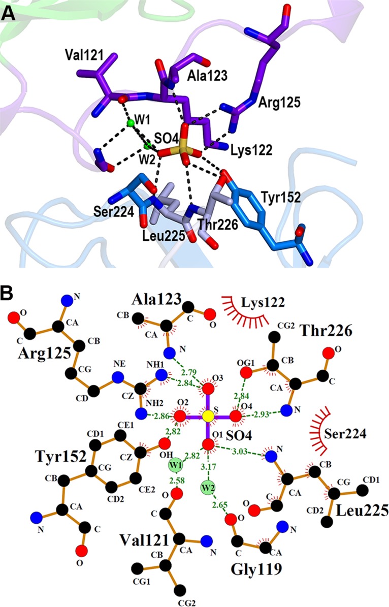
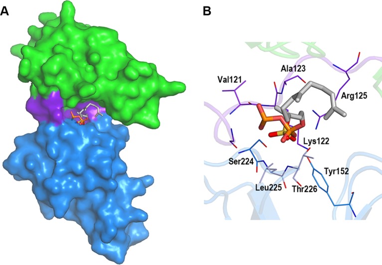
Nisin is a lantibiotic, a member of a family of polypeptides containing lanthionine with antimicrobial activity. Nisin-producing microorganisms require immunity proteins for self-protection from nisin itself. Lactococcus lactis, a microorganism that synthesizes nisin, has an integral NisFEG ABC transporter and an NisI lipoprotein that function in nisin immunity. Here, we present the crystal structure of the full length of NisI22-C, a lipid-free form of NisI, determined at 1.9-Å resolution. As with the nuclear magnetic resonance (NMR) structures of the N- and C-terminal domains of NisI, NisI22-C is composed of N- and C-terminal domains, both of which display a fold similar to that found in SpaI, a lipoprotein with immunity against subtilin in Bacillus subtilis The full-length structure of NisI22-c reveals a large, deep cleft by the interdomain association, one side of which is occupied by the residues important for immunity. Opposite the cleft, a shallow groove is found where nisin-interacting residues are distributed in the periphery composed of the C-terminal negative patch. Based on a sulfate ion found in the large and deep cleft, a model of NisI in complex with a farnesyl diphosphate backbone of lipid II is proposed, suggesting a mechanism for increasing the chances of encountering nisin.

Keywords: Lactococcus lactis; NisI; SpaI; X-ray crystallography; crystal structure; immunity protein; lantibiotics; nisin.


Figures

FIG 1
FIG 1
Structure-guided sequence alignment. Structure-guided sequence alignment of the N- and C-terminal domains of NisI22-C from L. lactis with those of SpaI from B. subtilis. Identical and similar residues are shown in bold, gaps are shown with hyphens, and internal disordered residues are underlined. The residue numbers are relative to those of the methionine residues in the prelipoproteins before processing of the N-terminal leader sequence. Secondary structures (β-strand, arrows; α-helix, rectangles) of NisI22-C are shown above the alignment and colored in the scheme as in Fig. 2A, and those of SpaI are shown in orange below the alignment. For the 21-amino-acid fragment of the C-terminal domain of NisI22-C, which includes the 5-amino-acid C-terminal fragment (pink), the loops of the secondary structures are also shown with thick lines. The numbers of the secondary structures are assigned only to β-strands comprising the β-barrel structure, and those of SpaI are based on the published report (29).
FIG 2
FIG 2
Structure of NisI22-C. (A) Ribbon diagram of NisI22-C. The region comprising the β-barrel structure (residues 22 to 114) and the link loop region (residues 115 to 136) of NisI22-C are displayed in green and purple, respectively. The C-terminal domain (residues 134 to 245) of NisI22-C is colored dark blue, except for the 21-amino-acid fragment, which is light gray, and the C-terminal 5 amino acids, which are magenta. The sulfate ion, which is derived from the crystallization solution, is shown in the stick model (oxygen in red and sulfur in yellow). Each secondary structure as described in Fig. 1 and the N- and C termini, are labeled. (B) Stereo view of the structural superposition of the N- and C-terminal domains of NisI22-C and SpaI (PDB accession number 2LVL). For clarity, the link loop region of the N-terminal domain of NisI22-C is not included. The ribbon diagrams of the N- and C-terminal domains of NisI22-C and SpaI are displayed in green, blue, and orange, respectively. The N- and C-terminal domains can be superimposed on those of SpaI with RMSDs of 3.1 Å and 2.8 Å over 87 and 87 Cα atoms, respectively. The domain boundaries of the N- and C-terminal domains of NisI22-C and the N- and C termini of SpaI are indicated with the N and C labels.
FIG 3
FIG 3
The 5-amino-acid fragment in the β-barrel core of the C-terminal domain. Closeup view of the interactions of the 5-amino-acid fragment with the neighboring residues in the β-barrel structure. Only three residues (Thr241, Lys242, and Val243) are shown. The last two C-terminal residues (Gly244 and Asn245) are invisible due to the poor electron density map. The residues belonging to the 5-amino-acid fragment (magenta), the interacting residues from β13 (gray), and the interacting residues from β10 (blue) are shown in the stick models. The dashed lines and red ovals indicate the hydrogen bonds and water molecules, respectively.
FIG 4
FIG 4
The cleft and groove of NisI22-C. The distribution of the electrostatic potentials of NisI22-C. The potentials are shown in the range of −80 kT/e (red, negative potential) to + 80 kT/e (blue, positive potential). The views are from the shallow groove (A) and from the deep cleft (B). The residues involved in the interaction with nisin (in blue rectangles) and residues belonging to the C-terminal 21-amino-acid fragment (in gray rectangle) are shown on the negative patch of the C-terminal domain. The orientation of the surface diagram in (B) is almost identical to that of Fig. 2A. The sulfate ion is also shown in the stick model. (C) Zoomed-in view of the deep cleft. The region marked with the rectangle in (B) is magnified. (D) View of cross section after clipping the molecules along the straight black line as in (B). The view is facing the N-terminal domain. The solvent-inaccessible areas are shown in gray. The sulfate ion and the water molecule below the sulfate ion are shown with the stick model and the green sphere, respectively. (E) The distribution of residues critical for the immunity. Residues belonging to the C-terminal 21-amino-acid fragment (light gray) and the C-terminal 5-amino-acid fragment (magenta), which is also a part of the C-terminal 21-amino-acid fragment, are plotted on the surface model of NisI22-C using the same color scheme as in Fig. 2A.
FIG 5
FIG 5
Sulfate ion in the deep cleft of NisI22-C. (A) Detailed view of the interactions of the sulfate ion with NisI22-C. The side chain and backbone atoms making contacts with a sulfate ion are shown with the stick models (carbon, as in Fig. 1B) (nitrogen in blue, oxygen in red, and sulfur in yellow). The hydrogen bonds are represented with dashed lines, and the green ovals indicate water-mediated interactions. (B) Two-dimensional view of the interaction. The diagram was produced using LigPlot (46). The hydrogen bonds are shown using dashed lines, and hydrophobic contacts are shown using an arc with spokes radiating to a binding partner. Water molecules (W1 and W2) are also included.
FIG 6
FIG 6
Simulated docking of a farnesyl diphosphate backbone variant of lipid II. The diphosphate group and 3 isoprene repeats were docked on the sulfate ion in the cleft. (A) Modeled farnesyl diphosphate variant and sulfate ion in the cleft of NisI22-C shown with the surface model (the N-terminal domain, green; the link region, purple; the C-terminal domain, dark blue). The farnesyl diphosphate variant and sulfate ion are shown with stick models (P in orange, S in yellow, O in red, and C in cyan). The view is from the deep cleft. (B) Zoomed-in view in the cleft. Residues making contacts with a sulfate ion are shown in stick models as in Fig. 5B.

References

    1. Cotter PD, Ross RP, Hill C. 2013. Bacteriocins—viable alternative to antibiotics? Nat Rev Microbiol 11:95–105. doi:10.1038/nrmicro2937. - DOI - PubMed
    1. Severina E, Severin A, Tomasz A. 1998. Antibacterial efficacy of nisin against multidrug-resistant Gram-positive pathogens. J Antimicrob Chemother 41:341–347. doi:10.1093/jac/41.3.341. - DOI - PubMed
    1. Cotter PD, Hill C, Ross RP. 2005. Bacterial lantibiotics: strategies to improve therapeutic potential. Curr Protein Pept Sci 6:61–75. doi:10.2174/1389203053027584. - DOI - PubMed
    1. Piper C, Cotter PD, Ross RP, Hill C. 2009. Discovery of medically significant lantibiotics. Curr Drug Discov Technol 6:1–18. doi:10.2174/157016309787581075. - DOI - PubMed
    1. Willey JM, van der Donk WA. 2007. Lantibiotics: peptides of diverse structure and function. Annu Rev Microbiol 61:477–501. doi:10.1146/annurev.micro.61.080706.093501. - DOI - PubMed

Publication types

MeSH terms

LinkOut - more resources