. 2018 Jan 30;115(5):E1061-E1068.
doi: 10.1073/pnas.1715489115. Epub 2018 Jan 8.

Detergent-extracted Volvox model exhibits an anterior-posterior gradient in flagellar Ca2+ sensitivity

Affiliations

Detergent-extracted Volvox model exhibits an anterior-posterior gradient in flagellar Ca2+ sensitivity

Noriko Ueki et al. Proc Natl Acad Sci U S A. .

Abstract

Volvox rousseletii is a multicellular spheroidal green alga containing ∼5,000 cells, each equipped with two flagella (cilia). This organism shows striking photobehavior without any known intercellular communication. To help understand how the behavior of flagella is regulated, we developed a method to extract the whole organism with detergent and reactivate its flagellar motility. Upon addition of ATP, demembranated flagella (axonemes) in the spheroids actively beat and the spheroids swam as if they were alive. Under Ca2+-free conditions, the axonemes assumed planar and asymmetrical waveforms and beat toward the posterior pole, as do live spheroids in the absence of light stimulation. In the presence of 10-6 M Ca2+, however, most axonemes beat three-dimensionally toward the anterior pole, similar to flagella in photostimulated live spheroids. This Ca2+-dependent change in flagellar beating direction was more conspicuous near the anterior pole of the spheroid, but was not observed near the posterior pole. This anterior-posterior gradient of flagellar Ca2+ sensitivity may explain the mechanism of V. rousseletii photobehavior.

Keywords: Volvox; calcium; flagella; phototaxis.


Conflict of interest statement

The authors declare no conflict of interest.

Figures
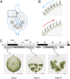
Fig. 1.
Fig. 1.
Life cycle and swimming style of V. rousseletii. (A) Schematic diagrams showing the directions of flagella-generated fluid streams (blue arrows) around a spheroid. The A–P current with a slight tilt causes the spheroid to swim while rotating bodily in a counterclockwise direction when viewed from the posterior pole. The dark gray circles in the spheroid represent gonidia that localize mainly in the posterior hemisphere. (B) Schematic diagrams showing flagellar beating in the somatic cells on the spheroid surface in an anterior region (box in A). Flagellar waveforms in posterior (Top) and anterior (Bottom) beating modes are shown. The blue and red arrows indicate the current generated in the two modes. Modified from ref. . Note that the cell closest to the anterior pole is depicted with a large eyespot (a red spot), with the eyespot size gradually decreasing with distance from the pole. (C) Asexual life cycle of V. rousseletii cultured on a 16-h/8-h light/dark cycle. Spheroids in three stages (termed stages I–III in this study) were used: Stage I represents newly hatched spheroids, in which gonidia (next-generation embryos) just start cell division (Left); stage II represents spheroids, in which gonidia are undergoing cleavage (Center); and stage III represents expanded spheroids, in which daughter spheroids have undergone inversion and are ready to hatch (Right). A, anterior pole; P, posterior pole. (Scale bar: 200 μm; note that the bar length differs in the three photographs.)
Fig. 2.
Fig. 2.
Detergent sensitivity of flagella along the A–P axis of spheroids at the three developmental stages. (A) Schematic diagrams showing the trapping method for demembranation of Volvox spheroids. (i–iii) Drop of culture containing several spheroids was placed on a glass slide. The medium was drained with a pipette, and washing solution (HMDEK; Materials and Methods) was added. (iv) After a few minutes, spheroids were withdrawn with a pipette and (v and vi) placed in a perfusion chamber. (vii) Top view of the chamber. Solution containing either Igepal or ATP was perfused. (B) Degree of demembranation in the three stages of spheroids treated with different concentrations of detergent. The demembranation index was calculated for the anterior or posterior hemisphere as follows: If visual inspection showed that all flagella in the examined area were stopped after detergent perfusion, the index was 1; if any flagella were moving, the index was 0. Index values were counted in three to 26 spheroids, with the average defined as the demembranation index. The arrowhead in stage I indicates the conditions used for Fig. 5, and the arrows in stage II indicate the conditions used for Figs. 3 and 4. The filled arrow and squares are located in the anterior hemisphere, and the open arrow and circles are located in the posterior hemisphere.
Fig. 3.
Fig. 3.
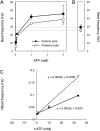
ATP-dependent beat frequency in the axonemes of detergent-extracted Volvox (DEV). (A) Beat frequency of axonemes near the anterior and posterior poles in DEVs at different ATP concentrations. Three to six axonemes were measured. (B) Beat frequency of flagella in live cells near the anterior and posterior poles. The average in six flagella is shown for each. (C) Double-reciprocal plot of the data in A. Intercepts yielded apparent maximal beat frequencies of 43.5 Hz (anterior region) and 48.8 Hz (posterior region) and apparent Michaelis constants of 0.10 mM (anterior) and 0.22 mM ATP (posterior).
Fig. 4.
Fig. 4.
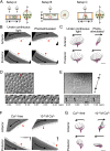
Ca2+-dependent changes in the direction of axonemal beating. (A) Experimental setups for observation of live or demembranated spheroids in a chamber. Glass slides and coverslips (gray), spacers (orange), the A–P (A↔P) direction of the spheroids, and the regions observed (red boxes) are indicated. (Left) In setup A, a spheroid was sandwiched between a glass slide and a coverslip, and regions near the anterior pole (A) and posterior pole (P) were observed. (Center) In setup B, a spheroid, after gentle pressing, was placed between the glass slide and a coverslip separated by thick spacers, and the region near the anterior pole was observed. (Right) In setup C, a spheroid was attached to the glass slide with 1% polyethyleneimine, and the region near the equator was observed. (B) Frames from high-speed recordings of regions near the anterior (Top) and posterior (Bottom) poles of a live spheroid. The observation using setup A was under stationary conditions in continuous light (Left) and after photostimulation (Right). The direction of the flagellar effective stroke (arrows) was determined by the direction of the typical hook shapes (red arrowheads) in the recovery stroke. After photostimulation, the direction of the flagellar effective stroke reversed in the anterior region. (Scale bar: 100 μm.) (C) Typical sequential flagellar waveforms in a single beating cycle under each condition. Waveforms recorded as in B were traced (time interval of 1/500 s). The typical hook-shaped waveforms appearing in recovery strokes are traced in magenta. (Scale bar: 10 μm.) Photographs show the time course of directional change in the flagellar effective stroke in live spheroids, near the anterior pole in setup B (D, Top) and near the equator in setup C (E, Top). (D and E, Bottom) Photographs show the time series of the change after photostimulation in the boxed cell. The direction of ciliary beating and the time after onset of illumination are shown. (Scale bars: Top, 100 μm; Bottom, 10 μm.) In D, the direction of the effective stroke was almost reversed at 0.27 s and recovered at 4.44 s. The red asterisk indicates a presumptive anterior pole. In E, the direction of the effective stroke rotated ∼90° at 0.72 s and recovered at 1.52 s. The two-headed arrow indicates the approximate A–P direction. (F) High-speed recording of regions near the anterior (Top) and posterior (Bottom) poles of DEVs reactivated in the presence of 0 or 10−6 M Ca2+ in setup A. The ATP concentration was 1 mM. In the presence of Ca2+, the axonemes in the anterior region showed anteriorly directed effective strokes, opposite to those in the absence of Ca2+. However, this change was not observed in the posterior region. Arrowheads indicate the typical hook-shaped waveforms. (Scale bar: 100 μm.) (G) Typical sequential axonemal waveforms traced for a single beating cycle under each condition (time interval of 1/500 s). The hook-shaped waveforms are shown in magenta. (Scale bar: 10 μm.)
Fig. 5.
Fig. 5.
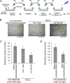
DEV swimming. (A) Schematic diagrams showing the strainer-scooping method for demembranation and reactivation. Spheroids were successively transferred from the culture medium to the washing, demembranation, and reactivation solutions, with ATP added during the final step to reactivate motility. (B) Swimming trajectories of spheroids in live and demembranated/reactivated spheroids. Movements of the spheroids were video recorded and tracked for 5 s. (Left) Live spheroids under continuous light before demembranation. (Center and Right) DEVs reactivated with 1 mM ATP in the absence and presence of 10−6 M Ca2+. (Scale bars: 5 mm.) (C) Swimming velocities (n = 20 for live spheroids, n = 30 for DEVs under each condition) calculated from the swimming trajectories for 5 s. DEVs that did not move at all were not counted. (D) Swimming velocities of live spheroids before and right after photostimulation calculated from the swimming trajectories for 3 s.

Similar articles

Cited by

References

    1. Höhn S, Hallmann A. There is more than one way to turn a spherical cellular monolayer inside out: Type B embryo inversion in Volvox globator. BMC Biol. 2011;9:89. - PMC - PubMed
    1. Solari CA, Michod RE, Goldstein RE. Volvox barberi, the fastest swimmer of the Volvocales (Chlorophyceae) J Phycol. 2008;44:1395–1398. - PubMed
    1. Ueki N, Matsunaga S, Inouye I, Hallmann A. How 5000 independent rowers coordinate their strokes in order to row into the sunlight: Phototaxis in the multicellular green alga Volvox. BMC Biol. 2010;8:103. - PMC - PubMed
    1. Foster KW, Smyth RD. Light antennas in phototactic algae. Microbiol Rev. 1980;44:572–630. - PMC - PubMed
    1. Bessen M, Fay RB, Witman GB. Calcium control of waveform in isolated flagellar axonemes of Chlamydomonas. J Cell Biol. 1980;86:446–455. - PMC - PubMed

Publication types

LinkOut - more resources