. 2018 Jan 23;115(4):768-773.
doi: 10.1073/pnas.1718709115. Epub 2018 Jan 8.

Structure of the C1r-C1s interaction of the C1 complex of complement activation

Affiliations

Structure of the C1r-C1s interaction of the C1 complex of complement activation

Jamal O M Almitairi et al. Proc Natl Acad Sci U S A. .

Abstract

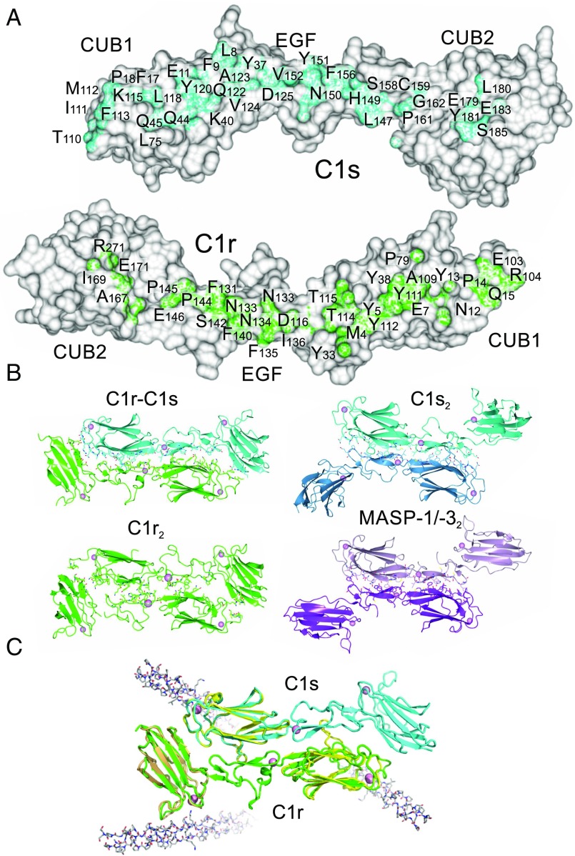
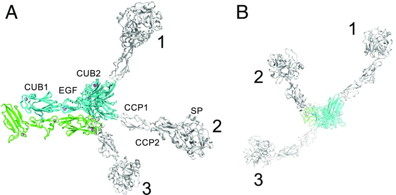
The multiprotein complex C1 initiates the classical pathway of complement activation on binding to antibody-antigen complexes, pathogen surfaces, apoptotic cells, and polyanionic structures. It is formed from the recognition subcomponent C1q and a tetramer of proteases C1r2C1s2 as a Ca2+-dependent complex. Here we have determined the structure of a complex between the CUB1-EGF-CUB2 fragments of C1r and C1s to reveal the C1r-C1s interaction that forms the core of C1. Both fragments are L-shaped and interlock to form a compact antiparallel heterodimer with a Ca2+ from each subcomponent at the interface. Contacts, involving all three domains of each protease, are more extensive than those of C1r or C1s homodimers, explaining why heterocomplexes form preferentially. The available structural and biophysical data support a model of C1r2C1s2 in which two C1r-C1s dimers are linked via the catalytic domains of C1r. They are incompatible with a recent model in which the N-terminal domains of C1r and C1s form a fixed tetramer. On binding to C1q, the proteases become more compact, with the C1r-C1s dimers at the center and the six collagenous stems of C1q arranged around the perimeter. Activation is likely driven by separation of the C1r-C1s dimer pairs when C1q binds to a surface. Considerable flexibility in C1s likely facilitates C1 complex formation, activation of C1s by C1r, and binding and activation of downstream substrates C4 and C4b-bound C2 to initiate the reaction cascade.

Keywords: X-ray crystallography; classical pathway; complement; structural biology.


Conflict of interest statement

The authors declare no conflict of interest.

Figures

Fig. 1.
Fig. 1.
Structure of the C1r-C1s heterodimer. (A) The traditional model first proposed in ref. . (B) Stacked tetramer model (7). Black dots indicate the positions of the binding sites for the collagen-like domains of C1q (10). (C) Gel filtration of CUB1-EGF-CUB2 fragments of C1s, C1r, and an equimolar mixture of C1r and C1s fragments in Ca2+ (solid line) and EDTA (black dotted line). Two different loading concentrations of C1r and C1s fragments are shown (1 and 0.5 mg/mL), and 50 µL was loaded in each case. Samples were separated on a Superdex 200 column (10/30) equilibrated in 20 mM Tris pH 7.4, containing 150 mM NaCl. Elution positions of aldolase (158 kDa), conalbumin (75 kDa), and ovalbumin (43 kDa) are shown. (D) Structure of the C1r-C1s heterodimer formed from the CUB1-EGF-CUB2 fragment of each protease. Ca2+ is shown as pink spheres; carbohydrates, as white sticks.
Fig. 2.
Fig. 2.
The C1r-C1s interface. (A) Residues buried at the heterodimer interface. (B) Dimers formed from the CUB1-EGF-CUB2 domains of C1r, C1s (PDB ID code 4LMF) and MASP-1/-3 (PDB ID code 3DEM). (C) The C1r-C1s dimers superposed with CUB1-collagen (PDB ID code 4LOR; yellow) and CUB2 collagen structures (PDB ID code 3POB; light brown), showing the position of the collagen-binding sites. Collagen is shown in gray.
Fig. 3.
Fig. 3.
Flexibility in C1s. Top (A) and side (B) views of the C1r-C1s dimer showing three different orientations of the CUB2 domain of C1s. (1) is from PDB ID code 4LMF. (2) and (3) show the two conformations of the C1s fragment in the two heterodimers of the asymmetric unit in the C1r-C1s crystals. The CUB1-EGF-CUB2 structures are overlaid with structures of the CUB2-CCP1 (PDB ID code 4LOS) and CCP1-CCP2-SP (PDB ID code 4J1Y) regions of C1s (white), to show the effects of flexibility at the EGF-CUB2 junction on the full-length C1s polypeptide.
Fig. 4.
Fig. 4.
Proposed mechanism of assembly of C1. (A) C1r2C1s2 adopts an extended structure in solution (Left), in which the two C1r-C1s dimers are linked by a central interaction between the catalytic domains of C1r. It folds up (Middle) to form a more compact structure to bind to the six collagenous stems of C1q (Right). Contacts between the catalytic domains of C1r prevent one C1r polypeptide from activating its partner. Black dots show the positions of the binding sites for the collagen-like domains of C1q (10). (B) Model of C1 generated by rigid-body fitting to SAXS data. C1q is in gray, C1r is in green, and C1s is in blue. (C) Rigid-body fit to scattering data SASDB38. I(s) is the intensity, and s is the scattering vector. The χ2 value for the fit is 2.9. The fit is shown as a solid line, and the residuals to the fit are shown below with a scale of ±0.5.

Comment in

  • Reply to Mortensen et al.: The zymogen form of complement component C1.
    Almitairi JOM, Venkatraman Girija U, Furze CM, Simpson-Gray X, Badakshi F, Marshall JE, Schwaeble WJ, Mitchell DA, Moody PCE, Wallis R. Almitairi JOM, et al. Proc Natl Acad Sci U S A. 2018 Apr 24;115(17):E3867-E3868. doi: 10.1073/pnas.1804497115. Epub 2018 Apr 13. Proc Natl Acad Sci U S A. 2018. PMID: 29654143 Free PMC article. No abstract available.
  • Models of the complement C1 complex.
    Mortensen SA, Sander B, Jensen RK, Pedersen JS, Golas MM, Thiel S, Andersen GR. Mortensen SA, et al. Proc Natl Acad Sci U S A. 2018 Apr 24;115(17):E3866. doi: 10.1073/pnas.1803577115. Epub 2018 Apr 13. Proc Natl Acad Sci U S A. 2018. PMID: 29654144 Free PMC article. No abstract available.

References

    1. Ricklin D, Hajishengallis G, Yang K, Lambris JD. Complement: A key system for immune surveillance and homeostasis. Nat Immunol. 2010;11:785–797. - PMC - PubMed
    1. Gaboriaud C, Ling WL, Thielens NM, Bally I, Rossi V. Deciphering the fine details of C1 assembly and activation mechanisms: “Mission impossible”? Front Immunol. 2014;5:565. - PMC - PubMed
    1. Wallis R, Mitchell DA, Schmid R, Schwaeble WJ, Keeble AH. Paths reunited: Initiation of the classical and lectin pathways of complement activation. Immunobiology. 2010;215:1–11. - PMC - PubMed
    1. Forneris F, Wu J, Gros P. The modular serine proteases of the complement cascade. Curr Opin Struct Biol. 2012;22:333–341. - PubMed
    1. Strang CJ, Siegel RC, Phillips ML, Poon PH, Schumaker VN. Ultrastructure of the first component of human complement: Electron microscopy of the crosslinked complex. Proc Natl Acad Sci USA. 1982;79:586–590. - PMC - PubMed

Publication types