. 2018 Jan 23;115(4):E620-E629.
doi: 10.1073/pnas.1715378115. Epub 2018 Jan 8.

Dysregulation of cotranscriptional alternative splicing underlies CHARGE syndrome

Affiliations

Dysregulation of cotranscriptional alternative splicing underlies CHARGE syndrome

Catherine Bélanger et al. Proc Natl Acad Sci U S A. .

Abstract

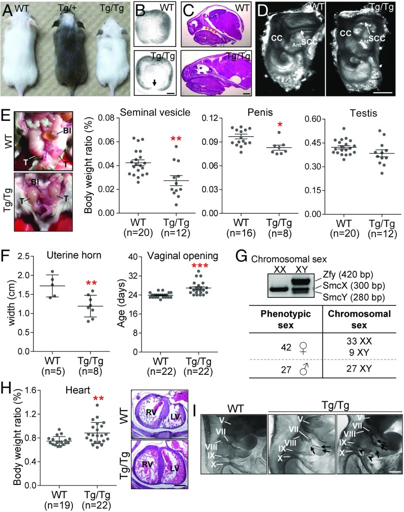
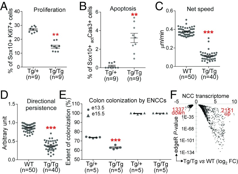
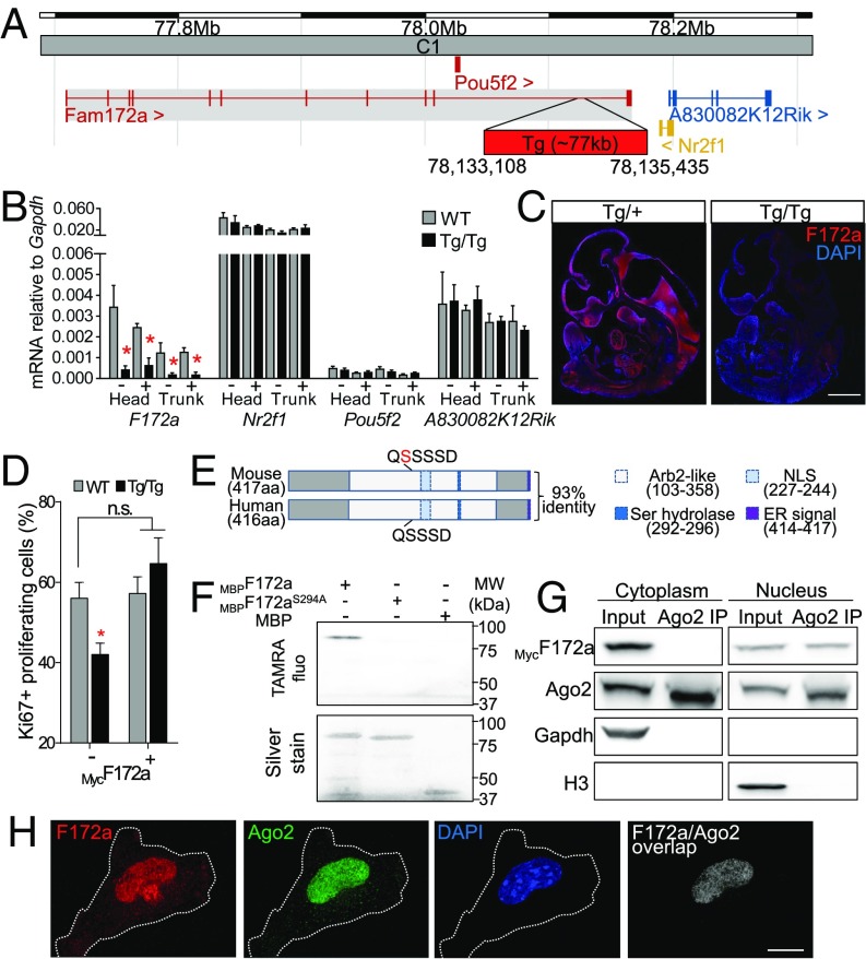
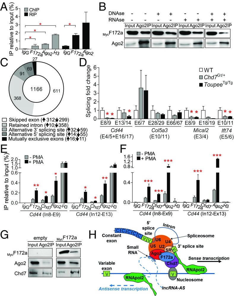
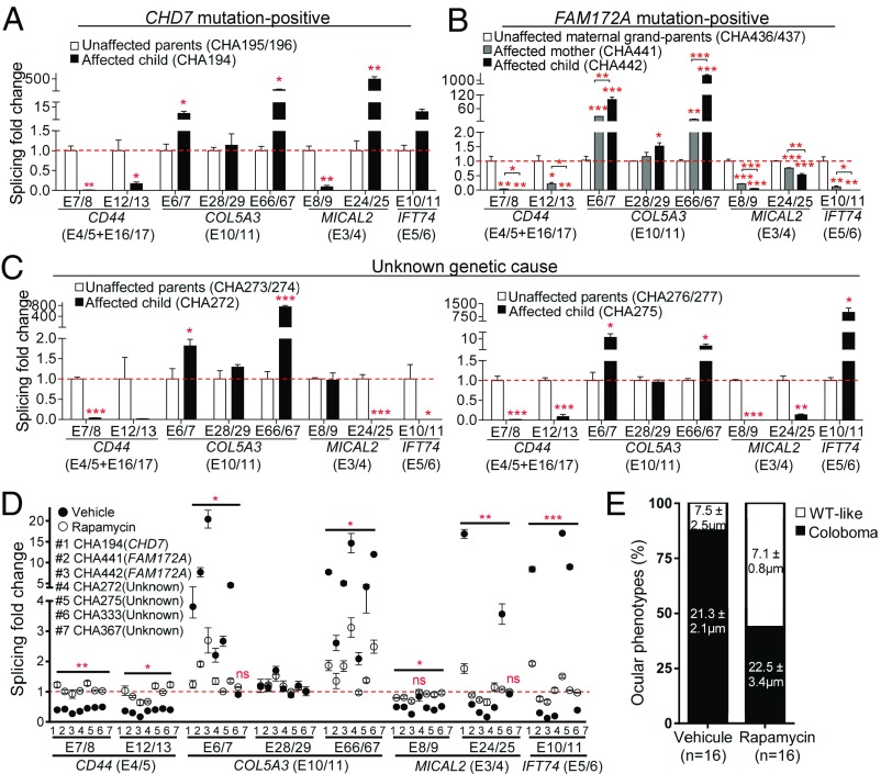
CHARGE syndrome-which stands for coloboma of the eye, heart defects, atresia of choanae, retardation of growth/development, genital abnormalities, and ear anomalies-is a severe developmental disorder with wide phenotypic variability, caused mainly by mutations in CHD7 (chromodomain helicase DNA-binding protein 7), known to encode a chromatin remodeler. The genetic lesions responsible for CHD7 mutation-negative cases are unknown, at least in part because the pathogenic mechanisms underlying CHARGE syndrome remain poorly defined. Here, we report the characterization of a mouse model for CHD7 mutation-negative cases of CHARGE syndrome generated by insertional mutagenesis of Fam172a (family with sequence similarity 172, member A). We show that Fam172a plays a key role in the regulation of cotranscriptional alternative splicing, notably by interacting with Ago2 (Argonaute-2) and Chd7. Validation studies in a human cohort allow us to propose that dysregulation of cotranscriptional alternative splicing is a unifying pathogenic mechanism for both CHD7 mutation-positive and CHD7 mutation-negative cases. We also present evidence that such splicing defects can be corrected in vitro by acute rapamycin treatment.

Keywords: CHARGE syndrome; Fam172a; alternative splicing; neural crest cells; sex reversal.


Conflict of interest statement

The authors declare no conflict of interest.

Figures

Fig. 1.
Fig. 1.
Major and minor features of CHARGE syndrome in ToupeeTg/Tg mice. (A) Comparison between WT, ToupeeTg/+, and ToupeeTg/Tg animals at P25. (B) Bright-field images of E12.5 eyes showing incomplete closure of the choroidal fissure in ToupeeTg/Tg embryos (n = 10 WT, n = 14 ToupeeTg/Tg). (C) H&E-stained sagittal sections of E18.5 heads (n = 10 WT, n = 14 ToupeeTg/Tg) with asterisks indicating cleft palate. (D) Bright-field images of Alizarin red- and Alcian blue-stained inner ears from P25 mice (n = 7 WT, n = 11 ToupeeTg/Tg). AntSCC, anterior semicircular canal; CC, common crus. (E) Overview of genital anomalies in P25 ToupeeTg/Tg males (Left, cryptorchidism; Middle, smaller androgen-sensitive seminal vesicles and penis; Right, normally sized testes). B1, bladder; T, testes. (F) ToupeeTg/Tg females present hypoplastic uterine horns at P25 (Left) and delayed opening of the vaginal cavity after P20 (Right). (G) PCR-based sexing of ToupeeTg/Tg animals revealed male-to-female sex reversal for 25% of XY animals. (H) A subset of ToupeeTg/Tg mice display heart malformation (Left, increased weight at P25; Right, hypertrophy of the left ventricle at E15.5). LV, left ventricle; RV, right ventricle. (I) Whole-mount staining of cranial nerves in E10.5 embryos using antineurofilament immunohistochemistry. ToupeeTg/Tg embryos exhibit supernumerary sprouting in the facial (VII) nerve (arrows) and extensive mingling between glossopharyngeal (IX) and vagal (X) nerves (arrowheads). *P ≤ 0.05, **P ≤ 0.01, ***P ≤ 0.001 (Student’s t test). [Scale bar: 50 µm (B); 1 mm (C and D); 500 µm (H and I).]
Fig. 2.
Fig. 2.
Global impairment of NCC development in ToupeeTg/Tg embryos. (A and B) Quantification of Ki67+ proliferating (A) and actCasp3+ apoptotic (B) NCCs (expressed in percentage of Sox10+ NCCs) in 30-μm transverse sections of E10.5 embryos at hindlimb level (ToupeeTg/+ vs. ToupeeTg/Tg). (C and D) Quantification of NCC migration speed (C) and movement persistence (D) in E10.5 embryos (WT;G4-RFP vs. ToupeeTg/Tg;G4-RFP). (E) Quantification of the extent of colon colonization by enteric NCCs (expressed in percentage of colon length from cecum to anus) at E13.5 and E15.5 (ToupeeTg/+;G4-RFP vs. ToupeeTg/Tg;G4-RFP). (F) Volcano plot summarizing a RNA-seq–based analysis of differential gene expression levels in E10.5 NCCs (WT;G4-RFP vs. ToupeeTg/Tg;G4-RFP). Only genes modulated at least 1.5-fold with a P value below 0.01 are displayed. **P ≤ 0.01 and ***P ≤ 0.001 (Student’s t test).
Fig. 3.
Fig. 3.
Functional characterization of Fam172a, the Toupee causative gene. (A) Schematic representation of the Toupee transgene insertion site in cytoband C1 of chromosome 13 (adapted from the Ensembl website), where ∼10 copies of a tyrosinase minigene are inserted in a 2,327-bp deletion in the last intron of Fam172a (red box). (B) RT-qPCR analysis of gene expression in E10.5 embryos (WT;G4-RFP vs. ToupeeTg/Tg;G4-RFP). Transcript levels of genes around the transgene insertion site were monitored in FACS-recovered NCCs (+) and non-NCCs (−) from the head and the trunk (n = 3 per condition). (C) Immunofluorescence labeling of the Fam172a protein (red) in sagittal sections of E10.5 mouse embryos (ToupeeTg/+ vs. ToupeeTg/Tg, n = 3 per genotype). DAPI (blue) was used to counterstain nuclei. (D) Quantification of proliferation in cultures of dissociated E10.5 embryos (WT vs. ToupeeTg/Tg; n = 3 per condition) after transfection with a MycFam172a-expressing vector. (E) Schematic representation of the functional domains of mouse Fam172a protein compared with its human ortholog. The serine in position 215 (highlighted in red) corresponds to the supernumerary amino acid between mouse (417 aa) and human (416 aa) sequences. Arb2-like, domain homologous to yeast Arb2 (Argonaute-binding protein 2); ER, endoplasmic reticulum retention signal; NLS, nuclear localization signal; Ser hydrolase, esterase-like serine hydrolase motif. (F) Hydrolase activity is demonstrated by covalent binding of a TAMRA-labeled fluorophosphonate probe on MBPFam172a protein (Upper). No binding is detected on MBPFam172a bearing a S294A mutation nor on the MBP tag alone. Lower panel shows that silver-stained MBP-tagged Fam172a proteins (∼90 kDa; slightly higher in the presence of fluorophosphonate probe) and MBP alone (42 kDa) were all present at the expected size in the same gel. (G) Co-IP assays using cytoplasmic (Gapdh+) and nuclear (H3+) fractions of Neuro2a cells transfected with a MycFam172a-expressing vector (n = 3). Inputs correspond to 10% of protein extracts used for IP. (H) Double immunofluorescence labeling of Fam172a and Ago2 in dissociated cells obtained from WT E10.5 embryos (n = 7) and counterstained with DAPI. (Right) The overlap of Fam172a and Ago2 signals (Pearson’s correlation coefficient of 0.82). *P ≤ 0.05 (Student’s t test). [Scale bar: 500 μm (C); 25 μm (H).]
Fig. 4.
Fig. 4.
Both Fam172a and Chd7 play a role in alternative splicing. (A) Untargeted ChIP and RIP assays in Neuro2A cells transfected with a Fam172a-expressing plasmid (n = 3). (B) Co-IP assays using RNase- and/or DNase-treated whole-cell extracts of Neuro2A cells transfected with a MycFam172a expression vector. Inputs correspond to 10% of protein extracts used for IP (n = 3 per condition). Impact of each treatment on the integrity of proteins, DNA, and RNA is shown in SI Appendix, Fig. S8G. (C) Donut chart showing the distribution of the 1,166 differentially modulated alternative splicing events (P < 0.01; variation in inclusion level ≥0.1) in ToupeeTg/Tg E10.5 NCCs. Upward- and downward-pointing arrows indicate splicing events that are over- and under-represented in ToupeeTg/Tg E10.5 NCCs, respectively. (D) RT-qPCR analysis of splicing events for Cd44, Col5a3, Mical2, and Ift74 in G4-RFP (WT), ToupeeTg/Tg;G4-RFP (ToupeeTg/Tg), and Chd7Gt/+ heads of E12.5 embryos (n = 5 per genotype). Expression levels of variable regions are normalized with levels of corresponding constant regions (indicated between parentheses). (E and F) ChIP (E) and RNA-ChIP (F) assays of the PMA-inducible Cd44 gene in Neuro2a cells. (G) Co-IP assays in Neuro2A cells transfected with empty or MycFam172a-expressing vector. Inputs correspond to 10% of protein extracts used for IP (n = 3 per condition). (H) Potential mode of action of Fam172a and Chd7 in Ago2-mediated alternative splicing (adapted from ref. 21). *P ≤ 0.05, **P ≤ 0.01, ***P ≤ 0.001 (Student’s t test).
Fig. 5.
Fig. 5.
Rapamycin-correctable dysregulation of alternative splicing in CHARGE syndrome patients. (AD) RT-qPCR analysis of splicing events for CD44, COL5A3, MICAL2, and IFT74 in lymphoblastoid cell lines. Expression levels of variable regions were normalized with levels of corresponding constant regions (indicated between parentheses). Results for unaffected parents were combined and used as reference value for calculation of splicing fold change (red dashed line). In AC, each graph depicts the results obtained for a given family (for each individual, n = 9 from three independent experiments). Results for other families can be found in SI Appendix, Fig. S12, while detailed information about each patient can be found in SI Appendix, Table S8. D depicts the results obtained after a 30-min treatment with rapamycin (10 µM) or vehicle only (ethanol), each vertically aligned pair of dots corresponding to a single CHARGE patient (for each individual, n = 6 from two independent experiments). In D, statistic tests refer to the difference between rapamycin and vehicle treatments. *P ≤ 0.05, **P ≤ 0.01, ***P ≤ 0.001, ns, not significant (Student’s t test). (E) Occurrence of coloboma in E12.5 ToupeeTg/Tg embryos (for each condition, n = 8 embryos/16 eyes) following a 3-d in utero exposition to rapamycin (1 mg/kg) or vehicle (20% ethanol). For each phenotypic group (coloboma and WT-like), the average width of choroidal fissure is indicated in their corresponding bar subdivision.

References

    1. Hsu P, et al. CHARGE syndrome: A review. J Paediatr Child Health. 2014;50:504–511. - PubMed
    1. Hale CL, Niederriter AN, Green GE, Martin DM. Response to correspondence to Hale et al. atypical phenotypes associated with pathogenic CHD7 variants and a proposal for broadening CHARGE syndrome clinical diagnostic criteria. Am J Med Genet A. 2016;170:3367–3368. - PubMed
    1. van Ravenswaaij-Arts CM, Blake K, Hoefsloot L, Verloes A. Clinical utility gene card for: CHARGE syndrome—Update 2015. Eur J Hum Genet. 2015 doi: 10.1038/ejhg.2011.45. - DOI - PMC - PubMed
    1. Delahaye A, et al. Familial CHARGE syndrome because of CHD7 mutation: Clinical intra- and interfamilial variability. Clin Genet. 2007;72:112–121. - PubMed
    1. Jongmans MC, et al. Familial CHARGE syndrome and the CHD7 gene: A recurrent missense mutation, intrafamilial recurrence and variability. Am J Med Genet A. 2008;146A:43–50. - PubMed

Publication types

MeSH terms