. 2018 Feb 9;80(2):247-259.
doi: 10.1292/jvms.17-0422. Epub 2018 Jan 1.

Visual evoked potential in rabbits' eyes with subretinal implantation by vitrectomy of Okayama University-type retinal prosthesis (OURePTM)

Affiliations

Visual evoked potential in rabbits' eyes with subretinal implantation by vitrectomy of Okayama University-type retinal prosthesis (OURePTM)

Toshihiko Matsuo et al. J Vet Med Sci. .

Abstract

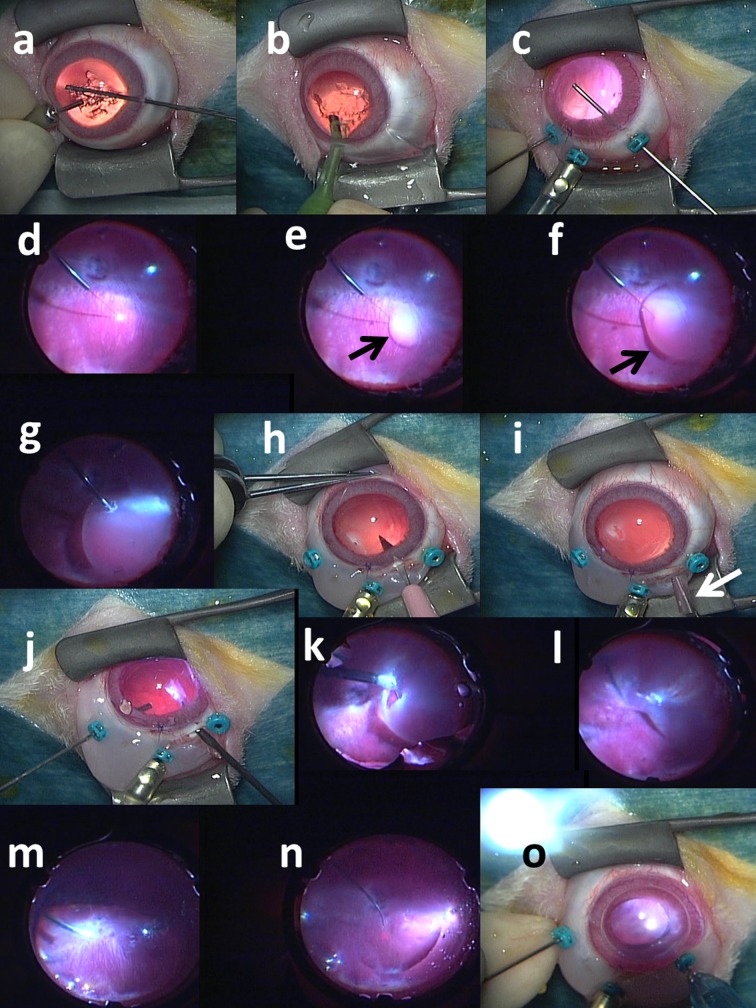
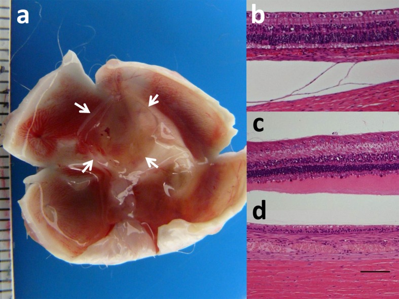
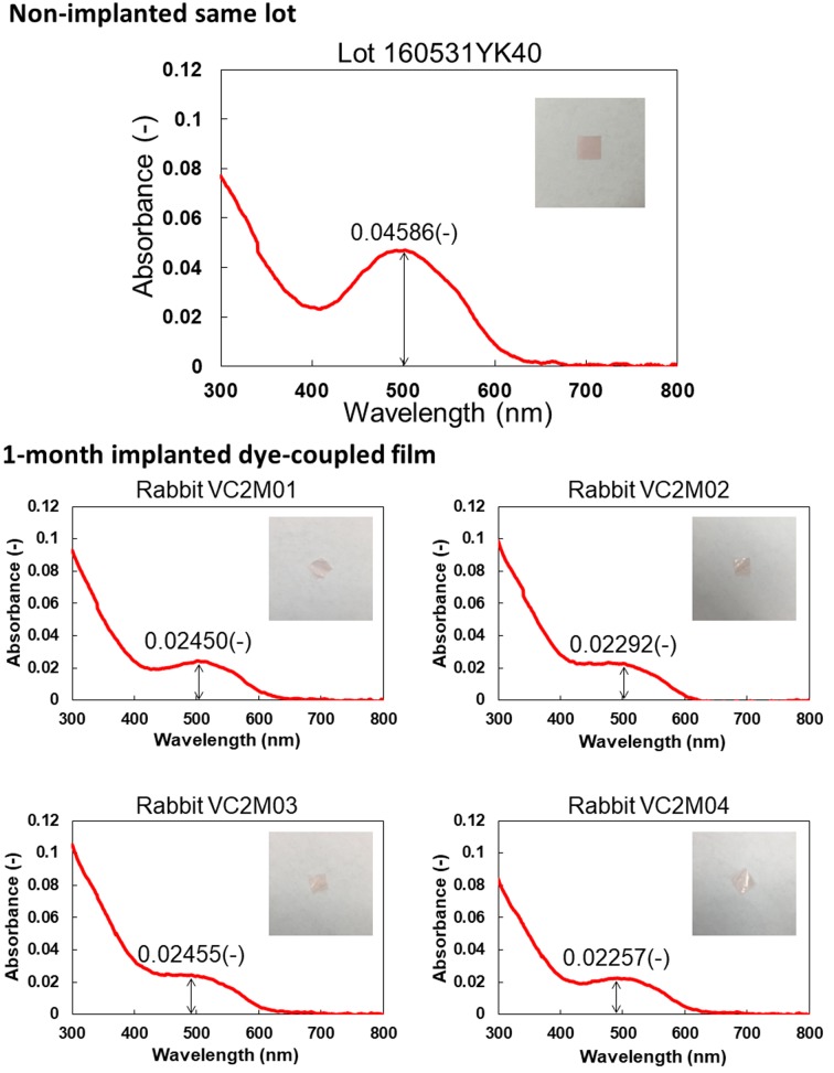
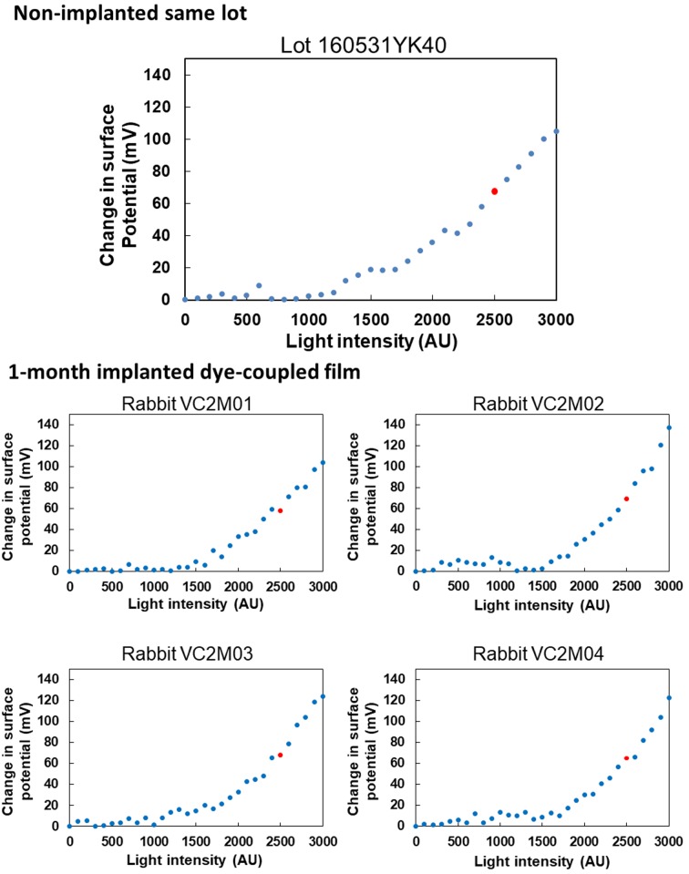
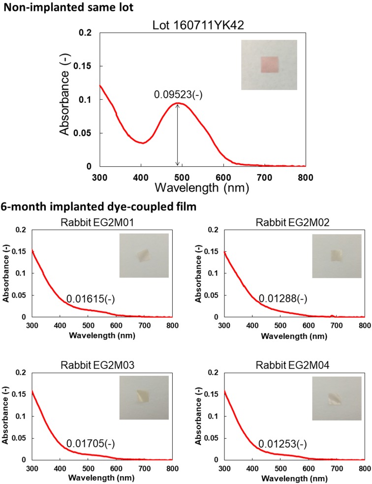
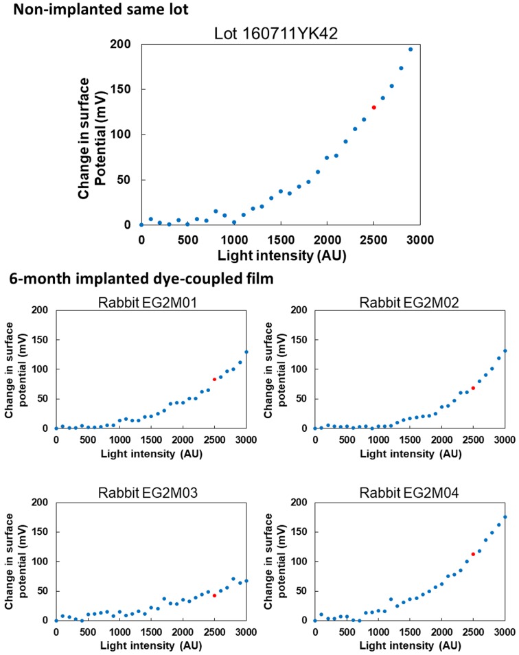
Okayama University-type retinal prosthesis (OURePTM) is a photoelectric dye-coupled polyethylene film which generates electric potential in response to light and stimulates nearby neurons. This study aims to test surgical feasibility for subretinal film implantation and to examine functional durability of films in subretinal space. Dye-coupled films were implanted subretinally by vitrectomy in the right eye of normal white rabbits: 8 rabbits for 1 month and 8 rabbits for 6 months. The implanted films were removed by vitrectomy in 4 of these 8 rabbits in 1-month or 6-month implantation group. The films were also implanted in 4 rhodopsin-transgenic retinal dystrophic rabbits. Visual evoked potential was measured before film implantation as well as 1 or 6 months after film implantation, or 1 month after film removal. The films were successfully implanted in subretinal space of retinal detachment induced by subretinal fluid injection with a 38G polyimide tip. The retina was reattached by fluid-air exchange in vitreous cavity, retinal laser coagulation, and silicone oil injection. The ratios of P2 amplitudes of visual evoked potential in the implanted right eye over control left eye did not show significant changes between pre-implantation and post-implantation or post-removal (paired t-test). In Kelvin probe measurements, 4 pieces each of removed films which were implanted for 1 or 6 months showed proportional increase of surface electric potential in response to increasing light intensity. The film implantation was safe and implanted films were capable of responding to light.

Keywords: dye-coupled thin film retinal prosthesis; rabbit; retinal dystrophy (retinitis pigmentosa); visual evoked potential; vitrectomy.


Figures

Fig. 1.
Fig. 1.
Surgical procedures to implant retinal prosthesis, OURePTM, in right eye of rhodopsin-transgenic white rabbit. a) Lens anterior capsule is cut with 25G vitreous cutter under irrigation with 25G infusion cannula in anterior chamber. b) Lens nucleus and cortex is aspirated with phaco-tip from corneal incision. c) Three 25G trocars are inserted over conjunctiva through sclera into vitreous at 2.5 mm from corneal limbus: a middle trocar is connected with infusion cannula, and the other two trocars are used for vitreous cutter and light guide. Posterior capsule is cut with vitreous cutter. d) After vitreous gel has been cut, subretinal fluid infusion is started with 38G tip. e) Bleb retinal detachment (arrow) is made by 38G tip infusion of BSS-Plus solution. f) Bleb (arrow) is enlarged with further infusion. g) Retinal tear is made by retinal coagulation with 25G bipolar diathermy. h) Scleral incision is made with 22.5° knife after conjunctival incision. i) Rolled-up dye-coupled film (arrow) is inserted through scleral incision with 20G subretinal forceps. j) Rolled-up film is inserted into vitreous with 20G subretinal forceps. k) Rolled-up film is inserted into subretinal space through retinal tear with 20G subretinal forceps. l) Film is now under detachment retina. m) Fluid-air exchange in vitreous cavity is accomplished with 25G vitreous cutter in aspiration mode to reattach the retina. n) Laser photocoagulation is applied around retinal tear. o) Silicone oil is injected in vitreous cavity with 25G tip. Finally, scleral and conjunctival incision is sutured and trocars are removed (not shown).
Fig. 2.
Fig. 2.
Macroscopic view of unfixed and dissected eye with subretinal square dye-coupled film (5 × 5 mm, arrows) in 1-month implantation (a). Light microscopic sections of the retina of the posterior pole near film implantation in normal rabbit with 1-month film implantation (b), rhodopsin-transgenic rabbit with 1-month film implantation (c), and normal rabbit with 1 month observation after removal of 6-month implanted film (d). The photoreceptors are at bottom of photographs. Separation between the choroid and sclera is artifact (b). Photoreceptor outer segments are relatively maintained in normal rabbit (b) while outer segments are shortened with eosin-stained serous fluid in rhodopsin-transgenic rabbit (c). Loss of retinal outer nuclear layer is noted 1 month after removal of 6-month implanted film in normal rabbit (d). Hematoxylin-eosin stain. Scale in ruler is 1 mm in a. Scale bar=100 µm in b, c and d.
Fig. 3.
Fig. 3.
Spectrophotometric absorbance spectra (4 bottom panels) of 4 pieces of dye-coupled films which have been implanted for 1 month in subretinal space of rabbits’ eyes and removed by vitrectomy. Top panel shows absorbance spectrum of the non-implanted same lot. Insets are photographs of films. Values in each panel represent maximum absorbance around the wavelength of 500 nm.
Fig. 4.
Fig. 4.
Surface electric potential in response to increasing light intensity on 4 pieces of dye-coupled films which have been implanted for 1 month in subretinal space of rabbits’ eyes and removed by vitrectomy. Top panel shows surface electric potential on the non-implanted same lot. Red dots on each panel represent electric potential at 2,500 arbitrary unit (AU) of light intensity which corresponds to 300 lux.
Fig. 5.
Fig. 5.
Spectrophotometric absorbance spectra (4 bottom panels) of 4 pieces of dye-coupled films which have been implanted for 6 months in subretinal space of rabbits’ eyes and removed by vitrectomy. Top panel shows absorbance spectrum of the non-implanted same lot. Insets are photographs of films. Values in each panel represent maximum absorbance around the wavelength of 500 nm.
Fig. 6.
Fig. 6.
Surface electric potential in response to increasing light intensity on 4 pieces of dye-coupled films which have been implanted for 6 months in subretinal space of rabbits’ eyes and removed by vitrectomy. Top panel shows surface electric potential on the non-implanted same lot. Red dots on each panel represent electric potential at 2,500 arbitrary unit (AU) of light intensity which corresponds to 300 lux.

References

    1. Alamusi M., Matsuo T., Hosoya O., Uchida T.2017. Visual evoked potential in RCS rats with Okayama University-type retinal prosthesis (OUReP™) implantation. J. Artif. Organs 20: 158–165. doi: 10.1007/s10047-016-0943-4 - DOI - PMC - PubMed
    1. Alamusi M., Matsuo T., Hosoya O., Tsutsui K. M., Uchida T.2015. Vision maintenance and retinal apoptosis reduction in RCS rats with Okayama University-type retinal prosthesis (OUReP™) implantation. J. Artif. Organs 18: 264–271. doi: 10.1007/s10047-015-0825-1 - DOI - PubMed
    1. Alamusi M., Matsuo T., Hosoya O., Tsutsui K. M., Uchida T.2013. Behavior tests and immunohistochemical retinal response analyses in RCS rats with subretinal implantation of Okayama-University-type retinal prosthesis. J. Artif. Organs 16: 343–351. doi: 10.1007/s10047-013-0697-1 - DOI - PubMed
    1. Al-Nawaiseh S., Thieltges F., Liu Z., Strack C., Brinken R., Braun N., Wolschendorf M., Maminishkis A., Eter N., Stanzel B. V.2016. A step by step protocol for subretinal surgery in rabbits. J. Vis. Exp. 115: 53927. - PMC - PubMed
    1. Asakawa K., Ishikawa H., Uga S., Mashimo K., Kondo M., Terasaki H.2016. Histopathological changes of inner retina, optic disc, and optic nerve in rabbit with advanced retinitis pigmentosa. Neuroophthalmology 40: 286–291. doi: 10.1080/01658107.2016.1229339 - DOI - PMC - PubMed