. 2017 Dec 19:11:706.
doi: 10.3389/fnins.2017.00706. eCollection 2017.

Optimizing Filter-Probe Diffusion Weighting in the Rat Spinal Cord for Human Translation

Affiliations

Optimizing Filter-Probe Diffusion Weighting in the Rat Spinal Cord for Human Translation

Matthew D Budde et al. Front Neurosci. .

Abstract

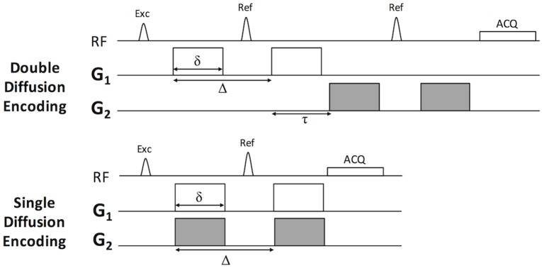
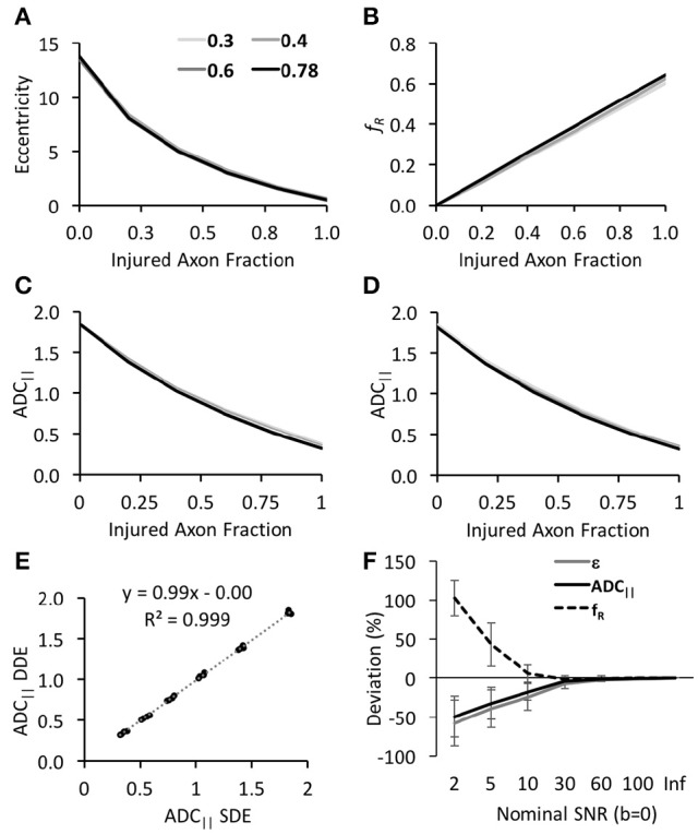
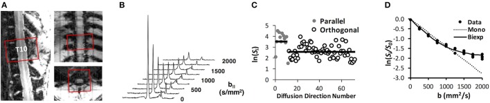
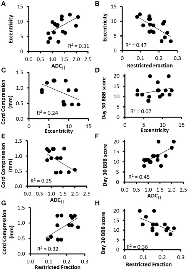
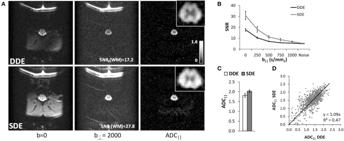
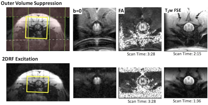
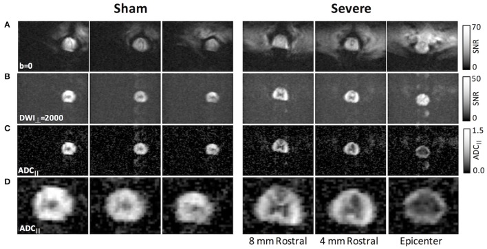
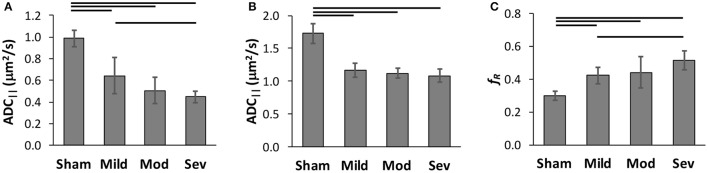
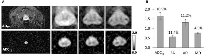
Diffusion tensor imaging (DTI) is a promising biomarker of spinal cord injury (SCI). In the acute aftermath, DTI in SCI animal models consistently demonstrates high sensitivity and prognostic performance, yet translation of DTI to acute human SCI has been limited. In addition to technical challenges, interpretation of the resulting metrics is ambiguous, with contributions in the acute setting from both axonal injury and edema. Novel diffusion MRI acquisition strategies such as double diffusion encoding (DDE) have recently enabled detection of features not available with DTI or similar methods. In this work, we perform a systematic optimization of DDE using simulations and an in vivo rat model of SCI and subsequently implement the protocol to the healthy human spinal cord. First, two complementary DDE approaches were evaluated using an orientationally invariant or a filter-probe diffusion encoding approach. While the two methods were similar in their ability to detect acute SCI, the filter-probe DDE approach had greater predictive power for functional outcomes. Next, the filter-probe DDE was compared to an analogous single diffusion encoding (SDE) approach, with the results indicating that in the spinal cord, SDE provides similar contrast with improved signal to noise. In the SCI rat model, the filter-probe SDE scheme was coupled with a reduced field of view (rFOV) excitation, and the results demonstrate high quality maps of the spinal cord without contamination from edema and cerebrospinal fluid, thereby providing high sensitivity to injury severity. The optimized protocol was demonstrated in the healthy human spinal cord using the commercially-available diffusion MRI sequence with modifications only to the diffusion encoding directions. Maps of axial diffusivity devoid of CSF partial volume effects were obtained in a clinically feasible imaging time with a straightforward analysis and variability comparable to axial diffusivity derived from DTI. Overall, the results and optimizations describe a protocol that mitigates several difficulties with DTI of the spinal cord. Detection of acute axonal damage in the injured or diseased spinal cord will benefit the optimized filter-probe diffusion MRI protocol outlined here.

Keywords: diffusion tensor imaging; double diffusion encoding; magnetic resonance imaging; spinal cord injury.


Figures

Figure 1
Figure 1
Pulse sequence for Double and Single Diffusion Encoding Techniques. The double diffusion encoding (DDE) sequence consists of two pairs of Stejskal-Tanner diffusion weighting gradients (G1 and G2) independent in their orientations, timing, and amplitude. The single diffusion encoding (SDE) consists of a single gradient pair. In this work, G1 and G2 were either parallel or perpendicular to one another and modulated relative to the laboratory frame of reference according to Table 1.
Figure 2
Figure 2
Simulation of DWI schemes and derived parameters. All estimated DWI parameters, including eccentricity (A), restricted fraction (B), and ADC|| from the DDE (C) or SDE (D), exhibited high sensitivity to the injured axon fraction (x-axis) with minimal differences between different intra-axonal volume fractions (shown as individual lines). Notably, ADC|| was nearly identical between the DDE and SDE (E) for uniformly-oriented fibers. The estimated parameters showed differential reliability under low SNR with ADC|| being most robust to noise (F).
Figure 3
Figure 3
DDE-PRESS Acquisition and Quantification. A voxel was positioned over the T10 vertebral segment (A) and aligned with the cord axis, shown here for a sham-injured animal. The integrated signal quantified as the area under the water peak (B) was obtained over the range ±2 ppm for subsequent analysis. For orientationally invariant DDE (C), the mean signal from parallel and orthogonal directions were compared to derive eccentricity, whereas for the filter-probe DDE, the diffusivity and restricted fraction (D) were derived from a mono- and bi-exponential fit (D) to the signals.
Figure 4
Figure 4
DDE-PRESS Relationship to Severity and Outcomes. Across all animals with a range of injury severities, both ADC|| and fR measured at the injury site were moderately correlated with eccentricity (A,B). The relationship with cord compression (C,E,G) were similar across all three parameters. The strength of the correlation with BBB score 30 days post-injury was lowest for eccentricity (D), greatest for ADC|| (F), and in between for fR (H).
Figure 5
Figure 5
Double and Single Filter-Probe Diffusion Encoding. Representative images (A) from the double (top) and single (bottom) diffusion encoding variants of the DWI sequence. As expected, the SDE had improved SNR compared to the DDE acquisition. ADC|| maps depict primarily the spinal cord white matter along with fat. Across 3 naïve animals, the white matter signal (B) in the SDE remains above noise floor even at b|| = 1,000 s/mm2, whereas the DDE signal was indistinguishable from the noise above b|| = 500 s/mm2. The measured ADC|| of the SDE and DDE were comparable (C), with a slight elevation in the SDE that could be attributable to the greater SNR. In the white matter voxels across all animals (D), ADC|| values from the DDE and SDE were strongly correlated with a slope of 1.09.
Figure 6
Figure 6
Reduced Field of View DWI and T2-weighted Imaging. rFOV using outer volume suppression (top) or 2DRF (bottom) provided high-quality images of the thoracic spine. Fold-over and chemical shift artifacts were evident in the OVS, which includes a separate fat-suppression module. The 2DRF with a slightly smaller FOV but an identical resolution offered similar SNR, minimal chemical shift artifacts (without separate fat suppression), and slight improvement in EPI distortion as shown by the b = 0 images and FA maps. In a T2-weighted fast spin echo, the smaller FOV allowed a reduction in acquisition time with a comparable SNR.
Figure 7
Figure 7
rFOV DWI in SCI with filter-probe SDE. Compared to the non-diffusion weighted images (A), the perpendicular diffusion-weighted images (B) were free from extraneous tissue signals, although some slight EPI ghosting was evident. The filtered ADC|| maps (C) reflected primarily the intra-axonal diffusivity. In an acute severe SCI (right), the ADC|| maps clearly demonstrated a reduction in the central region at the epicenter and in the dorsal columns rostral to the lesion. Magnification of filtered ADC|| maps are shown in (D).
Figure 8
Figure 8
Classification of Spinal Cord Injury Severity. Region of interest analysis of FP-SDE (A) was compared to DDE-PRESS (B,C) in the same animals across a range of injury severities. ADC|| from imaging with a whold-cord region of interest analysis (A) showed an effect of injury severity. ADC|| obtained from single voxel PRESS (B) was less sensitive to severity, while fR from the same voxel (C) better distinguished injury severity. Lines indicate significant group differences at p < 0.05.
Figure 9
Figure 9
Filter-Probe diffusion weighted imaging in Human Normal Cervical Spinal Cord. The filter-probe SDE scheme was implemented on a human 3T scanner by altering the diffusion gradient orientations of the commercially-available diffusion weighted imaging sequence. Compared with AD maps derived from DTI (A), FP-SDE provided comparable ADC|| maps but with almost complete attenuation of non-cord signals. Three of 10 slices are shown. Results from 3 healthy subjects (B) demonstrate similar coefficients of variation (indicated in text above bars) in the same subjects and regions of interest. ADC|| was significantly greater than AD from DTI. Bars indicate standard deviations.

References

    1. Basso D. M., Beattie M. S., Bresnahan J. C. (1995). A sensitive and reliable locomotor rating scale for open field testing in rats. J. Neurotrauma 12, 1–21. 10.1089/neu.1995.12.1 - DOI - PubMed
    1. Biering-Sorensen F., Alai S., Anderson K., Charlifue S., Chen Y., Devivo M., et al. . (2015). Common data elements for spinal cord injury clinical research: a National Institute for Neurological Disorders and Stroke project. Spinal Cord 53, 265–277. 10.1038/sc.2014.246 - DOI - PMC - PubMed
    1. Bottomley P. A. (1987). Spatial localization in NMR spectroscopy in vivo. Ann. N. Y. Acad. Sci. 508, 333–348. 10.1111/j.1749-6632.1987.tb32915.x - DOI - PubMed
    1. Budde M. D., Frank J. A. (2010). Neurite beading is sufficient to decrease the apparent diffusion coefficient after ischemic stroke. Proc. Natl. Acad. Sci. U.S.A. 107, 14472–14477. 10.1073/pnas.1004841107 - DOI - PMC - PubMed
    1. Budde M. D., Kim J. H., Liang H. F., Schmidt R. E., Russell J. H., Cross A. H., et al. . (2007). Toward accurate diagnosis of white matter pathology using diffusion tensor imaging. Magn. Reson. Med. 57, 688–695. 10.1002/mrm.21200 - DOI - PubMed

LinkOut - more resources