Review
. 2017 Dec 22:10:428.
doi: 10.3389/fnmol.2017.00428. eCollection 2017.

DFNB1 Non-syndromic Hearing Impairment: Diversity of Mutations and Associated Phenotypes

Affiliations
Review

DFNB1 Non-syndromic Hearing Impairment: Diversity of Mutations and Associated Phenotypes

Francisco J Del Castillo et al. Front Mol Neurosci. .

Abstract

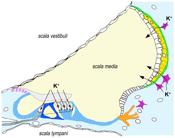
The inner ear is a very complex sensory organ whose development and function depend on finely balanced interactions among diverse cell types. The many different kinds of inner ear supporting cells play the essential roles of providing physical and physiological support to sensory hair cells and of maintaining cochlear homeostasis. Appropriately enough, the gene most commonly mutated among subjects with hereditary hearing impairment (HI), GJB2, encodes the connexin-26 (Cx26) gap-junction channel protein that underlies both intercellular communication among supporting cells and homeostasis of the cochlear fluids, endolymph and perilymph. GJB2 lies at the DFNB1 locus on 13q12. The specific kind of HI associated with this locus is caused by recessively-inherited mutations that inactivate the two alleles of the GJB2 gene, either in homozygous or compound heterozygous states. We describe the many diverse classes of genetic alterations that result in DFNB1 HI, such as large deletions that either destroy the GJB2 gene or remove a regulatory element essential for GJB2 expression, point mutations that interfere with promoter function or splicing, and small insertions or deletions and nucleotide substitutions that target the GJB2 coding sequence. We focus on how these alterations disrupt GJB2 and Cx26 functions and on their different effects on cochlear development and physiology. We finally discuss the diversity of clinical features of DFNB1 HI as regards severity, age of onset, inner ear malformations and vestibular dysfunction, highlighting the areas where future research should be concentrated.

Keywords: DFNB1; GJB2; GJB6; connexin-26; connexin-30; hearing impairment; inner ear.


Figures

Figure 1
Figure 1
Expression of connexin-26 (Cx26) and connexin-30 (Cx30) in the human cochlea. Cx26 and Cx30 are synthesized by all supporting cells types within the organ of Corti (blue), including inner and outer pillar cells (dark blue), as well as by root cells (orange), interdental cells (pink), fibrocytes from the underlying connective tissue (light blue) and basal (green) and intermediate cells (yellow) from the stria vascularis. For the sake of clarity, we have depicted only four fibrocytes (purple) in connective tissue. Arrows indicate the pathways for K+ influx in hair cells (bordered in red) and K+ secretion into endolymph through the stria vascularis. It must be noted that Cx26 and Cx30 seem to form distinct, homomeric plaques even when co-expressed in the same cell, although these plaques are closely associated. Some immuhistochemical staining experiments suggest that co-expression may not occur in all the cell types indicated above (e.g., Cx26 may not be synthesized in inner pillar cells and strial intermediate cells, whereas Cx30 may not be synthesized in strial basal cells), which might reflect specific functional associations with additional ion transporters (Liu et al., 2009, 2016).
Figure 2
Figure 2
Sequence of the GJB2 basal promoter and first exon, showing the location of the GC boxes (at -81 and -93), the TATA motif (at -24) and the transcription start site of the first canonical exon. The arrow indicates the position of the g.-77C>T promoter mutation.
Figure 3
Figure 3
Large deletions on chromosome 13q12 that are responsible for DFNB1 hearing impairment (HI). The proximal breakpoint of del131-kb and the distal breakpoint of del(GJB6-D13S1854) delimit the 95.4-kb sequence stretch where the cis-acting element regulating the expression of GJB2 must be situated. The two proximal breakpoints of deletions del(GJB6-D13S1830) and del(GJB6-D13S1854; vertical arrows) lie within GJB6.
Figure 4
Figure 4
(A) Topological organization of the Cx26 monomer with its different structural elements. Helices are represented as cylinders. Helices in light green line the hydrophilic pore of the channel, whereas helices in dark green are exposed to the hydrophobic environment of the lipid bilayer. Note the projection into the cytosol of helices M2 and M3. (B) Location of the truncating and missense mutations mentioned in the text within the different structural elements.

References

    1. Abe S., Usami S. I., Shinkawa H., Kelley P. M., Kimberling W. J. (2000). Prevalent connexin 26 gene (GJB2) mutations in Japanese. J. Med. Genet. 37, 41–43. 10.1136/jmg.37.1.41 - DOI - PMC - PubMed
    1. Ambrosi C., Walker A. E., Depriest A. D., Cone A. C., Lu C., Badger J., et al. . (2013). Analysis of trafficking, stability and function of human connexin 26 gap junction channels with deafness-causing mutations in the fourth transmembrane helix. PLoS One 8:e70916. 10.1371/journal.pone.0070916 - DOI - PMC - PubMed
    1. Anselmi F., Hernandez V. H., Crispino G., Seydel A., Ortolano S., Roper S. D., et al. . (2008). ATP release through connexin hemichannels and gap junction transfer of second messengers propagate Ca2+ signals across the inner ear. Proc. Natl. Acad. Sci. U S A 105, 18770–18775. 10.1073/pnas.0800793105 - DOI - PMC - PubMed
    1. Azaiez H., Chamberlin G. P., Fischer S. M., Welp C. L., Prasad S. D., Taggart R. T., et al. . (2004). GJB2: the spectrum of deafness-causing allele variants and their phenotype. Hum. Mutat. 24, 305–311. 10.1002/humu.20084 - DOI - PMC - PubMed
    1. Azaiez H., Smith R. J. H. (2007). In reference to temporal bone imaging in GJB2 deafness. Laryngoscope 117:1127. 10.1097/MLG.0b013e3180421729 - DOI - PubMed