. 2017 Dec 21:9:427.
doi: 10.3389/fnagi.2017.00427. eCollection 2017.

Changes in Blood Factors and Ultrasound Findings in Mild Cognitive Impairment and Dementia

Affiliations

Changes in Blood Factors and Ultrasound Findings in Mild Cognitive Impairment and Dementia

Kyoungjoo Cho et al. Front Aging Neurosci. .

Erratum in

Abstract

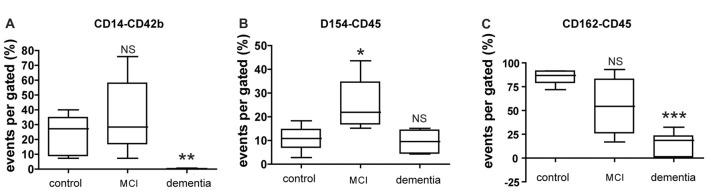
The present study aimed to assess the changes in blood factors and ultrasound measures of atherosclerosis burden patient with mild cognitive impairment (MCI) and dementia. Peripheral blood samples and ultrasonography findings were obtained for 53 enrolled participants. Flow cytometry was used to evaluate levels of activated platelets and platelet-leukocyte aggregates (PLAs). The number of platelets expressing p-selectin was correlated with intima media thickness (IMT) and plaque number in both the MCI and dementia groups. The number of platelets expressing p-selectin glycoprotein ligand (PSGL) was strongly correlated with IMT in patients with MCI, whereas the number of platelets expressing PGSL was correlated with plaque number rather than IMT in patients with dementia. PLAs was associated with both IMT and plaque number in patients with MCI but not in those with dementia. Our findings demonstrate that alterations in IMT and plaque number are associated with an increased risk of cognitive decline as well as conversion from MCI to dementia and that blood factor analysis may aid to detect the severity of cognitive decline.

Keywords: atherosclerosis; blood factor analysis; dementia; mild cognitive impairment; vascular disease.


Figures

Figure 1
Figure 1
The distribution of participants and atherosclerotic condition. (A) Of participant, patients groups are three. Without cognitive decline, patients with atherosclerosis only are 57.1% of total patients. With atherosclerosis, patients with mild cognitive impairment (MCI) are composed in 32.1% and patients with dementia are constituted in 14.3%. (B) Intima-media thickness (IMT) is shown as mean ± SD. Differences of IMT is significant between dementia and control group. Also it is significant between dementia and MCI. (C) Numbers of plaques are shown as mean ± SD. There are significant differences between control and MCI; control and dementia; and MCI and dementia. *p < 0.05 vs. control group, **p < 0.01 vs. control group.
Figure 2
Figure 2
Expression of circulating blood factors by fluorescence-activated cell sorting (FACS) analysis. (A) Circulating levels of monocyte aggregated with platelets in each group. (B) Circulating levels of leukocyte aggregated with CD40Ligand (CD154) in each group. (C) Circulating levels of leukocyte aggregated with p-selectin glycoprotein ligand (PSGL; CD162) in each group. The data presents with % as an event number per gate by FACS analysis. Bars represent median values, and the error bar, the standard deviation. *p < 0.05 vs. control group, **p < 0.01 vs. control group, ***p < 0.001 vs. control group. NS, not significant.
Figure 3
Figure 3
CD62p and CD162 expression in cell surface marker and soluble level. (A,B) Level of platelet surface expressing CD62p (p-selectin) or CD162 (PSGL) was evaluated by FACS analysis. (C,D) Level of CD62p or CD162 in plasma was evaluated by ELISA. The data of FACS represents with % as an event number per gate. The result of ELISA is presented as ng/ml. Bars represent median values, and the error bar, the standard deviation. *p < 0.05 vs. control group. NS, not significant.

Similar articles

Cited by

References

    1. Atiya M., Hyman B. T., Albert M. S., Killiany R. (2003). Structural magnetic resonance imaging in established and prodromal Alzheimer disease: a review. Alzheimer Dis. Assoc. Disord. 17, 177–195. 10.1097/00002093-200307000-00010 - DOI - PubMed
    1. Cardenas V. A., Reed B., Chao L. L., Chui H., Sanossian N., DeCarli C. C., et al. . (2012). Associations among vascular risk factors, carotid atherosclerosis, and cortical volume and thickness in older adults. Stroke 43, 2865–2870. 10.1161/STROKEAHA.112.659722 - DOI - PMC - PubMed
    1. Coley N., Andrieu S., Gardette V., Gillette-Guyonnet S., Sanz C., Vellas B., et al. . (2008). Dementia prevention: methodological explanations for inconsistent results. Epidemiol. Rev. 30, 35–66. 10.1093/epirev/mxn010 - DOI - PubMed
    1. Coutu J. P., Lindemer E. R., Konukoglu E., Salat D. H., Alzheimer’s Disease Neuroimaging Initiative (ADNI) . (2017). Two distinct classes of degenerative change are independently linked to clinical progression in mild cognitive impairment. Neurobiol. Aging 54, 1–9. 10.1016/j.neurobiolaging.2017.02.005 - DOI - PMC - PubMed
    1. de la Torre J. C. (2010). The vascular hypothesis of Alzheimer’s disease: bench to bedside and beyond. Neurodegener. Dis. 7, 116–121. 10.1159/000285520 - DOI - PubMed

LinkOut - more resources