Review
. 2017 Dec 19:8:701.
doi: 10.3389/fneur.2017.00701. eCollection 2017.

Optical Coherence Tomography in Alzheimer's Disease and Other Neurodegenerative Diseases

Affiliations
Review

Optical Coherence Tomography in Alzheimer's Disease and Other Neurodegenerative Diseases

Jonah Doustar et al. Front Neurol. .

Abstract

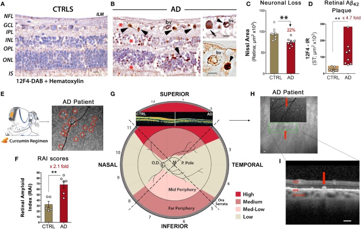
Over the past decade, a surge of evidence has documented various pathological processes in the retina of patients suffering from mild cognitive impairment, Alzheimer's disease (AD), Parkinson's disease (PD), and other neurodegenerative diseases. Numerous studies have shown that the retina, a central nervous system tissue formed as a developmental outgrowth of the brain, is profoundly affected by AD. Harboring the earliest detectable disease-specific signs, amyloid β-protein (Aβ) plaques, the retina of AD patients undergoes substantial ganglion cell degeneration, thinning of the retinal nerve fiber layer, and loss of axonal projections in the optic nerve, among other abnormalities. More recent investigations described Aβ plaques in the retina located within sites of neuronal degeneration and occurring in clusters in the mid- and far-periphery of the superior and inferior quadrants, regions that had been previously overlooked. Diverse structural and/or disease-specific changes were also identified in the retina of PD, Huntington's disease, and multiple sclerosis patients. The pathological relationship between the retina and brain prompted the development of imaging tools designed to noninvasively detect and monitor these signs in living patients. One such tool is optical coherence tomography (OCT), uniquely providing high-resolution two-dimensional cross-sectional imaging and three-dimensional volumetric measurements. As such, OCT emerged as a prominent approach for assessing retinal abnormalities in vivo, and indeed provided multiple parameters that allowed for the distinction between normal aged individuals and patients with neurodegenerative diseases. Beyond the use of retinal optical fundus imaging, which recently allowed for the detection and quantification of amyloid plaques in living AD patients via a wide-field view of the peripheral retina, a major advantage of OCT has been the ability to measure the volumetric changes in specified retinal layers. OCT has proven to be particularly useful in analyzing retinal structural abnormalities consistent with disease pathogenesis. In this review, we provide a summary of OCT findings in the retina of patients with AD and other neurodegenerative diseases. Future studies should explore the combination of imaging early hallmark signs together with structural-functional biomarkers in the accessible retina as a practical means of assessing risk, disease progression, and therapeutic efficacy in these patients.

Keywords: Huntington’s disease; Parkinson’s disease; alpha-synuclein; beta-amyloid; multiple sclerosis; optical coherence tomography; retinal imaging; spectral domain.


Figures

Figure 1
Figure 1
Noninvasive retinal imaging in Alzheimer’s patients: detecting Aβ deposits and nerve degeneration via scanning laser ophthalmoscopy and optical coherence tomography. (A,B) Retinal cross sections from superior quadrants of Alzheimer’s disease (AD) patients (n = 12) and matched healthy controls (CTRL; n = 8) stained with anti-Aβ42 mAbs (12F4) and peroxidase-based labeling (brown). Hematoxylin counterstain for nuclei (violet). Retinas of AD patients contained a multitude of Aβ deposits (arrowheads), especially in the ganglion cell layer (GCL). Marked loss of retinal cells is apparent in the GCL, inner nuclear layer (INL), and outer nuclear layer (ONL); areas of nuclei loss are indicated by red asterisks. The inner limiting membrane (ILM) and retinal nerve fiber layer (RNFL) are intact in CTRL in contrast to AD. Scale bars = 20 μm. Higher magnification images [(B), right panel] show Aβ deposits near and inside blood vessel walls (bv; arrowheads) and inside ganglion cell soma (arrow). Scale bars = 10 μm. (C) Quantitative Nissl neuronal area in AD patients (n = 9) and age- and gender-matched CTRL (n = 8) revealing a significant reduction in AD patients. (D) Quantitative analysis of 12F4-immunoreactive (IR) area of Aβ42-containing plaques in the superior quadrant of retinal flatmounts in a subset of definite AD patients (n = 8) and matched controls (n = 7) showing a significant increase of Aβ42 plaques in AD patients. (E) Schematic of a noninvasive retinal amyloid imaging method using curcumin (Longvida®) to label retinal Aβ in live human patients. Subjects’ retinas were imaged with a modified scanning-laser ophthalmoscope prior to and following curcumin intake. White spots marked by red circles are curcumin-positive amyloid plaques detected in the retina of a living AD patient. (F) Scatter bar plot displays retinal amyloid index (RAI) scores, a fully automated calculation of increased curcumin fluorescence representing amyloid deposits in the retina. AD patients (n = 6) showed a significant increase in RAI score in comparison to age-matched CTRL (n = 5). (G) Qualitative mapping of the “geometric hotspot” regions of Aβ deposits in retinal quadrants of AD patients. Schematic of OCT images from an AD patient vs. control are shown [adopted from: Coppola et al. (21)]. (H) OCT image of a selected curcumin-positive plaque (red arrow) in an AD patient with no maculopathy. Certain retinal amyloid plaque visualized by curcumin fluorescence fundography in insert. Green lines delineate region of OCT segmentation. (I) Retinal cross section by OCT reveals amyloid plaque in outer retinal layers; curcumin-positive deposit located above retinal pigment epithelium (RPE), along with intact RPE and Bruch’s membrane. Scale bars = 200 μm. Group means and SEMs are shown. *p < 0.05 and **p < 0.01, unpaired two-tailed Student’s t-test. Images and data of (A–I) panels, except for part of (E), are modified reprinted from Koronyo et al. (26).

References

    1. Byerly MS, Blackshaw S. Vertebrate retina and hypothalamus development. Wiley Interdiscip Rev Syst Biol Med (2009) 1:380–9.10.1002/wsbm.22 - DOI - PubMed
    1. Purves D, Augustine GJ, Fitzpatrick D, Katz LC, LaMantia A-S, McNamara JO, et al. The Retina. Sunderland, MA: Sinauer Associates; (2001).
    1. Trost A, Lange S, Schroedl F, Bruckner D, Motloch KA, Bogner B, et al. Brain and retinal pericytes: origin, function and role. Front Cell Neurosci (2016) 10:20.10.3389/fncel.2016.00020 - DOI - PMC - PubMed
    1. Crair MC, Mason CA. Reconnecting eye to brain. J Neurosci (2016) 36:10707–22.10.1523/JNEUROSCI.1711-16.2016 - DOI - PMC - PubMed
    1. Alzheimer’s A. 2016 Alzheimer’s disease facts and figures. Alzheimers Dement (2016) 12:459–509.10.1016/j.jalz.2016.03.001 - DOI - PubMed