

Biomimetic peptides protect cells from oxidative stress
- PMID: 29312503
- PMCID: PMC5752901
Biomimetic peptides protect cells from oxidative stress
Abstract
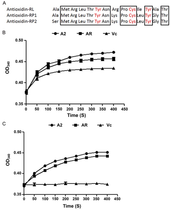
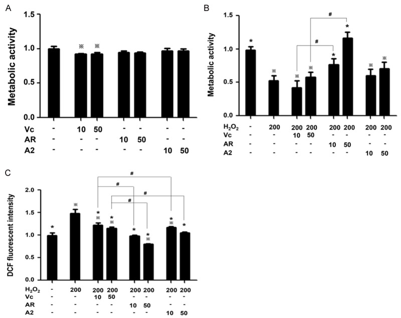
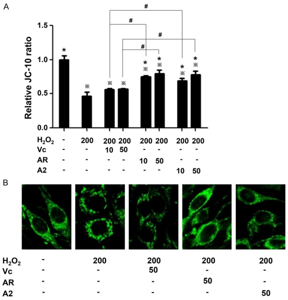
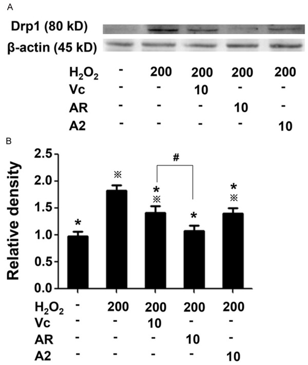
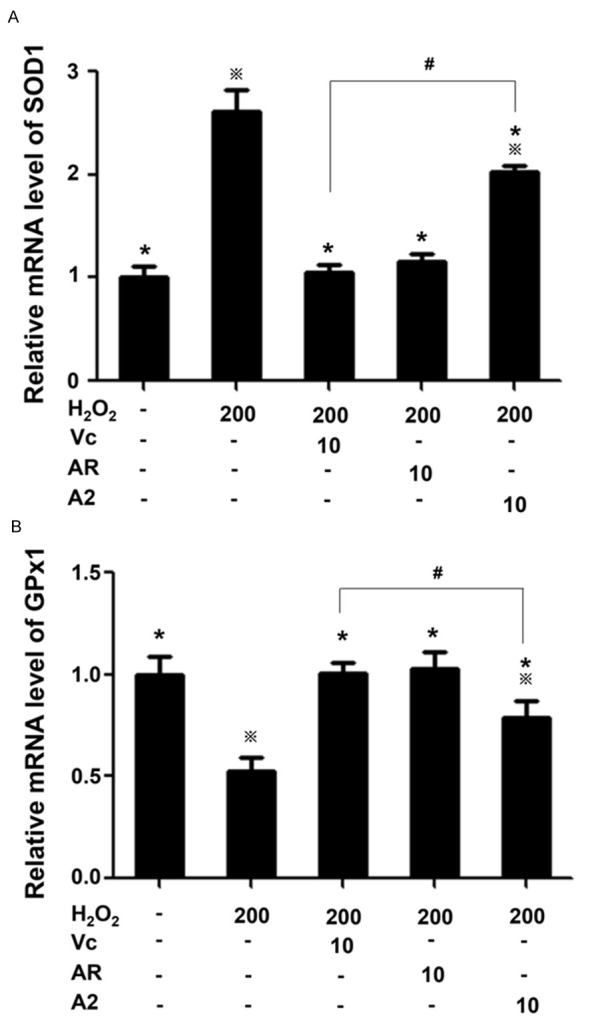
Most degenerative diseases are caused by free radicals. Antioxidin-RL peptide is a free radical scavenger found in the skin of plateau frog Odorrana livida, which is more stable than vitamin C as it resists light-induced degradation. However, whether and how antioxidin-RL protects cells from oxidative stress was not clear. Here we addressed this issue, and in addition, we designed a series of antioxidin cognates by adding tyrosine residues to enhance free radical-binding capability. We performed free radical-clearing assays in solution to screen the mutants, and found a mutant antioxidin-2 that was as stable as antioxidin-RL and cleared free radical faster. By using PC-12 cells as a model, we demonstrated that both antioxidin-2 and antioxidin-RL inhibited the accumulation of intracellular free radicals triggered by H2O2, reduced mitochondria membrane potential dissipation, maintained mitochondrial morphology, and decreased the expression of dynamin-related protein-1 in mitochondria, with antioxidin-RL more effective. Antioxidin-RL also attenuated the changes in SOD1 and GPx1 expression induced by H2O2. These findings provide insight into the anti-oxidative mechanisms of antioxidin-RL and its derivatives, which will provide rational basis for the development of more effective antioxidants to cure diseases.
Keywords: Antioxidin-RL; PC12 cell; metabolic activity; mitochondria; oxidative injury.
Conflict of interest statement
None.
Figures
Similar articles
-
Protective effects of antioxidin-RL from Odorrana livida against ultraviolet B-irradiated skin photoaging.Peptides. 2018 Mar;101:124-134. doi: 10.1016/j.peptides.2018.01.009. Epub 2018 Jan 16. Peptides. 2018. PMID: 29341894
-
Frog skins keep redox homeostasis by antioxidant peptides with rapid radical scavenging ability.Free Radic Biol Med. 2010 May 1;48(9):1173-81. doi: 10.1016/j.freeradbiomed.2010.01.036. Epub 2010 Feb 4. Free Radic Biol Med. 2010. PMID: 20138142
-
Structure and function of a novel antioxidant peptide from the skin of tropical frogs.Free Radic Biol Med. 2018 Feb 1;115:68-79. doi: 10.1016/j.freeradbiomed.2017.11.001. Epub 2017 Nov 21. Free Radic Biol Med. 2018. PMID: 29162516
-
Metal-induced hepatotoxicity.Semin Liver Dis. 1996 Feb;16(1):3-12. doi: 10.1055/s-2007-1007214. Semin Liver Dis. 1996. PMID: 8723319 Review.
-
Mitochondrial theory of aging matures--roles of mtDNA mutation and oxidative stress in human aging.Zhonghua Yi Xue Za Zhi (Taipei). 2001 May;64(5):259-70. Zhonghua Yi Xue Za Zhi (Taipei). 2001. PMID: 11499335 Review.
Cited by
-
Antioxidant Activity of Bioactive Peptide Fractions from Germinated Soybeans Conjugated to Fe3O4 Nanoparticles by the Ugi Multicomponent Reaction.Molecules. 2021 Sep 22;26(19):5726. doi: 10.3390/molecules26195726. Molecules. 2021. PMID: 34641270 Free PMC article.
-
The Role of Amphibian AMPs Against Oxidative Stress and Related Diseases.Antibiotics (Basel). 2025 Jan 25;14(2):126. doi: 10.3390/antibiotics14020126. Antibiotics (Basel). 2025. PMID: 40001370 Free PMC article. Review.
References
-
- Di Domenico F, Barone E, Perluigi M, Butterfield DA. Strategy to reduce free radical species in Alzheimer’s disease: an update of selected antioxidants. Expert Rev Neurother. 2015;15:19–40. - PubMed
-
- Liu C, Hong J, Yang H, Wu J, Ma D, Li D, Lin D, Lai R. Frog skins keep redox homeostasis by antioxidant peptides with rapid radical scavenging ability. Free Radic Biol Med. 2010;48:1173–81. - PubMed
LinkOut - more resources
Full Text Sources
Research Materials
Miscellaneous