. 2017 Dec 15;9(12):5548-5557.
eCollection 2017.

A comparison of apoptosis levels in keloid tissue, physiological scars and normal skin

Affiliations

A comparison of apoptosis levels in keloid tissue, physiological scars and normal skin

Ming-Zi Zhang et al. Am J Transl Res. .

Abstract

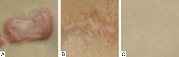
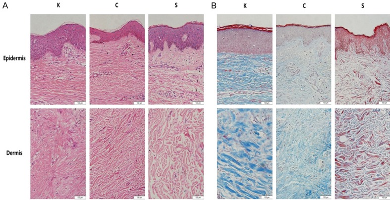
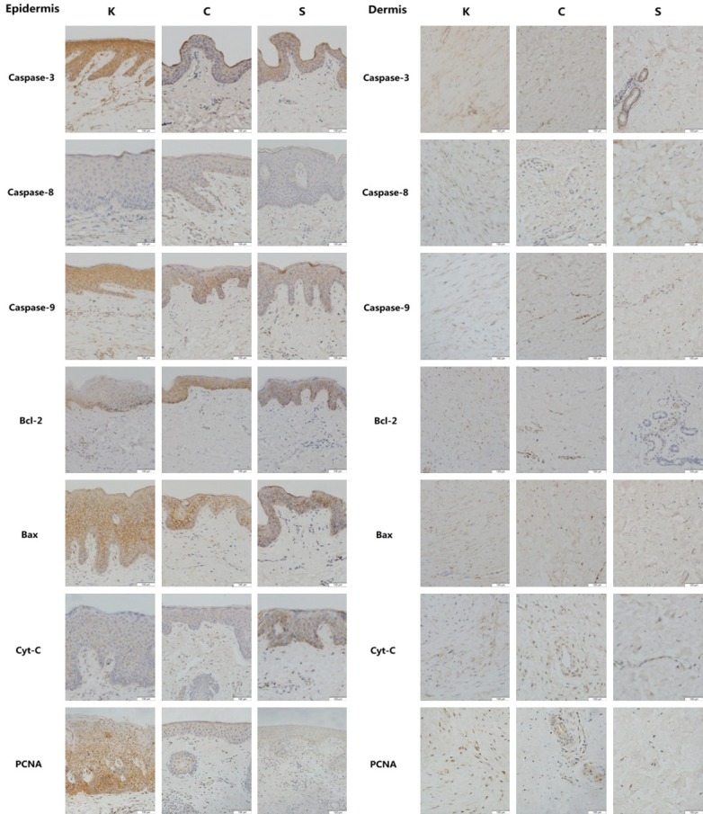
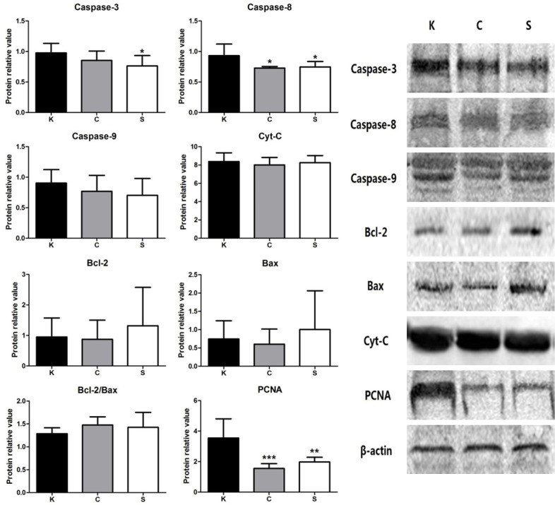
Apoptosis is a process of programmed cell death that occurs in multicellular organisms. The mitochondrial pathway plays a paramount role in apoptosis. In this study, the expression levels of key factors in the mitochondrial pathway and the cell proliferation factor (PCNA) were measured to evaluate the level of apoptosis and proliferation in keloid scars, physiological scars and normal skin tissue. Thirty samples were taken from 30 patients: 10 keloid patients, 10 physiological scar patients and 10 patients without obvious scarring. All 30 patients were selected randomly from the Department of Plastic Surgery at Peking Union Medical College Hospital from June 2016 to December 2016. Hematoxylin and eosin staining and Masson staining were used to observe the differences in histology and fiber tissue content. Mitochondrial pathway factors (caspase-3, caspase-8, caspase-9, Bcl-2, Bax, cytochrome-c) and PCNA expression levels were detected by immunohistochemistry and were analyzed as the percentage of positively stained cells in the epidermis and dermis. Relative protein expression levels were measured by western blotting. Compared with physiological scars and normal skin tissue, keloid tissue had an increase in fiber number and decrease in cell content. In our immunohistochemical and western blot analyses, all tissue types showed similar expression levels of the mitochondrial pathway factors. However, the percentage of PCNA-positive cells and the relative protein expression level of PCNA were significantly higher in keloid tissue. Keloid has a similar apoptosis level as physiological scars and normal skin but has a higher expression of PCNA, indicating that keloid scars have high levels of proliferation and normal apoptosis.

Keywords: Keloid; apoptosis; physiological scar; proliferation; skin.


Conflict of interest statement

None.

Figures

Figure 1
Figure 1
Sampling conditions of the 3 different groups of patients: A: Keloid tissue from keloid patients; B: Physiological scar tissue from scar patients; C: Normal skin tissue from patients without obvious scarring.
Figure 2
Figure 2
The results of epidermal and dermal H&E staining (A) and of Masson staining (B) in all groups (200×). In the K group, the epidermis was thicker, there were an increased number of bulky collagen fibrils intricately arranged in the dermis, and there was a reduction in cellular content. The C group had slender collagen fibrils which composed the majority of the dermis. In contrast, normal skin dermis showed increased cellular content and less fibrils, which were relatively loosely arranged compared with that of the K and C group.
Figure 3
Figure 3
Representative immunohistochemistry micrographs (200×) of epidermal and dermal skin tissue for caspase-3, caspase-8, caspase-9, Bcl-2, Bax, cyt-c and PNCA in all groups. Brown staining indicates the positive expression areas, and the shade of the color represents the expression level of the target protein. In the epidermal layer of the K group, caspase-3, caspase-9 and Bax showed higher expression levels; however, caspase-8, Bcl-2 and cyt-c expression levels were similar to those of the C and S group. PCNA expression was much higher in the keloid epidermal layer than in the non-keloid tissues. In the K dermal layer, Bcl-2 and Bax showed higher expression levels than those in the non-keloid tissues; however, caspase-3, caspase-8, caspase-9 and cyt-c expression levels were similar to those of the C and S group. Dermal PCNA expression was remarkably higher in the K group than in the non-keloid tissues.
Figure 4
Figure 4
Results of the percentage of positive cells. In both the epidermis and the dermis, the expression levels of most key factors of the mitochondrial pathway showed no significant differences between the K group and those of the other groups, except for caspase-9 in the epidermis and Bcl-2 in the dermis. The percentage of PCNA-positive cells in the keloid tissue was significantly higher than in the non-keloid tissues. Values are shown as the means ± SD; (n=10 in each group; *P<0.05, **P<0.01, ***P<0.001 vs. the K group).
Figure 5
Figure 5
Relative protein amounts for all target proteins. The expression levels of most key factors in the mitochondrial apoptosis pathway were not significantly different between the K group and the other groups, except caspase-3 and caspase-8. The highest expression level of PCNA was in the K group. Representative images of the western blots for caspase-3, caspase-8, caspase-9, Bcl-2, Bax, cyt-c and PCNA are shown on the right. Values are shown as the means ± SD; (n=10 in each group; *P<0.05, ***P<0.001 vs. the K group).

References

    1. Marneros AG, Krieg T. Keloids-clinical diagnosis, pathogenesis, and treatment options. J Dtsch Dermatol Ges. 2004;2:905–913. - PubMed
    1. Ogawa R. The most current algorithms for the treatment and prevention of hypertrophic scars and keloids. Plast Reconstr Surg. 2010;125:557–568. - PubMed
    1. Green DR. Means to an end: apoptosis and other cell death mechanisms. Cold Spring Harbor Laboratory Press; 2011.
    1. Brenner D, Mak TW. Mitochondrial cell death effectors. Curr Opin Cell Biol. 2009;21:871–877. - PubMed
    1. Chalah A, Khosravi-Far R. The mitochondrial death pathway. Adv Exp Med Biol. 2008;615:25–45. - PubMed

LinkOut - more resources