. 2017 Nov 24;8(65):109151-109160.
doi: 10.18632/oncotarget.22642. eCollection 2017 Dec 12.

Targeting of CD122 enhances antitumor immunity by altering the tumor immune environment

Affiliations

Targeting of CD122 enhances antitumor immunity by altering the tumor immune environment

Daniel O Villarreal et al. Oncotarget. .

Abstract

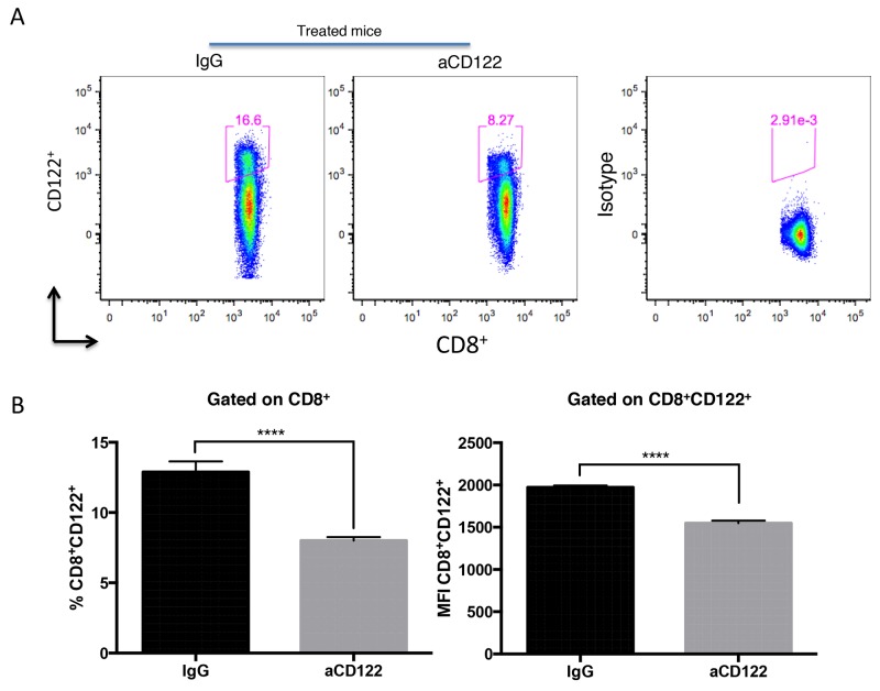
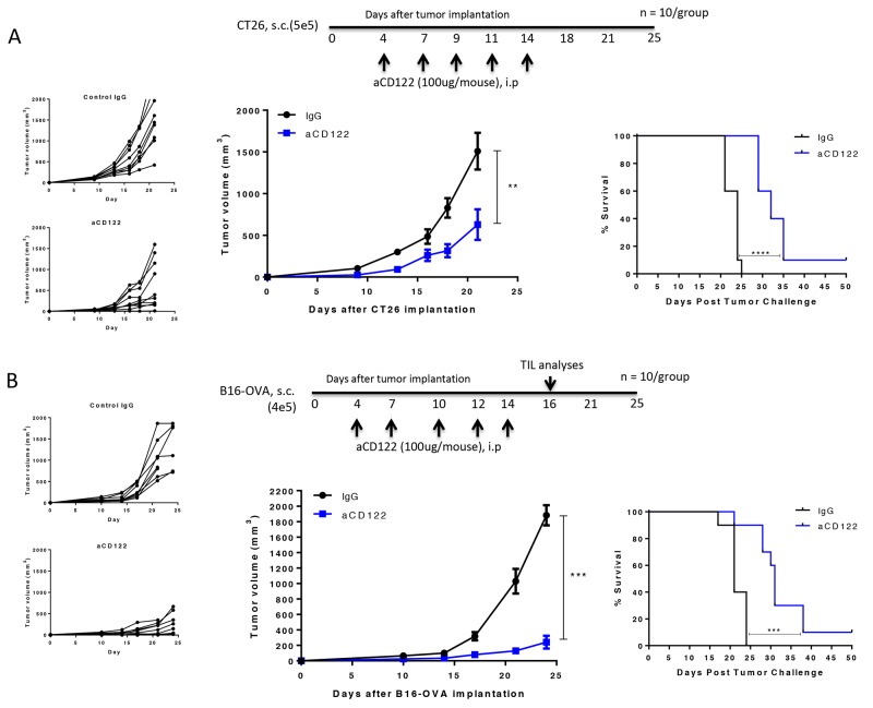
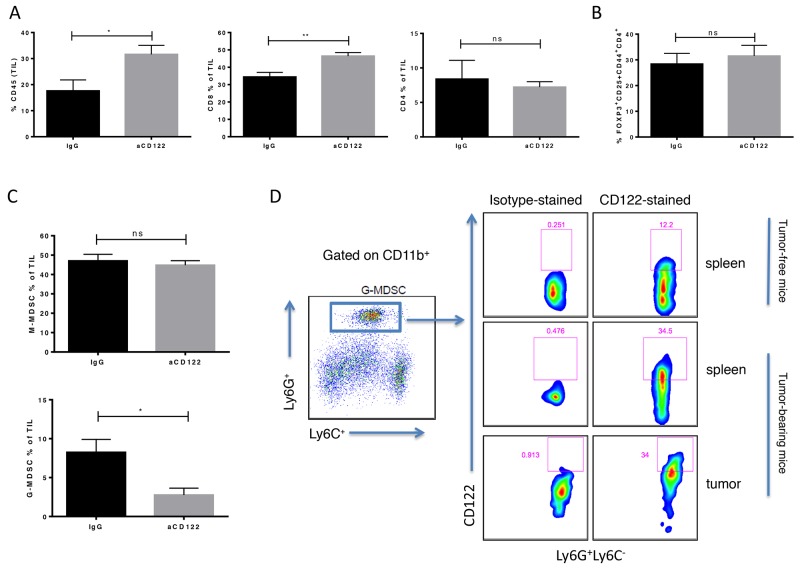
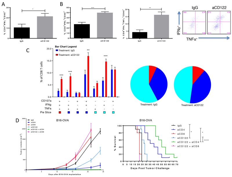
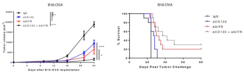
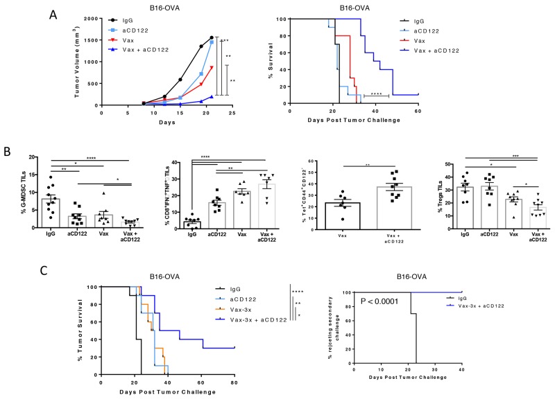
Mounting evidence demonstrates that CD8+CD122+ T cells have suppressive properties with the capacity to inhibit T cell responses. Therefore, these cells are rational targets for cancer immunotherapy. Here, we demonstrate that CD122 monoclonal antibody (mAb; aCD122) therapy significantly suppressed tumor growth and improved long-term survival in tumor-bearing mice. This therapeutic effect correlated with enhanced polyfunctional, cytolytic intratumoral CD8+ T cells and a decrease in granulocytic myeloid-derived suppressor cells (G-MDSCs). In addition, aCD122 treatment synergized with a vaccine to augment vaccine-induced antigen (Ag)-specific CD8+ T cell responses, reject established tumors and generate memory T cells. Furthermore, aCD122 mAb synergized with an anti-GITR (aGITR) mAb to confer significant control of tumor growth. These results suggest CD122 might be a promising target for cancer immunotherapy, either as a single agent or in combination with other forms of immunotherapy.

Keywords: CD122; CD8 T cells; GITR; immunotherapy; vaccines.


Conflict of interest statement

CONFLICTS OF INTEREST No potential conflicts of interest were disclosed by the authors.

Figures

Figure 1
Figure 1. Treatment with 5H4 mAb reduced CD8+CD122+ T cell population
Naïve B6 non-tumor bearing mice were injected i.p. with 100 ug of 5H4 anti-CD122 mAb or with the isotype (rat IgG2a) control. Three days after injection, splenocytes were analyzed for CD8+CD122+ T cells by flow cytometry. (A-B) Flow plot analysis and representative data showing the percentages of CD8+CD122+ T cells and the MFI of CD122 on CD8+CD122+ T cells in total splenic lymphocytes. Isotype indicates CD8+ splenocytes stained with the isotype control antibody for CD122 staining. Results are representative of 2 independent experiments. ****P<0.0001. Errors bars indicate SEM of n = 5/group.
Figure 2
Figure 2. CD122 mAb treatment suppressed CT26 and B16-OVA tumor growth and enhanced long-term survival
Treatment regimen, individual tumor growth, group tumor measurements, and survival of CT26 (A) and B16-OVA (B) implanted mice following treatment are indicated. Figures represent 2 (CT26) and 3 (B16-OVA) independent experiments. **P<0.01; ***P<0.001; ****P<0.0001.
Figure 3
Figure 3. CD122 mAb treatment altered the cellular composition of the tumor immune microenvironment in the B16-OVA melanoma model
TILs from tumors of B16-OVA mice were harvested 16 days after tumor implantation. (A) CD45+ leukocyte infiltrate and CD8+ and CD4+ TILs as percentage of total CD45+ cells are shown in treated versus untreated groups. TIL populations, including CD4+ Tregs (B) and M-MDSC (CD11b+Ly6C+Ly6G-) and G-MDSC (CD11b+Ly6C-Ly6G+) (C) were identified by flow cytometry. (D) CD122 expression by G-MDSC was shown by flow cytometry. M-MDSCs also expressed CD122 (data not shown). *P<0.05; **P<0.01; ns not significant. Error bars indicate SEM of n = 5/group.
Figure 4
Figure 4. CD122 mAb treatment of B16-OVA tumors increased the frequency and function of specific inflammatory cytokine production of tumor infiltrating T cells and efficacy is dependent on CD8+ T cells
(A) Summary data showing percentage of CD4+CD45+ TILs expressing IFNγ. (B-C) Representative and summary data showing single-, double- and triple positive CD8+ T cells releasing IFNγ, TNFα and/or co-expressing the degranulation marker, CD107a, following OVA257-264 and OVA323-339 peptide incubation with PMA/ION stimulation. (D) Tumor growth curves and survival over time for B16-OVA tumor-bearing mice treated aCD122 and with/without anti-CD4 or anti-CD8 mAbs. Experiments were repeated at least two times. *P<0.05; **P<0.01; ***P<0.001. Errors bars indicate SEM of n = 5/group (A-C); n= 10/group in (D).
Figure 5
Figure 5. Anti-CD122 mAb treatment synergizes with an aGITR mAb therapy
On day +4 post-implantation of B16-OVA, mice were treated with aCD122 or aGITR or the combination as indicated. Mean tumor growth and survival are depicted. Experiments were repeated at least two times. *P<0.05; **P<0.01; ***P<0.001; ****P<0.0001. Errors bars indicate SEM; n = 10/group.
Figure 6
Figure 6. CD122 mAb synergized with a tumor vaccine to inhibit the growth of B16-OVA tumors
(A) On day +7 post-implantation, mice were treated with aCD122 mAb (administered 5 times at 2-3 day intervals) or with peptide vaccine (1 dose) or the combination as indicated. Mean tumor growth and survival are depicted (B). TILs were harvested at day +16, followed by analysis of percent of tumor infiltrating G-MDSCs, IFNγ/TNFα cytokine secreting CD8+ T cells upon ex-vivo stimulation with OVA257-264 peptide, OVA tetramer-specific CD8+ TILs, and Tregs. (C) On day +7 post-implantation, mice were treated with aCD122 in combination with peptide vaccine (3 doses) as indicated. Survival curve on the left shows primary treatment results; curve on the right shows rechallenged mice that rejected tumors after aCD122/vaccine treatment. Data are representative of 2-3 independent experiments. *P<0.05; **P<0.01; ***P<0.001; ****P<0.0001. Error bars indicate SEM of n = 5/group for the bar graphs; n=10/group for the efficacy studies in B, and D.

References

    1. Rabinovich GA, Gabrilovich D, Sotomayor EM. Immunosuppressive strategies that are mediated by tumor cells. Annu Rev Immunol. 2007;25:267–96. - PMC - PubMed
    1. Sakaguchi S, Sakaguchi N, Shimizu J, Yamazaki S, Sakihama T, Itoh M, Kuniyasu Y, Nomura T, Toda M, Takahashi T. Immunologic tolerance maintained by CD25+Cd4+ regulaotry T cells: their common role in controlling autoimmunity, tumor immunity, and transplantation tolerance. Immunol Rev. 2001;182:18–32. - PubMed
    1. Josefowicz SZ, Lu LF, Rudensky AY. Regulatory T cells: Mechanisms of differentiation and function. Annu Rev immunol. 2012;30:531–564. - PMC - PubMed
    1. Kumar V, Patel S, Tcyganov E, Gabrilovich DI. The nature of myeloid-derived suppressor cells in the tumor microenvironment. Trends immunol. 2016;37:208–20. - PMC - PubMed
    1. Li S, Xie Q, Zeng Y, Zou C, Liu X, Wu S, Deng H, Xu Y, Li XC, Dai Z. A naturally occurring CD8+CD122+ T-cell subset as a memory-like Treg family. Cell Mol Immunol. 2014;11:326–31. - PMC - PubMed