. 2017 Nov 25;8(65):109271-109288.
doi: 10.18632/oncotarget.22669. eCollection 2017 Dec 12.

Androgen enhances the activity of ETS-1 and promotes the proliferation of HCC cells

Affiliations

Androgen enhances the activity of ETS-1 and promotes the proliferation of HCC cells

Hui Ren et al. Oncotarget. .

Abstract

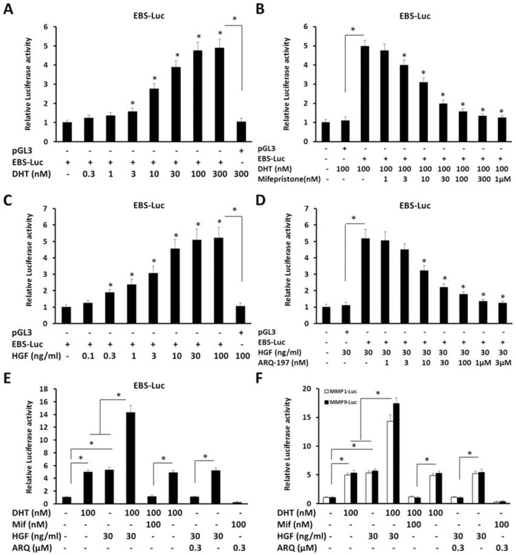
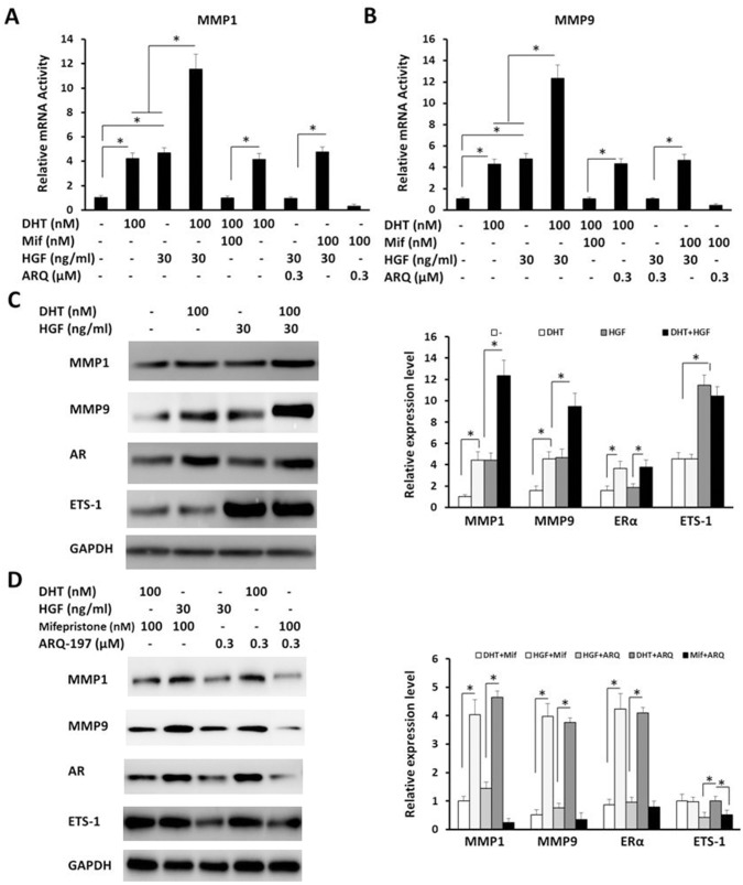
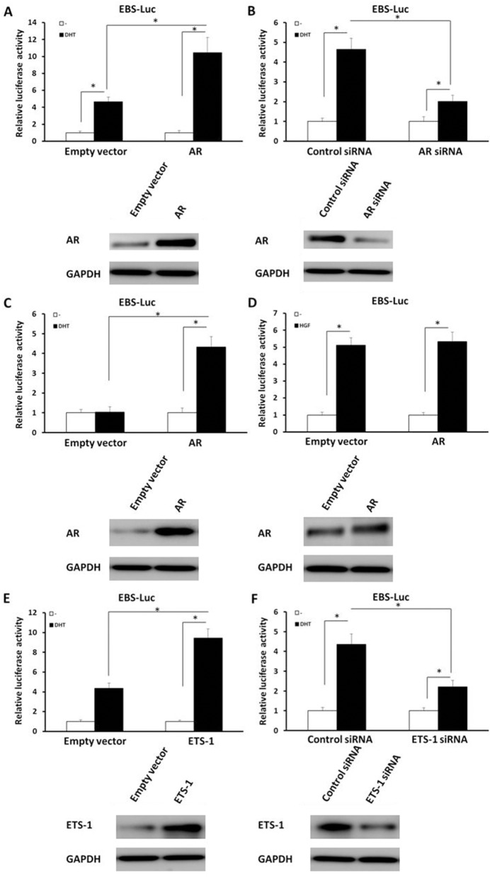
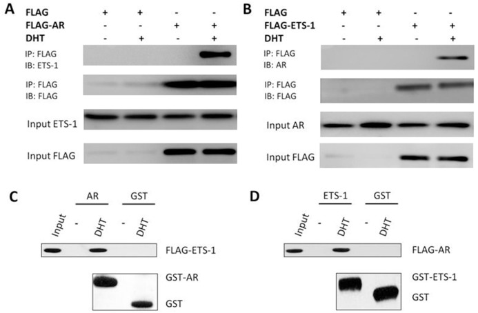
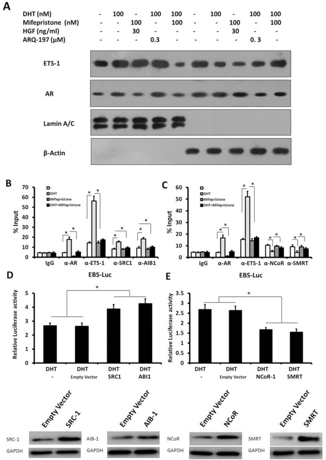
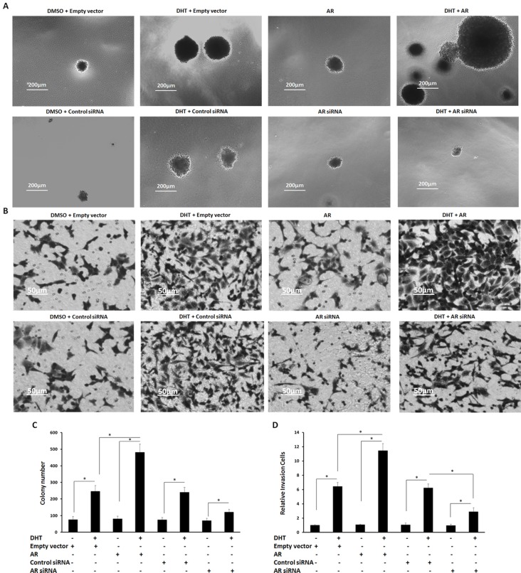
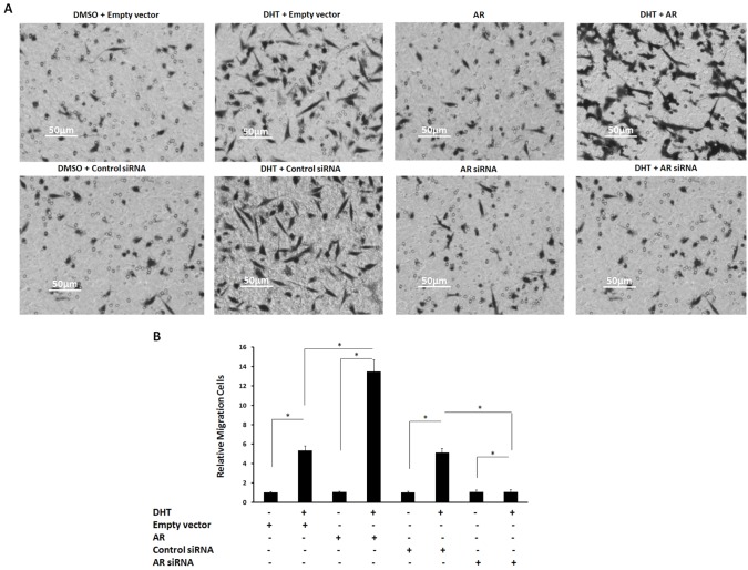
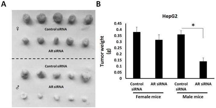
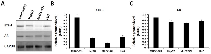
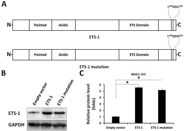
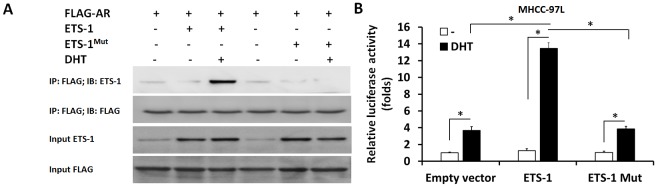
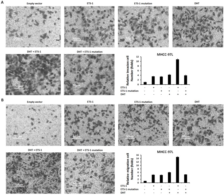
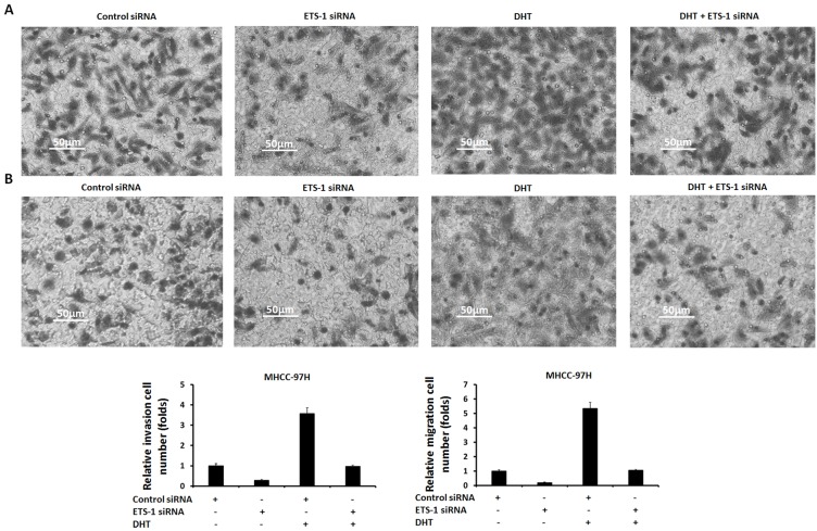
The expression of androgen receptor (AR) has been detected in hepatocellular cancer (HCC). However, there is no universal model detailing AR's function and mechanism in HCC. This study's results show that treatment with dihydrotestosterone (DHT), an endogenous androgen, promoted HCC cells' proliferation and up-regulated the transcription factor activity of ETS-1 (E26 transformation specific sequence 1), which mediates the migration and invasion of cancer cells via protein-protein interaction between AR and ETS-1. Results from luciferase assays showed that ETS-1's activity was significantly up-regulated following androgen treatment. AR mediated ETS-1's DHT-induced transcription factor activity. A potential protein-protein interaction between ETS-1 and AR was identified via glutathione S-transferase (GST) pull-down and co-immunoprecipitation assays. The mechanisms' data indicated that enhancing AR activity increases ETS-1's activity by modulating its cytoplasmic/nuclear translocation and recruiting ETS-1 to its target genes' promoter. Moreover, while overexpression of AR significantly increased the proliferation or in vitro migration or invasion of HepG2 cells in the presence of androgen, inhibiting AR's activity reduced these abilities. Thus, AR's function as a novel ETS-1 co-activator or potentially therapeutic target of HCC has been demonstrated.

Keywords: ETS-1; HCC; androgen; androgen receptor; proliferation.


Conflict of interest statement

CONFLICTS OF INTERSET The authors declare no conflicts of interest.

Figures

Figure 1
Figure 1. The dose-effect of androgen, mifepristone, HGF, or ARQ-197 on the transcription factor activity of ETS-1
(A-F) HepG2 cells, which were co-transfected with EBS-Luc, MMP1-Luc, or MMP9-Luc reporters, were treated with the indicated concentration (A, B, E, F) of DHT (the agonist of AR), (B, E, F) of mifepristone (the antagonist of AR), (C, D, E, F) of HGF (hepatocyte growth factor, the agonist of c-Met) or (D, E, F) of ARQ-197 (the antagonist of c-Met).Then, the cells were harvested and determined by Luciferase assays. The values are the mean ± SD from triplicate independent experiments. *P < 0.05.
Figure 2
Figure 2. The effect of AR on the expression of ETS-1-targeted genes
HepG2 cells were treated with the indicated concentration (ECmax/ICmax concentration) of DHT (A–D) mifepristone (A, B, D), HGF (A-D), or ARQ-197 (A, B, D). (A–B) Identification of ETS-1-targeted genes’ mRNA level was determined by real-time RT-PCR assays. (C-D) The protein level of AR, ETS-1, or its responses genes were identified by WB assays. GAPDH was used as the loading control. The values are the mean ± SD from triplicate independent experiments. *P < 0.05.
Figure 3
Figure 3. AR (but not HGF/c-Met) mediates the enhancement of androgen-induced ETS-1 activity
Cells were treated with 100 nM of DHT (A-C, E, F) or 30 ng/ml of HGF (D). HepG2 cells were stably transfected with empty vector (A, D, E), AR vectors (A, D), control siRNA (B, F), AR siRNA (B), ETS-1 vector (E), or ETS-1 siRNA (F), while PC-3 cells were stably transfected with empty vector (C) or AR vectors (C). Then, cells which were co-transfected with EBS-Luc reporters and harvested for Luciferase analysis. The expression of AR and ETS-1 were determined by immunoblots, and the results are shown in the panels at the bottom of the figure. The values are the mean ± SD from triplicate independent experiments. *P < 0.05.
Figure 4
Figure 4. AR can interact with ETS-1
(A–B) The interaction of endogenous AR or ETS-1 with exogenous FLAG-ETS1 or FLAG-AR. FLAG-tagged AR (A), FLAG-tagged ETS-1 (B), or FLAG empty vector (A–B) was transfected into HepG2 cells. Cell lysates were immunoprecipitated by an anti-FLAG monoclonal antibody, and the precipitates were then immunoblotted with anti-ETS-1 or anti-AR antibody. (C–D) In vitro interaction between ETS-1 and AR. Glutathione-Sepharose beads bound with GST-AR (C), GST-ETS1 (D), or GST (C–D) were incubated with purified FLAG-labeled ETS-1 or AR in the presence or absence of 100 nM DHT. After washing the beads, the bound proteins were eluted and subjected to SDS-PAGE and IB assays.
Figure 5
Figure 5. The mechanisms of AR’s effect on ETS-1’s activity
(A) HepG2 cells were treated with the indicated amount of DHT, mifepristone, HGF, or ARQ-197. Then, cells were fractionated into cytoplasmic and nuclear fractions. The fractions were detected with ETS-1 and ERα antibodies. Lamin A/C was the nuclear indicator. ß-actin was the cytoplasmic marker. (B) The recruitment of ETS-1, AR, SRC-1, and AIB-1 to the mmp1 promoter was detected by ChIP assay. (C) The recruitment of ETS-1, AR, NCoR, and SMRT to the mmp1 promoter was detected by ChIP assay. (D–E) HepG2 cells were stimulated with 100 nM DHT for 24 h. Cells were transfected with SRC-1 (D), AIB-1 (D), NCoR (E), or SMRT (E) expression vectors or empty vectors. Cells were then harvested for the luciferase assay. The values are the mean ± SD from triplicate independent experiments. Western blot (bottom) indicates the expression level of the proteins with anti-SRC1, anti-AIB1, anti-NCoR, or anti-SMRT antibodies. GAPDH was used as the loading control. *P < 0.05.
Figure 6
Figure 6. The effect of AR on HepG2 cells’ anchor-independent growth and invasion
(A, B) HepG2 cells, which were stably transfected with empty vectors, AR vectors, control siRNA, or AR siRNA, were treated with or without 100 nM of DHT. Cells were then measured by soft agar assay (A) or transwell assay (B). Colonies or invasion cells are shown in photographs A and B. (C, D) Data are mean ± SD of triplicate independent experiments and were repeated three times with similar numbers. *P < 0.05 versus Solvent control (DMSO) or DHT; *P < 0.05 versus empty vectors or AR vectors.
Figure 7
Figure 7. The effect of androgen/AR on HepG2 cells’ migration
(A) HepG2 cells, which were stably transfected with empty vectors, AR vectors, control siRNA, or AR siRNA, were treated with or without 100 nM of DHT. Then, the cells were measured by transwell assays (A). The migration cells are shown in the photograph (A). (B) Mean ± SD of triplicate independent experiments and have been repeated three times with similar numbers. *P < 0.05.
Figure 8
Figure 8. The effect of AR deletion on HepG2 cells’ in vivo growth
(A) HepG2 cells, which were stably transfected with control siRNA or AR siRNA, were seeded in female or male nude mice. Results are shown as photographs (A) or mean ± SD of tumor weight (B). *P < 0.05.
Figure 9
Figure 9. AR and ETS-1 are expressed in HCC cells
HCC cells (HepG2, MHCC-97H, Hu7, or MHCC-97L), were harvested for western blot analysis. Results are shown as photographs (A) or densitometric analysis (B and C). *P < 0.05.
Figure 10
Figure 10. The point mutation of ETS-1
The “LXXLL” motif located in the c-terminal of ETS-1 were mutated. The results are shown as bold, italicized font (A), bands of ETS-1 or GAPDH from western blot (B), or densitometric analysis (C). *P < 0.05.
Figure 11
Figure 11. The interaction between AR and ETS-1 or ETS-1 mutation
FLAG-tagged AR, wild-type ETS-1, or ETS-1 mutation was transfected into MHCC-97L cells. Cell lysates were immunoprecipitated by an anti-FLAG monoclonal antibody, and the precipitates were then immunoblotted with an anti-ETS-1 or anti-FLAG antibody (A). (B) MHCC-97L cells were harvested and analyzed by luciferase. *P < 0.05.
Figure 12
Figure 12. The effect of wild-type ETS-1 or ETS-1 mutation on HepG2 cells’ in vitro invasion or migration enhanced by DHT
MHCC-97L cells, which were stably transfected with empty vectors, wild-type ETS-1, or ETS-1 mutation vectors, were treated with or without 100 nM of DHT. Then, the cells were measured by transwell assays. The invasion (A) or migration (B) cells were shown as photographs or mean ± SD of triplicate independent experiments and were repeated three times with similar numbers. *P < 0.05.
Figure 13
Figure 13. The effect of ETS-1 knockdown on MHCC-97H cells’ in vitro invasion or migration enhanced by DHT
MHCC-97H cells, which were stably transfected with control siRNA or ETS-1 siRNA, were treated with without 100 nM. Then, the cells were measured by transwell assays. The invasion (A) or migration (B) cells were shown as photographs or mean ± SD of triplicate independent experiments and were repeated three times with similar numbers. *P < 0.05.

References

    1. Chen G, Nomura M, Morinaga H, Matsubara E, Okabe T, Goto K, Yanase T, Zheng H, Lu J, Nawata H. Modulation of androgen receptor transactivation by FoxH1. A newly identified androgen receptor corepressor. J Biol Chem. 2005;280:36355–36363. - PubMed
    1. Lu Y, Feng F, Yang Y, Gao X, Cui J, Zhang C, Zhang F, Xu Z, Qv J, Wang C, Zeng Z, Zhu Y, Yang Y. LINE-1 ORF-1p functions as a novel androgen receptor co-activator and promotes the growth of human prostatic carcinoma cells. Cell Signal. 2013;25:479–489. - PubMed
    1. Cui J, Yang Y, Zhang C, Hu P, Kan W, Bai X, Liu X, Song H. FBI-1 functions as a novel AR co-repressor in prostate cancer cells. Cell Mol Life Sci. 2011;68:1091–1103. - PMC - PubMed
    1. Tian YE, Xie XU, Lin Y, Tan G, Zhong WU. Androgen receptor in hepatocarcinogenesis: Recent developments and perspectives. Oncol Lett. 2015;9:1983–1988. - PMC - PubMed
    1. Chen PJ, Yeh SH, Liu WH, Lin CC, Huang HC, Chen CL, Chen DS, Chen PJ. Androgen pathway stimulates microRNA-216a transcription to suppress the tumor suppressor in lung cancer-1 gene in early hepatocarcinogenesis. Hepatology. 2012;56:632–643. - PubMed