

Role of the stromal cell derived factor-1/CXC chemokine receptor 4 axis in the invasion and metastasis of lung cancer and mechanism
- PMID: 29312699
- PMCID: PMC5757066
- DOI: 10.21037/jtd.2017.10.138
Role of the stromal cell derived factor-1/CXC chemokine receptor 4 axis in the invasion and metastasis of lung cancer and mechanism
Abstract
Background: Lung cancer is the most common tumor, and has the highest incidence and mortality rates among all malignant tumors. Since stromal cell derived factor-1 (SDF-1) and CXC chemokine receptor 4 (CXCR4) are specific to binding sites, they are more important than other members of the families for tumor invasion and metastasis. We herein aimed to investigate the role of the axis of chemokine SDF-1 and its receptor CXCR4 in the invasion and metastasis of lung cancer.
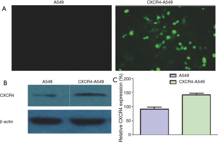
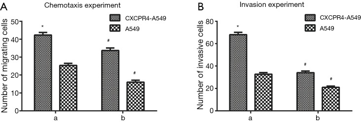
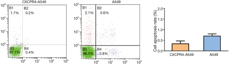
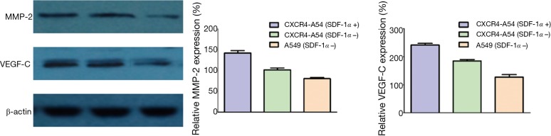
Methods: Sixty clinical non-small cell lung cancer (NSCLC) tissue samples were collected. The CXCR4 expressions in cancer, paracancerous and normal lung tissues were detected by immunocytochemical assay and PCR. Cells with CXCR4 overexpression (CXCR4-A549) were constructed. After induction with SDF-1, CXCR4-A549 and A549 cells were subjected to in vitro chemotaxis and invasion assays. Their proliferation and apoptosis were detected by flow cytometry. The activities of phosphoinositide 3-kinase/protein kinase B (AKT) and mitogen-activated protein kinase/extracellular signal-regulated kinase (ERK)-related signaling pathways were detected by Western blot. The downstream signaling molecules that may be activated by SDF-1/CXCR4 were analyzed. The expressions of vascular endothelial growth factor-C and matrix metalloproteinase-2 were detected by Western blot and PCR. A mouse model was established by subcutaneous inoculation of lung cancer cells. The effects of up-regulated CXCR4 expression on the migration of lung cancer cells in vitro and their tumorigenesis and metastasis in vivo were assessed.
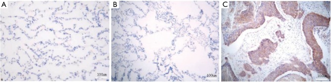
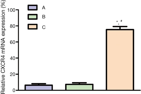
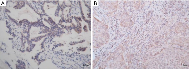
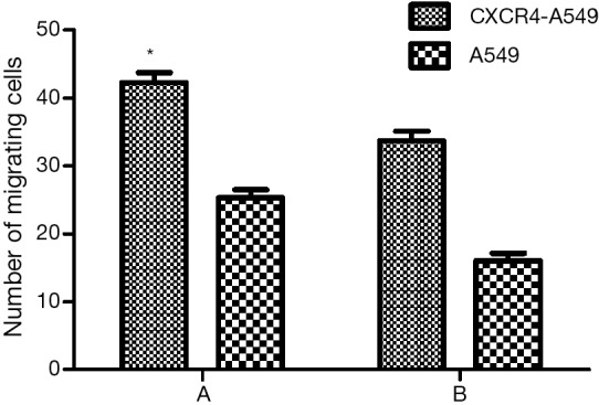
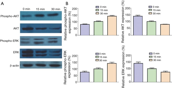
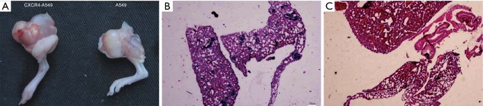
Results: There was no expression in normal or paracancerous tissues. The expression of CXCR4 mRNA in lung cancer tissues was 83.3% (50/60). The expressions of CXCR4 in lung squamous cell carcinoma and adenocarcinoma were similar (P>0.05). The expression of CXCR4 was 76.9% (10/13) in highly differentiated carcinoma, 82.1% (23/28) in moderately differentiated carcinoma and 84.2% (16/19) in lowly differentiated carcinoma (P>0.05). The expression of CXCR4 was 72.7% (8/11) in TNM stage I patients, 83.9% (26/31) in stage II patients, and 88.9% (16/18) in stage III patients, with significant correlations. After up-regulation of CXCR4, the invasion ability of CXCR4-A549 cells was increased 1.62-fold (P<0.05). ERK and AKT were significantly phosphorylated 30 min after SDF-1 treatment. The tumorigenic rates of six mice inoculated with CXCR4-A549 and A549 cells were both 100%, with the average tumor weights of (4.37±0.96 g) and (3.24±1.16 g) respectively (P<0.05). In the CXCR4-A549 group, metastatic tumors clearly formed in the lungs of 6 mice, but only 2 mice in the A549 group had tumor cell invasion.
Conclusions: SDF-1/CXCR4 played a key role in the invasion and metastasis of lung cancer. The interaction between SDF-1α and CXCR4 activated a series of downstream molecules by activating ERK and AKT.
Keywords: CXC chemokine receptor 4 (CXCR4); Stromal cell derived factor-1 (SDF-1); lung cancer; metastasis.
Conflict of interest statement
Conflicts of Interest: The authors have no conflicts of interest to declare.
Figures
References
LinkOut - more resources
Full Text Sources
Other Literature Sources
Miscellaneous