

Weight bearing cone beam CT scan versus gravity stress radiography for analysis of supination external rotation injuries of the ankle
- PMID: 29312872
- PMCID: PMC5756777
- DOI: 10.21037/qims.2017.12.02
Weight bearing cone beam CT scan versus gravity stress radiography for analysis of supination external rotation injuries of the ankle
Abstract
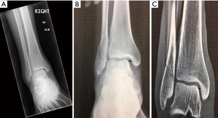
For AO 44-B2 ankle fractures of uncertain stability, the current diagnostic standard is to obtain a gravity stress radiograph, but some have advocated for the use of weight-bearing radiographs. The primary aim was to compare measures of medial clear space (MCS) on weight-bearing cone beam computed tomography (CBCT) scans versus gravity stress radiographs for determining the state of stability of ankle fractures classified as AO SER 44-B2 or Weber B. The secondary aim was to evaluate the details offered by CBCT scans with respect to other findings that may be relevant to patient care. Nine patients were enrolled in this cross-sectional study between April 2016 and February 2017 if they had an AO SER 44-B2 fracture of uncertain stability, had a gravity stress radiograph, and were able to undergo CT scan within seven days. The width of the MCS was measured at the level of the talar dome on all radiographs and at the mid coronal slice on CT. Wilcoxon signed-ranks tests were used to compare MCS between initial radiographs, gravity stress radiographs and weight-bearing CBCT scans. MCS on weight-bearing CBCT scan (1.41±0.41 mm) was significantly less than standard radiographs (3.28±1.63 mm, P=0.004) and gravity stress radiographs (5.82±1.93 mm, P=0.02). There was no statistically significant difference in MCS measured on standard radiographs versus gravity stress radiographs (P=0.11). Detailed review of the multiplanar CT images revealed less than perfect anatomical reduction of the fractures, with residual fibular shortening, posterior displacement, and fracture fragments in the incisura as typical findings. Similar to weight-bearing radiographs, weight-bearing CBCT scan can predict stability of AO 44-B2 ankle fractures by showing restoration of the MCS, and might be used to indicate patients for non-operative treatment. None of the fractures imaged in this study were perfectly reduced however, and further clinical research is necessary to determine if any of the detailed weight-bearing CBCT findings are related to patient outcomes.
Keywords: Weight bearing; ankle fracture; computed tomography (CT); gravity stress.
Conflict of interest statement
Conflicts of Interest: The authors have no conflicts of interest to declare.
Figures


References
LinkOut - more resources
Full Text Sources
Other Literature Sources
Miscellaneous