Review
. 2017 Dec 19:5:114.
doi: 10.3389/fcell.2017.00114. eCollection 2017.

The Multifaceted Role of the Lysosomal Protease Cathepsins in Kidney Disease

Affiliations
Review

The Multifaceted Role of the Lysosomal Protease Cathepsins in Kidney Disease

Pasquale Cocchiaro et al. Front Cell Dev Biol. .

Abstract

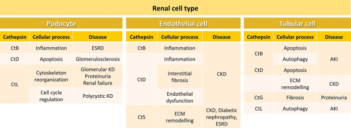
Kidney disease is worldwide the 12th leading cause of death affecting 8-16% of the entire population. Kidney disease encompasses acute (short-lasting episode) and chronic (developing over years) pathologies both leading to renal failure. Since specific treatments for acute or chronic kidney disease are limited, more than 2 million people a year require dialysis or kidney transplantation. Several recent evidences identified lysosomal proteases cathepsins as key players in kidney pathophysiology. Cathepsins, originally found in the lysosomes, exert important functions also in the cytosol and nucleus of cells as well as in the extracellular space, thus participating in a wide range of physiological and pathological processes. Based on their catalytic active site residue, the 15 human cathepsins identified up to now are classified in three different families: serine (cathepsins A and G), aspartate (cathepsins D and E), or cysteine (cathepsins B, C, F, H, K, L, O, S, V, X, and W) proteases. Specifically in the kidney, cathepsins B, D, L and S have been shown to regulate extracellular matrix homeostasis, autophagy, apoptosis, glomerular permeability, endothelial function, and inflammation. Dysregulation of their expression/activity has been associated to the onset and progression of kidney disease. This review summarizes most of the recent findings that highlight the critical role of cathepsins in kidney disease development and progression. A better understanding of the signaling pathways governed by cathepsins in kidney physiopathology may yield novel selective biomarkers or therapeutic targets for developing specific treatments against kidney disease.

Keywords: acute kidney injury; cathepsins; chronic kidney disease; lysosomal proteases; signaling pathways.


Figures

Figure 1
Figure 1
Nephron segments and their main physiological function. The nephron is the functional unit of the kidney and is composed by the renal corpuscle and the renal tubule. In the renal corpuscle, the glomerular filtrate is generated by filtration of water, ions, and small molecules from the bloodstream. The glomerular filtrate is transformed into urine by reabsorption and secretion of different molecules through the different sections of the renal tubule (proximal convoluted tubule, loop of Henle, and distal convoluted tubule) and the collecting duct system. Kidney and nephron image adapted from Smart Servier Medical Art under Creative Commons Attribution 3.0 Unported License.
Figure 2
Figure 2
Cellular processes and kidney diseases involving cathepsins in different renal cell types. In podocytes, CtB participates in inflammation during ESRD, CtD is involved in apoptosis in glomerulosclerosis, and CtL plays a role in cytoskeleton reorganization and cell cycle regulation during glomerular kidney disease, proteinuria, renal failure and polycystic kidney disease. In endothelial cells, CtB and CtD are involved in inflammation. In addition, CtD participates in interstitial fibrosis and endothelial dysfunction during CKD. CtS is important in CKD, diabetic nephropathy and ESRD. In tubular cells, CtsB is involved in apoptosis and autophagy during AKI, and CtD in apoptosis and ECM remodeling during CKD. CtG participates in fibrosis during proteinuria, and CtL in autophagy in AKI. ESRD, end-stage renal disease; KD, kidney disease; CKD, chronic kidney disease; ECM, extracellular matrix; AKI, acute kidney injury.

References

    1. Abd-Elgaliel W. R., Cruz-Monserrate Z., Wang H., Logsdon C. D., Tung C. H. (2013). Pancreatic cancer-associated Cathepsin E as a drug activator. J. Control Release 167, 221–227. 10.1016/j.jconrel.2013.02.007 - DOI - PMC - PubMed
    1. Adamson J. W. (1996). Regulation of red blood cell production. Am. J. Med. 101, 4S−6S. 10.1016/S0002-9343(96)00160-X - DOI - PubMed
    1. Aikawa E., Aikawa M., Libby P., Figueiredo J. L., Rusanescu G., Iwamoto Y., et al. . (2009). Arterial and aortic valve calcification abolished by elastolytic cathepsin S deficiency in chronic renal disease. Circulation 119, 1785–1794. 10.1161/CIRCULATIONAHA.108.827972 - DOI - PMC - PubMed
    1. Aisa M. C., Cappuccini B., Barbati A., Orlacchio A., Baglioni M., Di Renzo G. C. (2016). Biochemical parameters of renal impairment/injury and surrogate markers of nephron number in intrauterine growth-restricted and preterm neonates at 30-40 days of postnatal corrected age. Pediatr. Nephrol. 31, 2277–2287. 10.1007/s00467-016-3484-4 - DOI - PubMed
    1. Alghamdi T. A., Majumder S., Thieme K., Batchu S. N., White K. E., Liu Y., et al. . (2017). Janus Kinase 2 regulates transcription factor EB expression and autophagy completion in glomerular podocytes. J. Am. Soc. Nephrol. 28, 2641–2653. 10.1681/ASN.2016111208 - DOI - PMC - PubMed

LinkOut - more resources