Review
. 2016 May 27;1(1):30-46.
doi: 10.1002/btm2.10004. eCollection 2016 Mar.

Biomimetic strategies for targeted nanoparticle delivery

Affiliations
Review

Biomimetic strategies for targeted nanoparticle delivery

Diana Dehaini et al. Bioeng Transl Med. .

Abstract

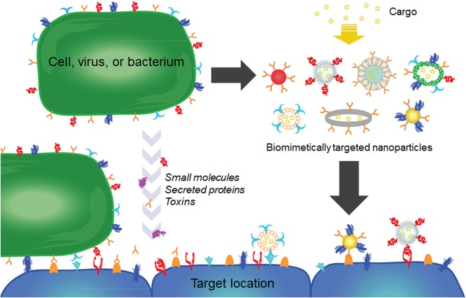
Nanoparticle-based drug delivery and imaging platforms have become increasingly popular over the past several decades. Among different design parameters that can affect their performance, the incorporation of targeting functionality onto nanoparticle surfaces has been a widely studied subject. Targeted formulations have the ability to improve efficacy and function by positively modulating tissue localization. Many methods exist for creating targeted nanoformulations, including the use of custom biomolecules such as antibodies or aptamers. More recently, a great amount of focus has been placed on biomimetic targeting strategies that leverage targeting interactions found directly in nature. Such strategies, which have been painstakingly selected over time by the process of evolution to maximize functionality, oftentimes enable scientists to forgo the specialized discovery processes associated with many traditional ligands and help to accelerate development of novel nanoparticle formulations. In this review, we categorize and discuss in-depth recent works in this growing field of bioinspired research.

Keywords: bioinspired; biomimetic nanoparticle; drug delivery; imaging; nanomedicine; targeting.


Figures

Figure 1
Figure 1
Schematic of biomimetic targeting strategies. Targeting affinities exist among cells, small molecules, proteins, and toxins. These can be directly leveraged to create targeted nanoparticle formulations for therapeutics and imaging
Figure 2
Figure 2
Examples of targeting strategies using small molecules. (a–c) Targeting of red blood cell membrane‐coated nanoparticles (RBC‐NPs) with folate using a membrane‐anchoring approach. (a) Schematic of a folate molecule conjugated to a lipid‐PEG tether along with a flow cytometry histogram demonstrating that targeted RBC‐NPs display increased affinity to folate receptor‐overexpressing cells. The targeting is abrogated in the presence of free folate. (b) Quantification of mean fluorescence intensities from the experiment in (a). (c) Fluorescent imaging demonstrating targeting capability of targeted RBC‐NPs. Red: dye labeled nanoparticles; blue: DAPI‐stained nuclei. Scale bar = 25 µm. (d) Heparin‐folate‐paclitaxel nanoparticles (HFT‐Ts) target folate receptors in a KB xenograft tumor model in vivo. Particles were labeled with Cy5.5 dye for in vivo fluorescence imaging. (a–c) Adapted with permission from ref. 10. Copyright 2013 by The Royal Society of Chemistry. (d) Adapted with permission from ref. 30. Copyright 2011 by American Chemical Society
Figure 3
Figure 3
Examples of targeting strategies using carbohydrates. (a, b) Galactose‐functionalized micelles for liver targeting. (a) Schematic of micelles functionalized with and without galactose‐PEG ligands. Galactose‐targeted particles are localized in the liver, and nontargeted particles are localized in the tumor. (b) In vivo biodistribution of galactose‐targeted and nontargeted particles 48 hr after intravenous administration. Bioluminescence originates from luciferase‐expressing tumor cells. (c, d) Glycan‐functionalized gold nanoparticles (AuNPs) for dendritic cell targeting. (c) Glycomimetic α‐fucosylamide functionalization scheme of AuNPs, these nanoparticles can be taken up preferentially by dendritic cells via DC‐SIGN lectins. (d) Internalization of α‐fucosylamide functionalized AuNPs (with either 0, 15, 30, or 50% α‐fucosylamide linked ligands on the surface) by dendritic cells. Increasing the amount of α‐fucosylamide on the surface increases percent of nanoparticles internalized by the dendritic cells. (a, b) Adapted with permission from ref. 49. Copyright 2014 by John Wiley & Sons, Inc. (c, d) Adapted with permission from ref. 60. Copyright 2014 by American Chemical Society
Figure 4
Figure 4
Examples of targeting strategies using peptides. (a, b) Chlorotoxin (CTX)‐functionalized iron oxide nanoparticles for glioma targeting. (a) Schematic showing conjugation scheme for CTX functionalization of PEGylated iron oxide nanoparticles. (b) MRI images of CTX nanoprobe localization in tumor xenograft. CTX‐targeted nanoparticles showed higher accumulation in tumors than a control nanoparticle, NP‐PEG‐SIA. (c, d) TAT‐functionalized liposomes for enhanced tumor entry. (c) Schematic of nanoformulation functionalized with both TAT peptide and transferrin (TF) using cholesterol‐PEG (Cho‐PEG) conjugates. TF enables cell targeting while the TAT enhances cellular entry. (d) Fluorescently labeled liposome uptake by tumors in an in vivo xenograft model. Formulations, from top to bottom, were liposomes with TAT and TF, liposomes with TAT only, liposomes with TF only, and bare liposomes. (a, b) Adapted with permission from ref. 92. Copyright 2008 by John Wiley & Sons, Inc. (c, d) Adapted with permission from ref. 107. Copyright 2013 by Elsevier
Figure 5
Figure 5
Examples of targeting strategies using proteins. (a, b) Lactoferrin (Lf)‐functionalized iron oxide nanoparticles for delivery across the BBB. (a) Schematic of Lf‐coated iron oxide nanoparticles demonstrating the interactions between the nanoparticles and brain endothelial cells. (b) In vivo MRI image showing brain uptake of Lf‐coated iron oxide nanoparticles. Red dotted lines encircle areas of contrast where nanoparticles are present. (c, d) HDL nanoparticles for delivery to atherosclerotic plaque. (c) Schematic (left) and transmission electron microscopy (TEM) image (right) of statin‐loaded recombinant HDL nanoparticles ([S]‐rHDL). TEM image shows [S]‐rHDL were 26 nm in diameter. (d) Fluorescent imaging shows colocalization between the [S]‐rHDL particles labeled with dye and macrophages (CD68) at sites of plaque in an aorta. Scale bars = 400 μm for main panel and 100 μm for inset. (a, b) Adapted with permission from ref. 135. Copyright 2012 by American Chemical Society. (c, d) Adapted with permission from ref. 147. Copyright 2014 by Macmillan Publishers Ltd.
Figure 6
Figure 6
Examples of targeting strategies using pathogen‐derived particles. (a, b) Canine parvovirus‐like particles (CVP‐VLPs) for delivery to tumors. (a) Model of the protein capsid with a single subunit depicted on the inset. (b) CVP‐VLPs were internalized by HeLa cells, as shown by colocalization of antibody‐labeled CVP‐VLPs (green) and dye‐conjugated transferrin (red). Scale bar = 25 µm. (c, d) Bacteria ghosts for delivery to tumors. (c) Schematic showing formation of bacterial ghosts through evacuation of the intrabacterial contents and loading with therapeutic or imaging agents. (d) Doxorubicin‐loaded bacterial ghosts as targeted drug delivery vehicles. Microscopy images show bacterial ghosts (bright field) and doxorubicin (red) overlaid. (a, b) Adapted with permission from ref. 163. Copyright 2006 by BioMed Central Ltd. (c) Adapted with permission from ref. 168. Copyright 2014 by Medknow. (d) Adapted with permission from ref. 166. Copyright 2009 by Elsevier
Figure 7
Figure 7
Examples of targeting strategies using mammalian cell membranes. (a, b) Macrophage exosomes for delivery to the brain. (a) Schematic of macrophage exosome derivation and application. Catalase‐loaded macrophage exosomes were designed as a brain‐targeted Parkinson's disease (PD) therapy. (b) Exosome formulations were tested for uptake by PC12 neuronal cells compared with control particles. Macrophage‐derived exosomes (I and II) showed greatly enhanced uptake compared to nontargeted polymeric PLGA particles (III) and liposomes (IV) at 24 hr. Scale bar = 20 μm. (c–e) Platelet membrane‐coated nanoparticles (PNPs) for delivery to bacteria and damaged vasculature. (c) Schematic of fabrication and targeting abilities of PNPs. (d) Pseudocolored scanning electron microscopy (SEM) image of PNPs (orange) naturally targeting MRSA252 bacteria (gold). Scale bar = 400 nm. (e) Targeting of fluorescently labeled PNPs (red) to intact carotid artery (top) and damaged artery (bottom). PNPs show preferential binding to damaged areas. Scale bar = 500 μm. (a, b) Adapted with permission from ref. 180. Copyright 2015 by Elsevier. (c–e) Adapted with permission from ref. 189. Copyright 2015 by Macmillan Publishers Ltd.

References

    1. Wang AZ, Langer R, Farokhzad OC. Nanoparticle delivery of cancer drugs. Annu Rev Med. 2012;63:185–198. - PubMed
    1. Tibbitt MW, Dahlman JE, Langer R. Emerging frontiers in drug delivery. J Am Chem Soc. 2016;138:704–717. - PubMed
    1. Bao G, Mitragotri S, Tong S. Multifunctional nanoparticles for drug delivery and molecular imaging. Annu Rev Biomed Eng. 2013;15:253–282. - PMC - PubMed
    1. Farokhzad OC, Langer R. Impact of nanotechnology on drug delivery. ACS Nano. 2009;3:16–20. - PubMed
    1. Maeda H. Toward a full understanding of the EPR effect in primary and metastatic tumors as well as issues related to its heterogeneity. Adv Drug Deliv Rev. 2015;91:3–6. - PubMed