Review
. 2016 Jun 20;1(1):47-62.
doi: 10.1002/btm2.10005. eCollection 2016 Mar.

Engineering nanoparticles to overcome barriers to immunotherapy

Affiliations
Review

Engineering nanoparticles to overcome barriers to immunotherapy

Randall Toy et al. Bioeng Transl Med. .

Abstract

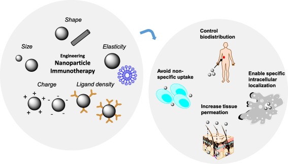
Advances in immunotherapy have led to the development of a variety of promising therapeutics, including small molecules, proteins and peptides, monoclonal antibodies, and cellular therapies. Despite this wealth of new therapeutics, the efficacy of immunotherapy has been limited by challenges in targeted delivery and controlled release, that is, spatial and temporal control on delivery. Particulate carriers, especially nanoparticles have been widely studied in drug delivery and vaccine research and are being increasingly investigated as vehicles to deliver immunotherapies. Nanoparticle-mediated drug delivery could provide several benefits, including control of biodistribution and transport kinetics, the potential for site-specific targeting, immunogenicity, tracking capability using medical imaging, and multitherapeutic loading. There are also a unique set of challenges, which include nonspecific uptake by phagocytic cells, off-target biodistribution, permeation through tissue (transport limitation), nonspecific immune-activation, and poor control over intracellular localization. This review highlights the importance of understanding the relationship between a nanoparticle's size, shape, charge, ligand density and elasticity to its vascular transport, biodistribution, cellular internalization, and immunogenicity. For the design of an effective immunotherapy, we highlight the importance of selecting a nanoparticle's physical characteristics (e.g., size, shape, elasticity) and its surface functionalization (e.g., chemical or polymer modifications, targeting or tissue-penetrating peptides) with consideration of its reactivity to the targeted microenvironment (e.g., targeted cell types, use of stimuli-sensitive biomaterials, immunogenicity). Applications of this rational nanoparticle design process in vaccine development and cancer immunotherapy are discussed.

Keywords: cancer immunotherapy; drug delivery; intracellular delivery; targeted nanoparticles; tissue permeation; vaccines.


Figures

Figure 1
Figure 1
Engineering nanoparticle immunotherapy. Nanoparticles of unique size, shape, elasticity, charge, and ligand density can be formulated to enhance the delivery of immunotherapies. Through an understanding of the effect of each of these parameters on biotransport and immunogenicity, nanoparticles can be designed to control the biodistribution of immunotherapies, evade nonspecific uptake by phagocytic cells, increase tissue permeation, and enable specific localization to targeted cellular compartments
Figure 2
Figure 2
Shape affects the internalization of nanoparticles. Cellular‐uptake kinetics of different shape‐specific nanoparticles in various cell lines. (A) HeLa cells, (B) HEK 293 cells, (C) BMDCs, and (D) HUVEC cells. In A–D, red lines are for nanodiscs (hollow for 325 × 100‐nm disks, dashed for 220 × 100‐nm disks, and solid for 80 × 70‐nm disks), and blue lines are for nanorods (dashed for 400 × 100 × 100‐nm rods and solid for 800 × 100 × 100‐nm rods). Error bars are SD with n = 3 for each data point. (E, F) Normalized median particle uptake per cell (indicates relative number of particles internalized by cells when normalized to 100 particles of 80 × 70‐nm disks) at the maximum internalization time point (72 hr for HeLa and BMDC, 48 hr for HEKs, and 24 hr for endothelial cells). Reproduced with permission from ref. 95
Figure 3
Figure 3
iRGD enhances the endothelial permeation of indocyanine‐labeled liposomes. The binding and penetration of iRGD–ICG‐LPs or ICG‐LPs to angiogenic endothelial cells were assessed with intravital and histological examination. The tumor vascular images were captured at 10 min after injecting 40 kDa FITC‐Dextran. The frozen sections were examined under a confocal microscope. Green represented the blood vessels labeled by FITC‐Dextran and red represented ICG‐loaded nanoparticles (Scale bar, 50 μm). Reproduced with permission from ref. 37
Figure 4
Figure 4
Viral peptides enhance the endosomal escape of nanoparticles. (A) Illustration of the R8‐MEND (left) and KALA‐MEND (right). (B) The R8‐MEND and KALA‐MEND encapsulating a conventional pDNA (pcDNA3.1‐Luc; opened bar) or CpG‐free pDNA (pCpGfree‐Luc(0); closed bar) were transfected to BMDCs. Data were presented as the mean ± SD of three independent experiments. Statistical differences were evaluated by one‐way ANOVA, followed by Student's t test (**p < .01). (C) The transfection activity of KALA‐MENDs encapsulating a pDNA with various set of backbone and inserts was also evaluated. Data were presented as the mean ± SD of three independent experiments. Statistical analyses were performed by one‐way ANOVA, followed by Bonferroni test. **p < .01 versus pCpGfree‐Luc(0). Reproduced with permission from ref. 56
Figure 5
Figure 5
Nanoparticles to treat immune suppression. (A) Preparation of siCTLA‐4‐encapsulated nanoparticles (NPsiCTLA‐4) with poly(ethylene glycol)‐block‐poly(d,l‐lactide) and a cationic lipid BHEM‐Chol by double emulsification. (B) Enhancing T‐cell‐mediated immune responses by blocking CTLA‐4 using NPsiCTLA‐4. CTLA‐4 plays a strong inhibitory role in T‐cell activation and proliferation, which significantly curbs T‐cell‐mediated tumor rejection. NPsiCTLA‐4‐mediated CTLA‐4 knockdown enhanced the activation and proliferation of T‐cells, which inhibited the overall growth of tumors. Reproduced with permission from ref. 76
Figure 6
Figure 6
Targeted multivalent aptamers deliver biotherapy to anaplastic large cell lymphoma. (A) Schema showing receptor oligomerization inducing downstream signaling. CD30‐associated signaling is activated by its ligand through trimerization of the receptor, leading to varied outcomes that range from apoptosis to proliferation. (B) CD30‐positive and ‐negative cells were incubated without any treatment; in presence of control streptavidin, monomeric aptamer C2NP, and multimeric aptamer C2NP, for 72 hr to detect aptamer‐mediated CD30 signal transduction. A multivalent CD30 aptamer was made using biotinylated C2NP (3×) with streptavidin (1×). (C) The multivalent CD30 aptamer‐induced signaling, resulting in a higher percentage of dead cells in CD30‐positive ALCL (K299 cells), and had no effect on cell death in CD30‐negative (HL60) cells. Reproduced with permission from ref. 81

Similar articles

Cited by

References

    1. Adams JL, Smothers J, Srinivasan R, Hoos A. Big opportunities for small molecules in immuno‐oncology. Nat Rev Drug Discov. 2015;14(9):603–622. - PubMed
    1. Weiner LM, Surana R, Wang S. Monoclonal antibodies: versatile platforms for cancer immunotherapy. Nat Rev Immunol. 2010;10(5):317–327. - PMC - PubMed
    1. Mahoney KM, Rennert PD, Freeman GJ. Combination cancer immunotherapy and new immunomodulatory targets. Nat Rev Drug Discov. 2015;14(8):561–584. - PubMed
    1. Lizotte PH, Wen AM, Sheen MR, et al. In situ vaccination with cowpea mosaic virus nanoparticles suppresses metastatic cancer. Nat Nanotechnol. 2016;11(3):295–303. - PMC - PubMed
    1. June CH, Maus MV, Plesa G, et al. Engineered T cells for cancer therapy. Cancer Immunol Immunother CII. 2014;63(9):969–975. - PMC - PubMed