. 2018 Apr;31(2):127-138.
doi: 10.1007/s13577-017-0198-2. Epub 2018 Jan 8.

CD146 positive human dental pulp stem cells promote regeneration of dentin/pulp-like structures

Affiliations

CD146 positive human dental pulp stem cells promote regeneration of dentin/pulp-like structures

Mikiko Matsui et al. Hum Cell. 2018 Apr.

Abstract

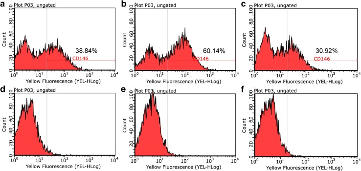
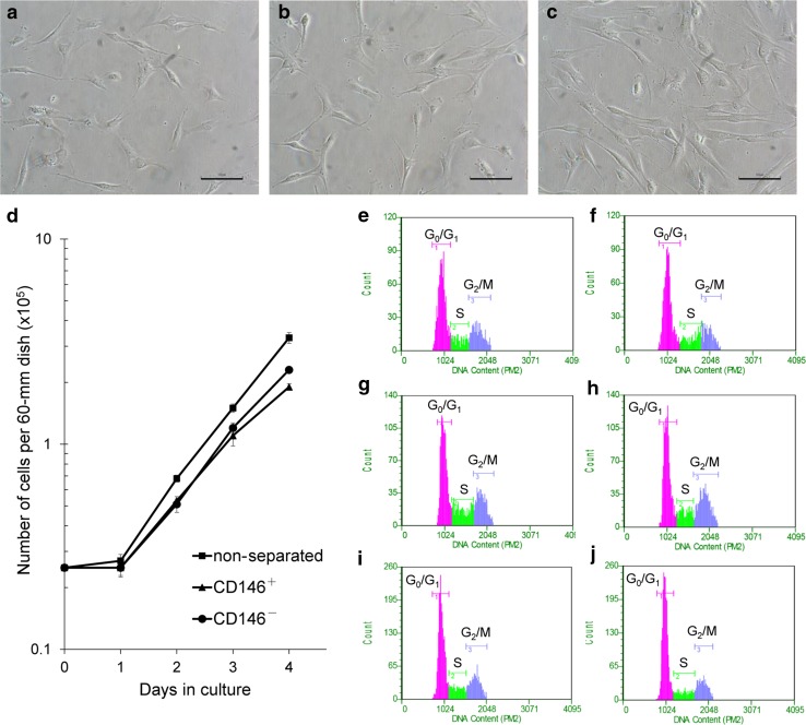
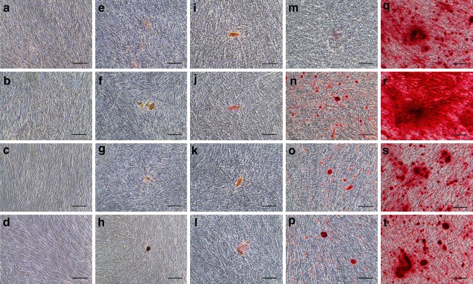
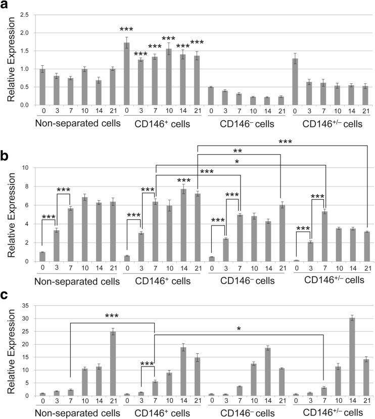
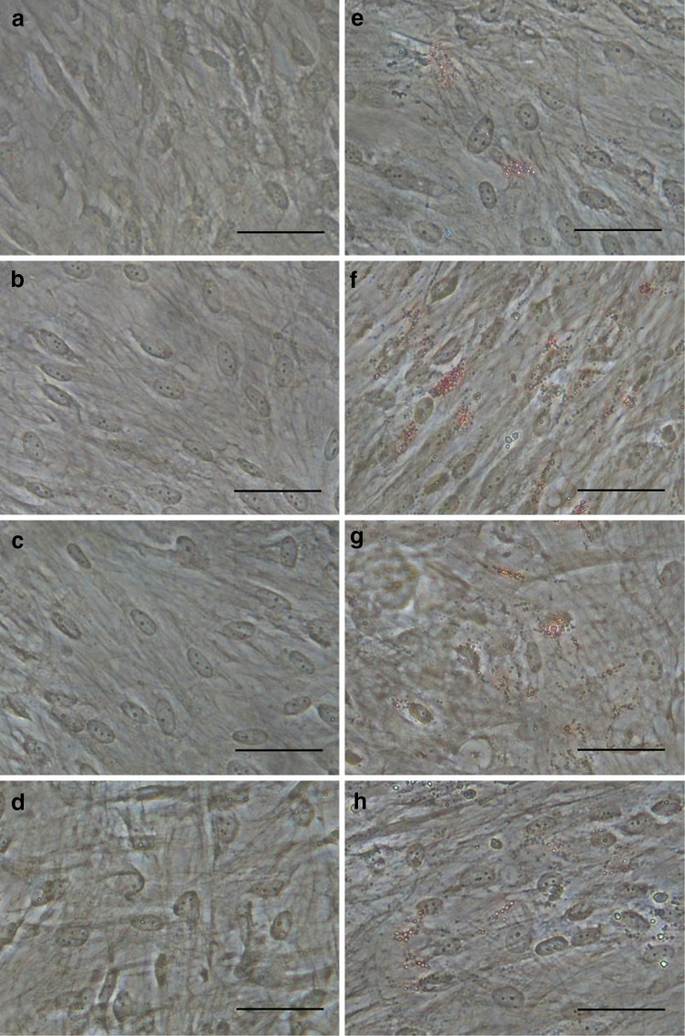
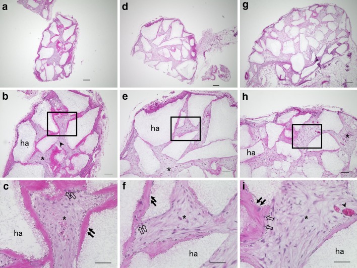
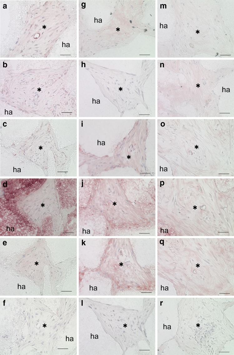
CD146 and STRO-1 are endothelial biomarkers that are co-expressed on the cellular membranes of blood vessels within human dental pulp tissue. This study characterized the percentage of dentin-like structures produced by CD146-positive (CD146+) human dental pulp stem cells (DPSCs), compared with their CD146-negative (CD146-) counterparts. DPSC populations were enriched using magnetic-activated cell sorting (MACS), yielding CD146+ and CD146- cells, as well as mixtures composed of 25% CD146+ cells and 75% CD146- cells (CD146+/-). Cell growth assays indicated that CD146+ cells exhibit an approximate 3-4 h difference in doubling time, compared with CD146- cells. Cell cycle distributions were determined by flow cytometry analysis. The low percentage of CD146+ cells' DNA content in G0/G1 phase were compared with CD146- and non-separated cells. In contrast to CD146- and non-separated cells, prompt mineralization was observed in CD146+ cells. Subsequently, qRT-PCR revealed high mRNA expression of CD146 and Alkaline phosphatase in mineralization-induced CD146+ cells. CD146+ cells were also observed high adipogenic ability by Oil red O staining. Histological examinations revealed an increased area of dentin/pulp-like structures in transplanted CD146+ cells, compared with CD146- and CD146+/- cells. Immunohistochemical studies detected dentin matrix protein-1 (DMP1) and dentin sialophosphoprotein (DSPP), as well as human mitochondria, in transplanted DPSCs. Co-expression of CD146 and GFP indicated that CD146 was expressed in transplanted CD146+ cells. CD146+ cells may promote mineralization and generate dentin/pulp-like structures, suggesting a role in self-renewal of stem cells and dental pulp regenerative therapy.

Keywords: CD146; Human dental pulp stem cells; Mineralization; Regenerative therapy; Transplantation.


Conflict of interest statement

The authors declare that they have no conflict of interest.

Figures

Fig. 1
Fig. 1
Flow cytometry analysis of the proportion of CD146 in each cell group (ac), compared with isotype control (df). CD146 of non-separated (a), CD146+ (b), and CD146 cells (c). Isotype control of non-separated (d), CD146+ (e), and CD146 cells (f)
Fig. 2
Fig. 2
Comparisons of cell morphology, cell growth, and cell cycle among cell groups. Morphology of non-separated (a), CD146+ (b), and CD146 cells (c). d Cell growth curve of each cell group. Cell numbers are presented as the mean ± SD from three independent experiments. Cell cycle analyses of non-separated (e, f), CD146+ (g, h), and CD146 cells (i, j) at 2 day (e, g, i) and 3 day (f, h, j) post-seeding. Scale bar = 100 µm (a, b, c)
Fig. 3
Fig. 3
Alizarin red S staining of mineralization potential (at) following induction with differentiation medium for 3 days (ad), 7 (eh), 10 days (il), 14 days (mp), and 21 days (qt). Non-separated (a, e, i, m, q), CD146+ (b, f, j, n, r), CD146 (c, g, k, o, s), and CD146+/− cells (d, h, l, p, t). Scale bars = 100 µm (at)
Fig. 4
Fig. 4
qRT-PCR analysis of CD146 mRNA (a), Alkaline phosphatase (ALP) mRNA (b), and Osteocalcin mRNA (c) expression in differentiation medium. Each group was analyzed after induction with differentiation medium for 0, 3, 7, 10, 14, and 21 days. All data were compared with non-separated cells at 0 days that were 80–100% confluent. Three statistical analyses were performed using a one-way ANOVA with Tukey’s post-test. The data are expressed as mean ± SD of three tests. *0.01 ≤ p < 0.05, **0.001 ≤ p < 0.01, ***p < 0.001
Fig. 5
Fig. 5
Oil red O staining of adipogenic potential (ah) following induction with differentiation medium for 7 days (ad) and 14 days (eh). Non-separated (a, e), CD146+ (b, f), CD146 (c, g), and CD146+/− cells (d, h). Scale bars = 50 µm (ah)
Fig. 6
Fig. 6
Cross sections are representative of transplanted CD146+ (a, b, c), CD146 (d, e, f), and CD146+/− cells (g, h, i). Low magnification of transplants (a, d, g). High magnification of the box region in (b, e, h) shows dentin/pulp-like structures (c, f, i). ha, HA/TCP carriers; asterisk, connective tissue; arrowhead, blood vessels; black arrow, dentin-like structure; open arrow, odontoblast-like cells. Dotted lines (c, f, i) denote area selected for analysis of DSA. Scale bars = 200 (a, d, g), 100 (b, e, h), and 50 µm (c, f, i)
Fig. 7
Fig. 7
Histological and immunohistochemical analysis of transplantation (ar). CD146+ (af), CD146 (gl), and CD146+/− cells (mr). Immunohistochemical staining with antibodies against CD146 (a, g, m), GFP (b, h, n), human mitochondria (c, i, o), DMP1 (d, j, p), DSPP (e, k, q), and no antibody (negative control; f, l, r). ha, HA/TCP carriers; asterisk, connective tissue. Scale bars = 50 µm (ar)

References

    1. Rohban R, Pieber TR. Mesenchymal stem and progenitor cells in regeneration: tissue specificity and regenerative potential. Stem Cells Int. 2017;2017:5173732. doi: 10.1155/2017/5173732. - DOI - PMC - PubMed
    1. Gronthos S, Mankani M, Brahim J, Robey PG, Shi S. Postnatal human dental pulp stem cells (DPSCs) in vitro and in vivo. Proc Natl Acad Sci USA. 2000;97:13625–13630. doi: 10.1073/pnas.240309797. - DOI - PMC - PubMed
    1. Harkness L, Zaher W, Ditzel N, Isa A, Kassem M. CD146/MCAM defines functionality of human bone marrow stromal stem cell populations. Stem Cell Res Ther. 2016;7:4. doi: 10.1186/s13287-015-0266-z. - DOI - PMC - PubMed
    1. Lehmann JM, Holzmann B, Breitbart EW, Schmiegelow P, Riethmüller G, Johnson JP. Discrimination between benign and malignant cells of melanocytic lineage by two novel antigens, a glycoprotein with a molecular weight of 113,000 and a protein with a molecular weight of 76,000. Cancer Res. 1987;47:841–845. - PubMed
    1. Shi S, Bartold PM, Miura M, Seo BM, Robey PG, Gronthos S. The efficacy of mesenchymal stem cells to regenerate and repair dental structures. Orthod Craniofac Res. 2005;8:191–199. doi: 10.1111/j.1601-6343.2005.00331.x. - DOI - PubMed