. 2017 Oct 19;61(4):2833.
doi: 10.4081/ejh.2017.2833.

Neurodegeneration in zebrafish embryos and adults after cadmium exposure

Affiliations

Neurodegeneration in zebrafish embryos and adults after cadmium exposure

Antonio Monaco et al. Eur J Histochem. .

Abstract

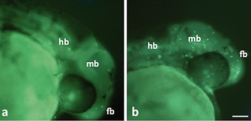
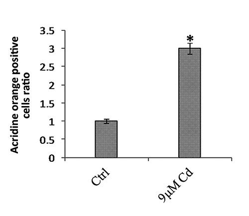
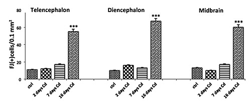
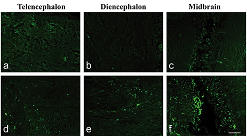
Cadmium is a biologically non-essential metal. It is also toxic to many organs including the brain. The aim of this study was to analyse the neurodegenerative effects of this metal in embryos and adults of zebrafish exposed to sub-lethal concentrations of cadmium. The study was performed by cytochemical stainings. Six hours after fertilisation (hpf) zebrafish embryos were treated for 24 hours with 9 μM of cadmium and subsequently stained with Acridine orange in whole mount to detect apoptosis in the brain. Adult zebrafish were treated for 16 days with the same concentration of cadmium, and cell death in the brain was detected by Fluoro-Jade B staining at 2, 7 and 16 days of treatment. An increase in cell death was observed only at 16 days of treatment in adults, while an increase in apoptotic events was revealed in the brain of embryos after 24 h of treatment. This evidence is indicative that cadmium, even at a sub-lethal concentration, induces cell death in the brain of embryos but also in adults of zebrafish in which the phenomenon appears time-dependent.Â.

Keywords: Cadmium; brain; cell death; fish; neurodegeneration; zebrafish..


Figures

Figure 1.
Figure 1.
Acridine orange stain in whole-mount zebrafish embryos. a) Control embryos. b) Embryos after 24-hour treatment; note an increase in the number of fluorescent cells in all brain areas. fb, forebrain; mb, midbrain; hb, hindbrain. Scale bar: 75 µm.
Figure 2.
Figure 2.
Average of the positive fluorescent cells in the embryos after 24-hour treatment with 9 µM of Cd and in the control embryos by Acridine orange staining in whole-mount. A 3-fold increase in apoptotic cells was observed in the treated embryos compared with the controls. *P>0.05.
Figure 3.
Figure 3.
Graphical representation of the mean numbers of Fluoro-Jade B positive cells/0.1 mm2 in the area of the telencephalon, diencephalon and midbrain in zebrafish adults after 2, 7 and 16 days of treatment with 9 µM of Cd and in control fish. Fish treated at 2 and 7 days showed few fluorescent signals similar to those revealed in control brains. A diffused fluorescent signal was particularly revealed at 16 days of treatment and the diencephalon appeared to be the area with more positivity than the others. ***P>0.001.
Figure 4.
Figure 4.
Fluoro-Jade B staining. Sagittal sections: a) telencephalon, b) diencephalon and c) midbrain in control adult fish; d) telencephalon, e) diencephalon and f ) midbrain of adults after 16 days treated with 9 µM of Cd. Note the increase in fluorescence in treated fish compared with the control. Fluorescence was especially present at the level of cell bodies and few nervous processes were stained. Scale bar: 50 µm.

References

    1. Fulda S, Gorman AM, Osamu H, Samali A. Cellular stress responses: Cell survival and cell death. Int J Cell Biol 2010;2010:214074. - PMC - PubMed
    1. Ichim G, Tauszig-Delamasure S, Mehlen P. Neurotrophins and cell death. Exp Cell Res 2012;318:1221-8. - PubMed
    1. Vaarmann A, Kovac S, Holmström KM, Gandhi S, Abramov AY. Dopamine protects neurons against glutamate-induced excitotoxicity. Cell Death Dis 2013;4: e455. - PMC - PubMed
    1. Weber JT. Altered calcium signaling following traumatic brain injury. Front Pharmacol 2012;3:60. - PMC - PubMed
    1. Mattson MP. Apoptosis in neurodegenerative disorders. Nat Rev Mol Cell Biol 2000;1:120-9. - PubMed