Review
. 2018 May;154(1):3-20.
doi: 10.1111/imm.12888. Epub 2018 Feb 27.

Human dendritic cell subsets: an update

Affiliations
Review

Human dendritic cell subsets: an update

Matthew Collin et al. Immunology. 2018 May.

Abstract

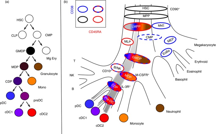
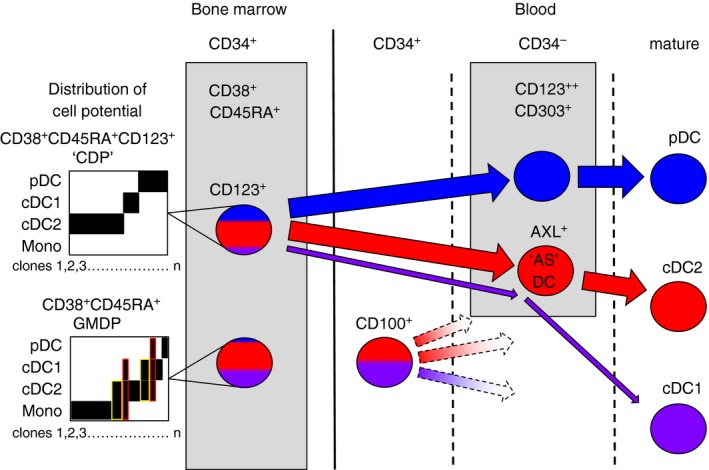
Dendritic cells (DC) are a class of bone-marrow-derived cells arising from lympho-myeloid haematopoiesis that form an essential interface between the innate sensing of pathogens and the activation of adaptive immunity. This task requires a wide range of mechanisms and responses, which are divided between three major DC subsets: plasmacytoid DC (pDC), myeloid/conventional DC1 (cDC1) and myeloid/conventional DC2 (cDC2). Each DC subset develops under the control of a specific repertoire of transcription factors involving differential levels of IRF8 and IRF4 in collaboration with PU.1, ID2, E2-2, ZEB2, KLF4, IKZF1 and BATF3. DC haematopoiesis is conserved between mammalian species and is distinct from monocyte development. Although monocytes can differentiate into DC, especially during inflammation, most quiescent tissues contain significant resident populations of DC lineage cells. An extended range of surface markers facilitates the identification of specific DC subsets although it remains difficult to dissociate cDC2 from monocyte-derived DC in some settings. Recent studies based on an increasing level of resolution of phenotype and gene expression have identified pre-DC in human blood and heterogeneity among cDC2. These advances facilitate the integration of mouse and human immunology, support efforts to unravel human DC function in vivo and continue to present new translational opportunities to medicine.

Keywords: antigen presentation/processing; dendritic cell; transcriptomics.


Figures

Figure 1
Figure 1
Ontological overview and functional specialization of human dendritic cells (DC). (a) DC are often depicted as a single ‘all purpose’ cell in diagrams of T‐cell differentiation but each subset is specialized to make specific responses to pathogen or danger signals. Depending on the context, many different responses may be observed and selected principal functions of human plasmacytoid DC (pDC), conventional DC1 (cDC1) and cDC2 are depicted. (b) Ontological basis of DC, monocyte and macrophage classification. Haematopoietic stem cells (HSC) give rise to DC and monocyte‐derived cells by distinct routes marked by differences in the relative expression of interferon regulatory factor 8 (IRF8) and IRF4 as shown in schematic bivariate plots beneath. Monocytes are IRF4/8 low but can be induced to differentiate into monocyte‐derived DC (mo‐DC). Monocyte‐derived macrophages are also ontologically distinct from many populations of long‐lived resident macrophages derived from early myeloid progenitors (EMP).
Figure 2
Figure 2
Classical and revised models of human haematopoiesis. (a) In classical models of haematopoiesis, cell potential partitions by successive bifurcations descending from the apex where common lymphoid and common myeloid progenitors (CLP; CMP) arise from the haematopietic stem cell (HSC). Each progenitor population has homogeneous differentiation potential such that every cell has an equal probability of two mutually exclusive fates. Hence, dendritic cells (DC) were proposed to arise in the sequence: CMPs, granulocyte–macrophage DC progenitor (GMDP), macrophage DC progenitor (MDP), common DC progenitor (CDP) with a final pre‐DC stage leading to conventional DC1 (cDC1) and cDC2. Each population is given a uniform colour to indicate homogeneous potential. (b) Experimental data support several revisions to the classical model. First lineage is primed in early progenitors so that most populations contain only cells with a single potential. Second, lymphoid and myeloid potential run together originating as the lymphoid primed multi‐potent progenitor (LMPP) that separates from megakaryocyte and erythroid potential (MkE) at the apex. Hence the gates defined by CD38 (blue borders) and CD45RA (red borders) contain phenotypically related cells but with restricted potentials, indicated by bands of colour each corresponding to a discrete lineage.
Figure 3
Figure 3
Segregation of human dendritic cell (DC) potential in late precursor compartments. The CD34+ CD38+ CD45RA+ human granulocyte–macrophage DC progenitor (GMDP) contains only a minority of progenitor cells with bi‐ or tri‐potential indicated in yellow and red, respectively in the diagrams of cell potential of several hundred individual progenitors differentiated in vitro (schematic redrawn from data of Lee et al.32). A small minority of progenitors in the GMDP qualify as MDP [except without plasmacytoid DC (pDC) potential] or common DC progenitor (CDP; all three DC potentials). The CD123+ fraction of GMDP has been described as a CDP; although it does not contain monocyte potential, trilineage common DC progenitors are not found in this gate. In the blood, a CD34+ CD123 precursor fraction is found to contain conventional DC1 (cDC1) and cDC2 potential9 together with a CD34 CD123+ AXL+ SIGLEC6+ pre‐DC with mainly cDC2 potential.9, 10 Traces of cDC1 potential are also found in CD123+ cells. It is not known how the CD34+ CD123 blood precursor relates to GMDP, if it is a physiological route of cDC differentiation, or whether this occurs via AXL+ precursors. Broken lines indicate unconfirmed relationships.
Figure 4
Figure 4
Features of the principal human dendritic cell (DC) subsets. Diagrams of the main surface markers, pathogen sensors and responses of (a) plasmacytoid DC (pDC); (b) conventional DC1 (cDC1) and (c) cDC2. Data are principally drawn from observations on freshly isolated blood DC and do not capture the full range of responses possible following inflammatory activation.

References

    1. Guilliams M, Ginhoux F, Jakubzick C, Naik SH, Donna N, Schraml BU et al Dendritic cells, monocytes and macrophages: a unified nomenclature based on ontogeny. Nat Rev Immunol 2014; 14:571–8. - PMC - PubMed
    1. Guilliams M, Dutertre CA, Scott CL, McGovern N, Sichien D, Chakarov S et al Unsupervised high‐dimensional analysis aligns dendritic cells across tissues and species. Immunity 2016; 45:669–84. - PMC - PubMed
    1. Heidkamp GF, Sander J, Lehmann CHK, Heger L, Eissing N, Baranska A et al Human lymphoid organ dendritic cell identity is predominantly dictated by ontogeny, not tissue microenvironment. Sci Immunol 2016; 1:eaai7677. - PubMed
    1. Granot T, Senda T, Carpenter DJ, Matsuoka N, Weiner J, Gordon CL et al Dendritic cells display subset and tissue‐specific maturation dynamics over human life. Immunity 2017; 46:504–15. - PMC - PubMed
    1. Dzionek A, Fuchs A, Schmidt P, Cremer S, Zysk M, Miltenyi S et al BDCA‐2, BDCA‐3, and BDCA‐4: three markers for distinct subsets of dendritic cells in human peripheral blood. J Immunol 2000; 165:6037–46. - PubMed

Publication types