. 2018 Jun;58(6):736-744.
doi: 10.1165/rcmb.2017-0329OC.

Bacterial-derived Neutrophilic Inflammation Drives Lung Remodeling in a Mouse Model of Chronic Obstructive Pulmonary Disease

Affiliations

Bacterial-derived Neutrophilic Inflammation Drives Lung Remodeling in a Mouse Model of Chronic Obstructive Pulmonary Disease

Bradley W Richmond et al. Am J Respir Cell Mol Biol. 2018 Jun.

Abstract

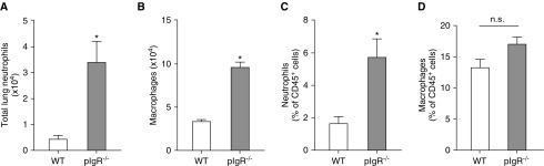
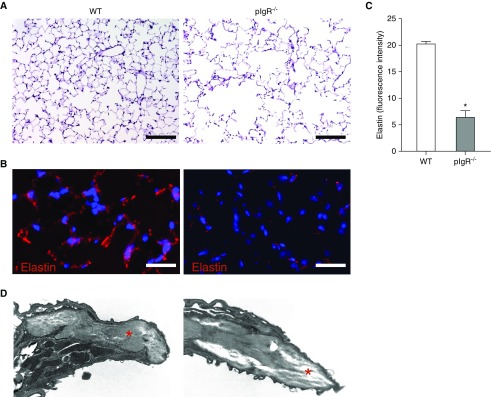
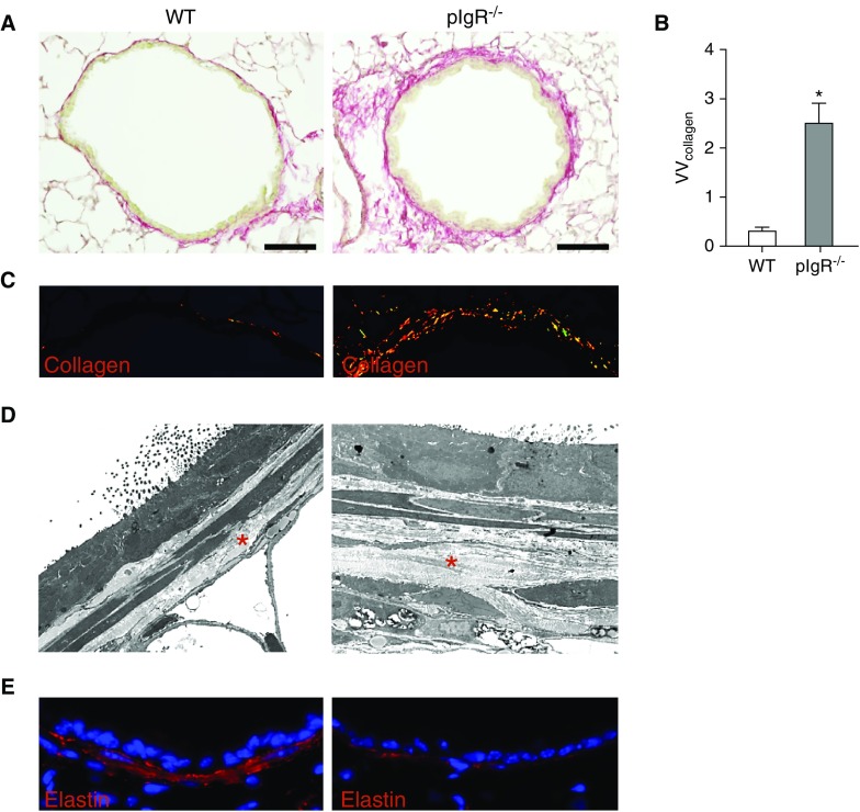
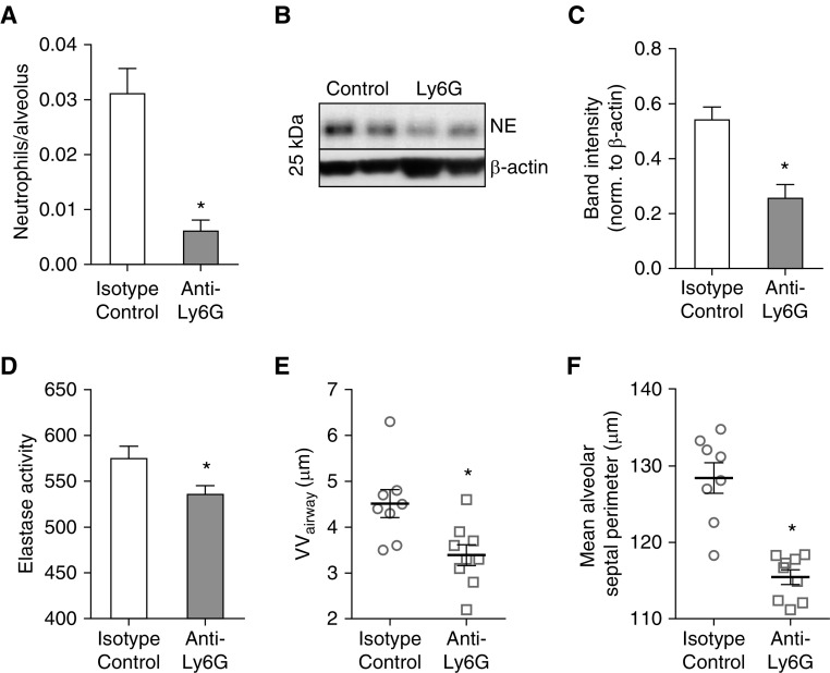
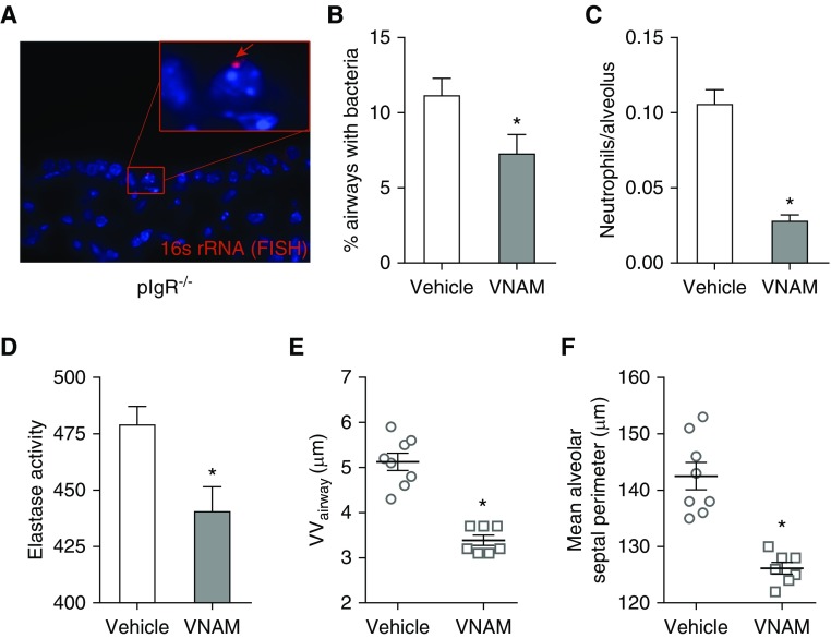
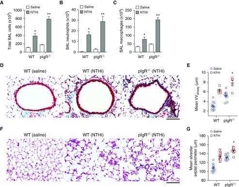
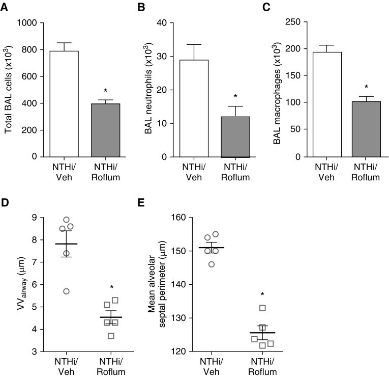
Loss of secretory IgA is common in the small airways of patients with chronic obstructive pulmonary disease and may contribute to disease pathogenesis. Using mice that lack secretory IgA in the airways due to genetic deficiency of polymeric Ig receptor (pIgR-/- mice), we investigated the role of neutrophils in driving the fibrotic small airway wall remodeling and emphysema that develops spontaneously in these mice. By flow cytometry, we found an increase in the percentage of neutrophils among CD45+ cells in the lungs, as well as an increase in total neutrophils, in pIgR-/- mice compared with wild-type controls. This increase in neutrophils in pIgR-/- mice was associated with elastin degradation in the alveolar compartment and around small airways, along with increased collagen deposition in small airway walls. Neutrophil depletion using anti-Ly6G antibodies or treatment with broad-spectrum antibiotics inhibited development of both emphysema and small airway remodeling, suggesting that airway bacteria provide the stimulus for deleterious neutrophilic inflammation in this model. Exogenous bacterial challenge using lysates prepared from pathogenic and nonpathogenic bacteria worsened neutrophilic inflammation and lung remodeling in pIgR-/- mice. This phenotype was abrogated by antiinflammatory therapy with roflumilast. Together, these studies support the concept that disruption of the mucosal immune barrier in small airways contributes to chronic obstructive pulmonary disease progression by allowing bacteria to stimulate chronic neutrophilic inflammation, which, in turn, drives progressive airway wall fibrosis and emphysematous changes in the lung parenchyma.

Keywords: chronic obstructive pulmonary disease; emphysema; mucosal immunity; polymeric Ig receptor; secretory IgA.


Figures

Figure 1.
Figure 1.
Increased neutrophils and macrophages in lungs of polymeric Ig receptor null (pIgR−/−) mice. Absolute number of (A) neutrophils and (B) macrophages in lungs of wild-type (WT) and pIgR−/− mice; n = 3–6 mice/group; *P < 0.05 (t test). Percentage of (C) neutrophils and (D) macrophages relative to CD45+ immune/inflammatory cells in lungs of 8- to 9-month-old WT and pIgR−/− mice; n = 3–6 mice/group; *P < 0.05 (t test). From CD45+ cells, alveolar macrophages were defined as CD11chiF4/80hiCD103-CD11b and neutrophils were defined as CD11b+/Ly6G+. n.s. = not significant.
Figure 2.
Figure 2.
Elastin degradation in lung parenchyma of pIgR−/− mice. (A) Hematoxylin and eosin staining of lung sections shows emphysematous lung destruction in a pIgR−/− mouse (12-mo-old) relative to an age-matched WT control. Scale bars: 50 μm. (B) Immunostaining for elastin from a WT mouse (12-mo-old) and a pIgR−/− mouse. Scale bars: 100 μm. (C) Quantification of fluorescent intensity of elastin staining reported as actual pixel density for each field of lung parenchyma (captured at ×40 objective); n = 6 mice/group; *P < 0.0001 (t test). (D) Transmission electron microscopy image of an interalveolar septum from a WT and pIgR−/− mouse (×50,000). The red stars denote extracellular matrix.
Figure 3.
Figure 3.
Collagen deposition and subepithelial elastin degradation in small airways of pIgR−/− mice. (A) Picosirius red staining to detect collagen in a small airway from a WT mouse (12-mo-old) and an age-matched pIgR−/− mouse. Scale bars: 50 μm. (B) Quantification of collagen content in small airway walls normalized to basement membrane length (VVcollagen); n = 6 mice/group; *P < 0.001 (t test). (C) High-power magnification of picosirius red staining under polarized light in the same airways. (D) Transmission electron microscopy image of a small airway wall in a WT mouse and an age-matched pIgR−/− mouse (×10,000). The red stars denote subepithelial collagen. (E) Immunostaining for elastin in small airway wall from a WT mouse (12-mo-old) and an age-matched pIgR−/− mouse.
Figure 4.
Figure 4.
Neutrophil depletion in pIgR−/− mice blocks small airway wall fibrosis and emphysema. pIgR−/− mice were treated with anti-Ly6G or anti-IgG2a isotype control antibodies between 4 and 8 months of age. (A) Quantification of parenchymal neutrophil numbers after immunostaining lung sections with neutrophil elastase–specific antibodies; n = 8–9 mice/group; *P < 0.001 (t test). (B) Western blot and (C) densitometry for neutrophil elastase (NE; 26 kD) in lung tissue, normalized to β-actin; n = 6–7 mice/group; *P < 0.001 (t test). (D) Elastase activity in whole-lung lysates; n = 4–5 mice/group; *P < 0.05 (t test). (E) Morphometric analysis of small airway wall thickness (VVairway); n = 8–9 mice/group; *P < 0.01 (t test). (F) Morphometric analysis of emphysema (mean alveolar septal perimeter); n = 8–9 mice/group; *P < 0.0001 (t test).
Figure 5.
Figure 5.
Treatment with broad-spectrum antibiotics inhibits small airway wall remodeling and emphysema in pIgR−/− mice. pIgR−/− mice received an antibiotics cocktail dissolved in drinking water (vancomycin, neomycin, ampicillin, and metronidazole [VNAM]) or regular drinking water only between 9 and 12 months of age. (A) Representative image of a bacterium invading the mucosa in a small airway from an untreated pIgR−/− mouse. The bacterium (red arrow) is labeled by a fluorescent in situ hybridization (FISH) probe targeting prokaryotic 16s rRNA. (B) Quantification of the percentage of airways/mouse with luminal bacteria visualized by FISH staining for bacterial 16s rRNA; n = 7–8 mice/group; *P < 0.05 (t test). (C) Quantification of neutrophil numbers in lung parenchyma after immunostaining with neutrophil elastase–specific antibodies; n = 7–8 mice/group; *P < 0.0001 (t test). (D) Elastase activity in whole-lung lysates; n = 6 mice/group; *P < 0.05 (t test). (E) Morphometric analysis of VVairway; n = 7–8 mice/group; *P < 0.0001 (t test). (F) Morphometric analysis of emphysema (mean alveolar septal perimeter); n = 7–8 mice/group; *P < 0.0001 (t test).
Figure 6.
Figure 6.
Increased lung inflammation in pIgR−/− mice treated with nontypeable Haemophilus influenzae (NTHi). WT and pIgR−/− mice were treated with NTHi lysates via nebulization once weekly from 2 to 6 months of age. (AC) Total cells, neutrophils, and macrophages in BAL fluid in WT and pIgR−/− mice treated with NTHi or saline only, as indicated; n = 5–7 mice/group; *P < 0.05 compared with saline-treated WT mice; **P < 0.05 compared with all other groups (ANOVA). (D) Representative images of subepithelial collagen (stained blue) around the small airways of a saline-treated WT mouse or NTHi-treated WT or pIgR−/− mouse as indicated. Masson’s trichrome stain; scale bar: 50 μm. (E) Morphometric analysis of VVairway in WT and pIgR−/− mice as shown in D; n = 5–12 mice/group; *P = 0.05 compared with NTHi-treated WT mice and P < 0.0001 compared with all other groups (ANOVA). (F) Representative images of emphysema in the lungs of a saline-treated WT mouse or NTHi-treated WT or pIgR−/− mouse, as indicated. Hematoxylin and eosin; scale bar: 50 μm. (G) Morphometric analysis of emphysema (mean alveolar septal perimeter) in WT and pIgR−/− mice as shown in F; n = 11–12 mice/group; *P < 0.01 compared with all other groups (ANOVA).
Figure 7.
Figure 7.
Roflumilast treatment inhibits inflammation and blocks lung remodeling in pIgR−/− mice after repetitive exposure to NTHi lysate. pIgR−/− mice were treated by oral gavage with daily roflumilast or vehicle (Veh) concurrent with weekly NTHi nebulizations from 2 to 6 months of age. (AC) Total cells, neutrophils, and macrophages in BAL fluid; n = 6–7 mice/group; *P < 0.05 (t test). (D) Morphometric analysis of small VVairway; n = 5 mice/group; *P < 0.01 (t test). (E) Morphometric analysis of emphysema (mean alveolar septal perimeter); n = 5 mice/group; *P < 0.0001.

Comment in

References

    1. Hogg JC, Chu F, Utokaparch S, Woods R, Elliott WM, Buzatu L, et al. The nature of small-airway obstruction in chronic obstructive pulmonary disease. N Engl J Med. 2004;350:2645–2653. - PubMed
    1. Polosukhin VV, Cates JM, Lawson WE, Zaynagetdinov R, Milstone AP, Massion PP, et al. Bronchial secretory immunoglobulin a deficiency correlates with airway inflammation and progression of chronic obstructive pulmonary disease. Am J Respir Crit Care Med. 2011;184:317–327. - PMC - PubMed
    1. Polosukhin VV, Richmond BW, Du RH, Cates JM, Wu P, Nian H, et al. Secretory IgA deficiency in individual small airways is associated with persistent inflammation and remodeling. Am J Respir Crit Care Med. 2017;195:1010–1021. - PMC - PubMed
    1. Wright JL, Lawson LM, Pare PD, Wiggs BJ, Kennedy S, Hogg JC. Morphology of peripheral airways in current smokers and ex-smokers. Am Rev Respir Dis. 1983;127:474–477. - PubMed
    1. Rutgers SR, Postma DS, ten Hacken NH, Kauffman HF, van Der Mark TW, Koëter GH, et al. Ongoing airway inflammation in patients with COPD who do not currently smoke. Thorax. 2000;55:12–18. - PubMed

Publication types

MeSH terms