. 2018 Mar;12(3):322-338.
doi: 10.1002/1878-0261.12165. Epub 2018 Jan 24.

Crosstalk between hepatitis B virus X and high-mobility group box 1 facilitates autophagy in hepatocytes

Affiliations

Crosstalk between hepatitis B virus X and high-mobility group box 1 facilitates autophagy in hepatocytes

Sha Fu et al. Mol Oncol. 2018 Mar.

Abstract

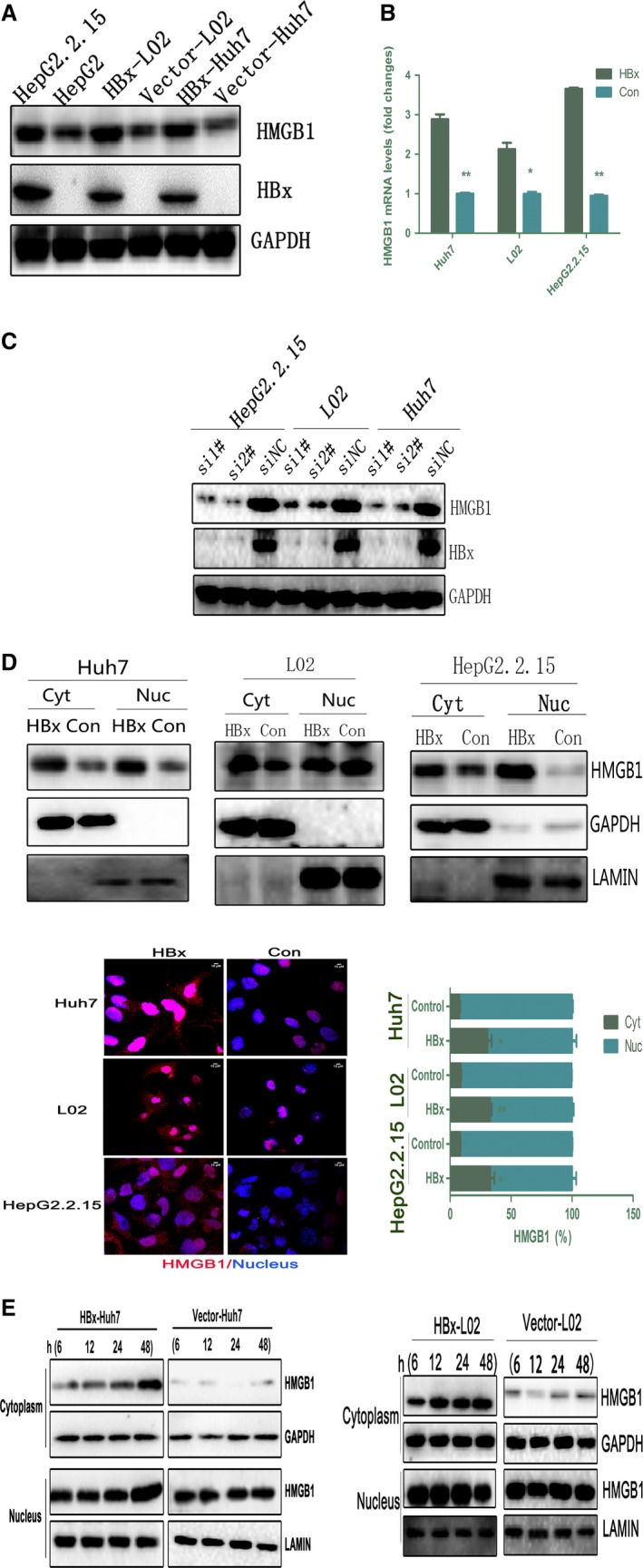
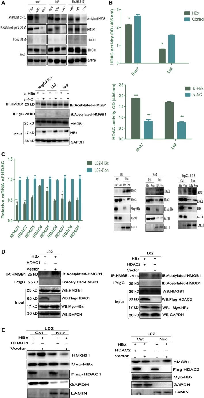
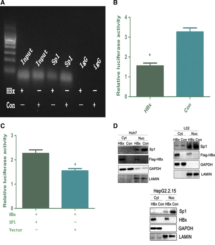
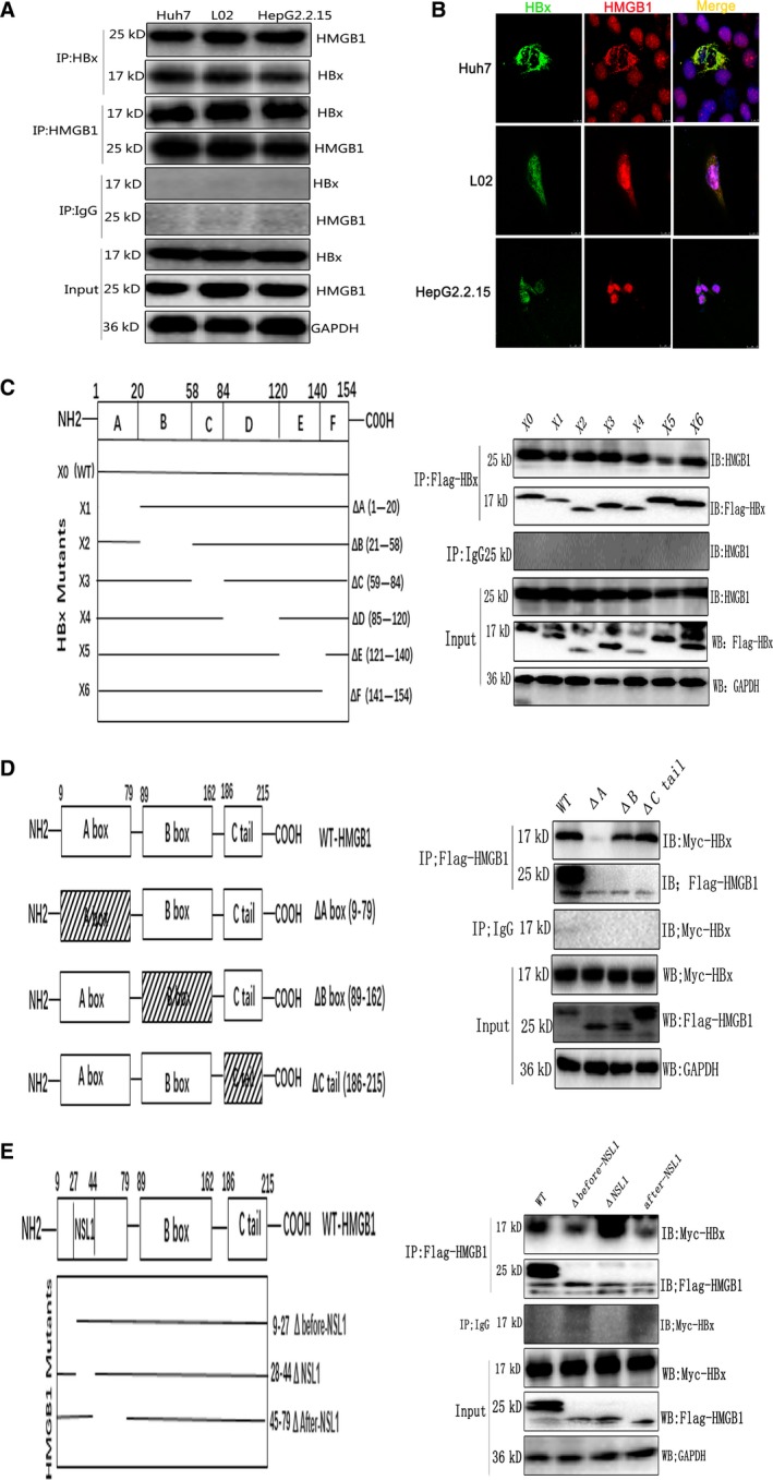
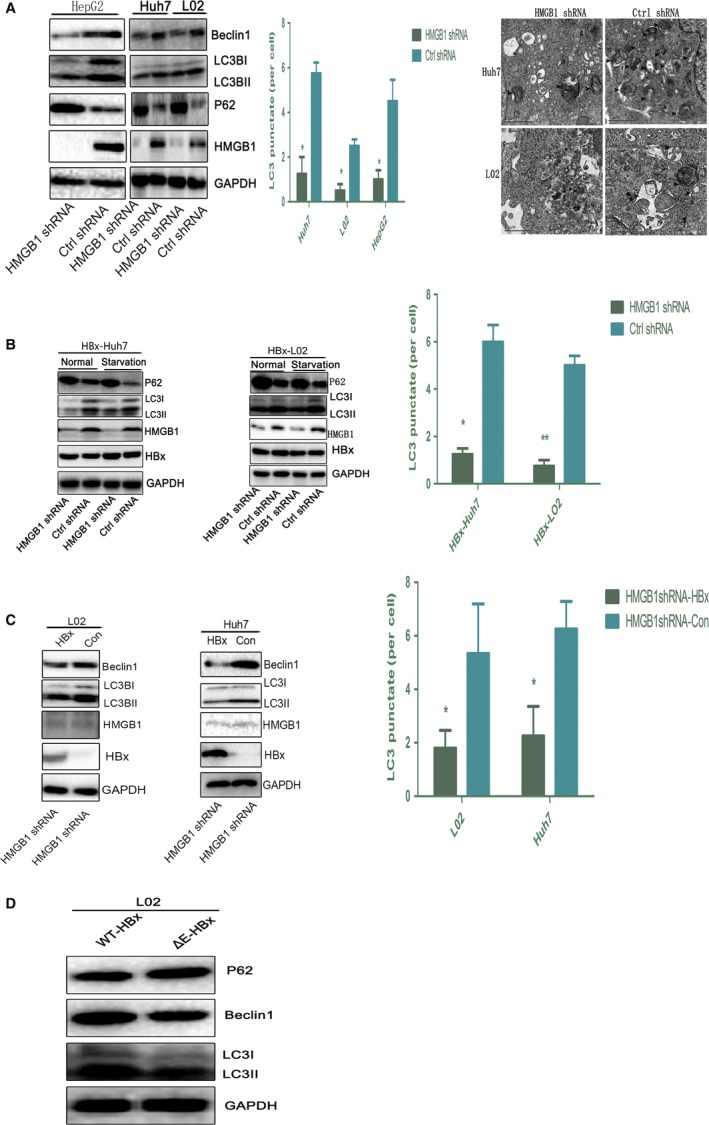
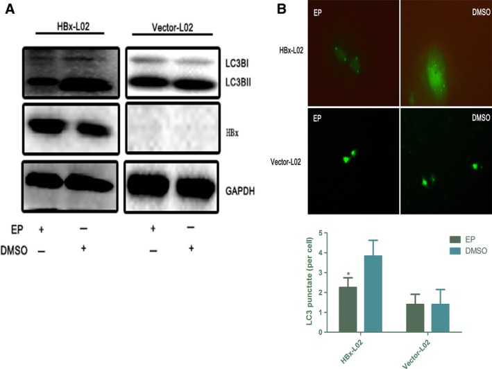
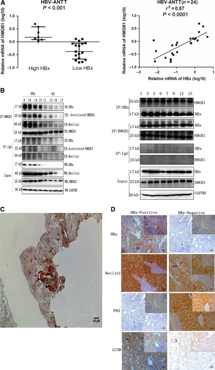
Hepatitis B virus (HBV) X (HBx) protein is a pivotal regulator of HBV-triggered autophagy. However, the role of HBx-induced epigenetic changes in autophagy remains largely unknown. The cytoplasmic (Cyt) high-mobility group box 1 (HMGB1) has been identified as a positive regulator of autophagy, and its Cyt translocation is closely associated with its acetylation status. Here, we evaluated the function of HMGB1 in HBx-mediated autophagy and its association with histone deacetylase (HDAC). Using cell lines with enforced expression of HBx, we demonstrated that HBx upregulated the expression of HMGB1 and promoted its Cyt translocation by acetylation to facilitate autophagy. We further identified the underlying mechanism by which decreased nuclear HDAC activity and expression levels contribute to the HBx-promoted hyperacetylation and subsequent translocation of HMGB1. We also identified the HDAC1 isoform as a critical factor in regulating this phenomenon. In addition, HBx bound to HMGB1 in the cytoplasm, which triggered autophagy in hepatocytes. Pharmacological inhibition of HMGB1 Cyt translocation with ethyl pyruvate prevented HBx-induced autophagy. These results demonstrate a novel function of acetylated HMGB1 in HBx-mediated autophagy in hepatocytes.

Keywords: acetylation; autophagy; hepatitis B virus; high-mobility group box 1; histone deacetylases; protein protein interaction.


Figures

Figure 1
Figure 1
HBx upregulates HMGB1 expression and promotes its Cyt translocation. (A,B) HMGB1 expression levels in HepG2.2.15, HBx‐Huh7, and HBx‐L02 cells were analyzed by immunoblot and RTPCR. *< 0.05, **< 0.01 compared with empty vector, n = 3. (C) The indicated cells were pretreated with HBx‐specific siRNA for 72 h and immunoblotted with an HMGB1‐specific antibody. (D) Nuclear/Cyt HMGB1 expression in cells stably transfected with HBx (HBx‐Huh7, HBx‐L02, and HepG2.2.15) was analyzed by immunoblot and immunofluorescence. LAMIN was used as a nuclear fraction control, and GAPDH was used as a Cyt fraction control. The mean nuclear (Nuc) and Cyt HMGB1 intensity per cell was determined by immunofluorescence. *< 0.05, **< 0.01 versus the empty vector (Control) group; = 3. Representative images are depicted on the left, HMGB1 (red) and DAPI (blue). Scale bar, 10 μm. (E) Immunoblot analysis of nuclear/Cyt HMGB1 expression in cells (Huh7 and L02) transiently transfected with HBx (3 μg) for the indicated time periods (6, 12, 24, and 48 h).
Figure 2
Figure 2
HBx induces HMGB1 acetylation by regulating HDAC activity and expression. (A) HBx promotes the acetylation of HMGB1. TSA (1 μm, 24 h) treatment was used as a positive control. Whole lysates of HBx‐L02, HBx‐Huh7, and HepG2.2.15 cells were immunoprecipitated with an acetylated lysine or HMGB1 antibody and then subjected to immunoblot analysis with the indicated antibodies. Samples were pulled down with anti‐HMGB1 and immunoblotted with anti‐acetylated lysine following HBx knockdown via siRNA treatment for 72 h. (B) HDAC activity was determined by colorimetric assay after HBx overexpression or knockdown. *< 0.05, **< 0.01, n = 3. (C) RTPCR analysis of HDAC1–9 expression in HBx‐L02 and Vector‐L02 cells. *< 0.05, **< 0.01, n = 3. Immunoblot analysis of nuclear/Cyt HDAC1 or HDAC2 expression in HBx‐L02 and Vector‐L02 cells. (D and E) L02 cells were cotransfected with Myc‐tagged HBx (1.5 μg) and Flag‐tagged HDAC1 (1.5 μg) or Flag‐tagged HDAC2 (1.5 μg) for 48 h. Whole lysates were immunoprecipitated with anti‐HMGB1 and immunoblotted with anti‐acetylated lysine. Nuclear/Cyt HMGB1 distribution was then analyzed by western blot.
Figure 3
Figure 3
HBx regulates HDAC1 expression via SP1. (A). A ChIP assay demonstrates a decrease in SP1 binding to the HDAC1 promoter following HBx overexpression. (B,C). 293Tcells were transfected with reporter constructs derived from the pGL3‐Basic vector containing SP1‐binding sites in the promoter of human HDAC1 gene as indicated. *< 0.05, n = 3. HDAC1 promoter activity was reversed after cotransfection with HBx (1.5 μg) and SP1 (1.5 μg). (D). Immunoblot analysis of the subcellular distribution of SP1 in HBx‐Huh7, HBx‐L02, and HepG2.2.15 cells.
Figure 4
Figure 4
HBx binds HMGB1 in the cytoplasm. (A). Whole‐cell extracts of HBx‐Huh7, HBx‐L02, and HepG2.2.15 cells were immunoprecipitated with the indicated antibodies, and the immunoprecipitates were analyzed by immunoblotting. (B) Colocalization of HBx and HMGB1 was analyzed by confocal microscopy. (C) Scheme of HBx mutants. The 154‐amino‐acid WT HBx sequence (X0) was divided into six regions (A–F). The line indicates the portion of HBx present in the mutant, with the deleted region shown as a gap. 293T cells were transfected with Flag‐tagged full‐length HBx and deletion (Δ) mutants (X1–X6) of HBx. Cell lysates were immunoprecipitated with anti‐Flag and probed by western blot with anti‐HMGB1. (D) Scheme of HMGB1 mutants. The 215‐amino‐acid WT HMGB1 sequence was divided into three regions, including the A box, B box, and C‐tail. The box with hatch marks indicates the deleted region of HMGB1. 293T cells were transfected with Myc‐HBx and Flag‐tagged WT HMGB1 or the A box, B box, or C‐tail deletion of HMGB1. Cell lysates were immunoprecipitated with anti‐Flag and probed by western blot with anti‐Myc. (E) The 70‐amino‐acid A box in HMGB1 was further divided into three regions based on the location of NSL1, that is, one NSL1‐containing domain, one domain before NSL1 and one domain after NSL1. The line indicates the portion of HMGB1 present in the mutant, with the deleted region shown as a gap. 293T cells were transfected with Myc‐HBx and Flag‐tagged WT HMGB1 or the NSL1, before‐NSL1, or after‐NSL1 deletion of HMGB1. Cell lysates were immunoprecipitated with anti‐Flag and probed by western blot with anti‐Myc.
Figure 5
Figure 5
HBx induces autophagy in an HMGB1‐dependent manner. (A) The expression of the indicated proteins and LC3 puncta was detected in cells (Huh7, L02, and HepG2) transfected with HMGB1 shRNA or Ctrl shRNA by immunoblot and immunofluorescence, respectively. *< 0.05, **< 0.01, n = 3. Ultrastructural features in the indicated cells were observed by electron microscopy. (B) Immunoblot detection of the indicated proteins in cells treated with HMGB1 shRNA and Ctrl shRNA and transfected with HBx under basal conditions and in response to starvation. LC3 puncta formation was assayed by immunofluorescence under basal conditions, *< 0.05, **< 0.01, n = 3. (C) Western blot analysis of the indicated proteins in cells (L02 and Huh7) treated with HMGB1 shRNA and transfected with HBx or empty vector for 48 h. LC3 puncta formation was assayed by immunofluorescence. *< 0.05, n = 3. (D) The indicated proteins were analyzed by immunoblot in L02 cells transfected with WT HBx (3 μg) and an HBx mutant with deletion of the E domain (3 μg).
Figure 6
Figure 6
Cyt HMGB1 is required for HBx‐induced autophagy. (A,B) Analysis of LC3 processing by immunoblot and immunofluorescence in HBx‐L02 and Vector‐L02 cells after EP or DMSO (0.25 mm, 24 h) treatment. Quantitative GFPLC3 data are shown on the right. *< 0.05, n = 3.
Figure 7
Figure 7
Interaction of HBx and HMGB1 in vivo. (A) mRNA levels of intrahepatic HBx and HMGB1 were examined by RTPCR, and the correlation between HBx and HMGB1 was assessed by linear regression (r 2 = 0.57, < 0.0001). ANTT: Adjacent nontumor tissue. (B) HBx/HMGB1, HMGB1/Beclin1, and HMGB1/acetylated lysine complexes immunoprecipitated from liver samples from HBx‐transgenic mice or HBV patients. (C,D) Double‐stained liver tissue sections from HBV patients showing HBx/HMGB1 expression and colocalization. Brown: HBx, Red: HMGB1. Arrows indicate the overlap of HBx and HMGB1. Immunohistochemical analysis of the indicated proteins in the livers of patients in the HBx‐positive and HBx‐negative expression groups.

References

    1. Barqasho B, Nowak P, Abdurahman S, Walther‐Jallow L and Sonnerborg A (2010) Implications of the release of high‐mobility group box 1 protein from dying cells during human immunodeficiency virus type 1 infection in vitro . J Gen Virol 91(Pt 7), 1800–1809. - PubMed
    1. Belloni L, Pollicino T, De Nicola F, Guerrieri F, Raffa G, Fanciulli M, Raimondo G and Levrero M (2009) Nuclear HBx binds the HBV minichromosome and modifies the epigenetic regulation of cccDNA function. Proc Natl Acad Sci USA 106, 19975–19979. - PMC - PubMed
    1. Bonaldi T, Talamo F, Scaffidi P, Ferrera D, Porto A, Bachi A, Rubartelli A, Agresti A and Bianchi ME (2003) Monocytic cells hyperacetylate chromatin protein HMGB1 to redirect it towards secretion. EMBO J 22, 5551–5560. - PMC - PubMed
    1. Chen S, Dong Z, Yang P, Wang X, Jin G, Yu H, Chen L, Li L, Tang L, Bai S et al (2017) Hepatitis B virus X protein stimulates high mobility group box 1 secretion and enhances hepatocellular carcinoma metastasis. Cancer Lett 394, 22–32. - PubMed
    1. Chen R, Hou W, Zhang Q, Kang R, Fan XG and Tang D (2013) Emerging role of high‐mobility group box 1 (HMGB1) in liver diseases. Mol Med 19, 357–366. - PMC - PubMed

Publication types

MeSH terms

LinkOut - more resources