Review
. 2018 Apr;7(8):e1700939.
doi: 10.1002/adhm.201700939. Epub 2018 Jan 9.

Exploiting Advanced Hydrogel Technologies to Address Key Challenges in Regenerative Medicine

Affiliations
Review

Exploiting Advanced Hydrogel Technologies to Address Key Challenges in Regenerative Medicine

Daniel A Foyt et al. Adv Healthc Mater. 2018 Apr.

Abstract

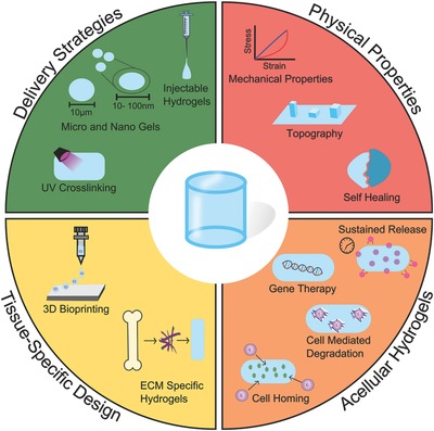
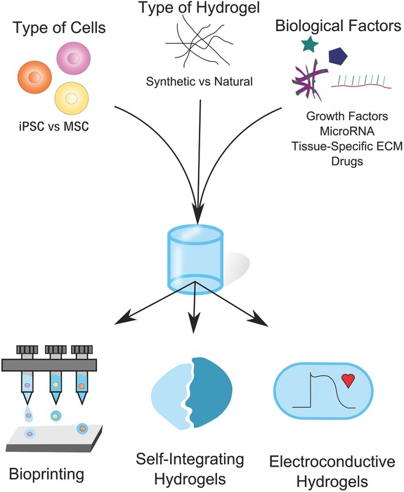
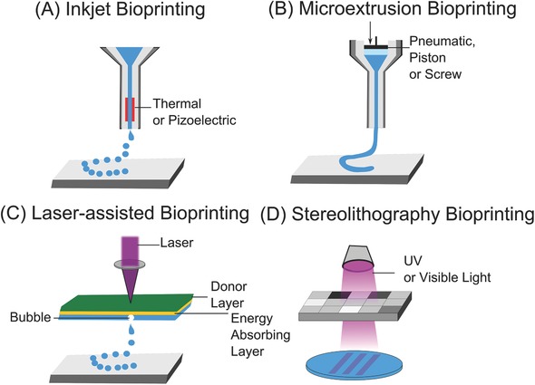
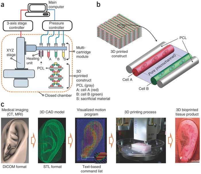
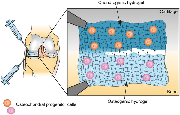
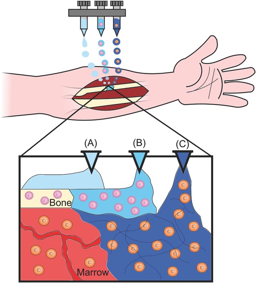
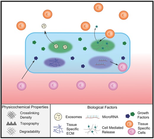
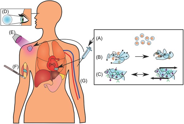
Regenerative medicine aims to tackle a panoply of challenges from repairing focal damage to articular cartilage to preventing pathological tissue remodeling after myocardial infarction. Hydrogels are water-swollen networks formed from synthetic or naturally derived polymers and are emerging as important tools to address these challenges. Recent advances in hydrogel chemistries are enabling researchers to create hydrogels that can act as 3D ex vivo tissue models, allowing them to explore fundamental questions in cell biology by replicating tissues' dynamic and nonlinear physical properties. Enabled by cutting edge techniques such as 3D bioprinting, cell-laden hydrogels are also being developed with highly controlled tissue-specific architectures, vasculature, and biological functions that together can direct tissue repair. Moreover, advanced in situ forming and acellular hydrogels are increasingly finding use as delivery vehicles for bioactive compounds and in mediating host cell response. Here, advances in the design and fabrication of hydrogels for regenerative medicine are reviewed. It is also addressed how controlled chemistries are allowing for precise engineering of spatial and time-dependent properties in hydrogels with a look to how these materials will eventually translate to clinical applications.

Keywords: advanced therapies; biomaterials; bioprinting; hydrogels; regenerative medicine; tissue engineering.


Conflict of interest statement

The authors declare no conflict of interest.

Figures

Figure 1
Figure 1
Summary of state‐of‐the‐art strategies for hydrogel design and fabrication and their applications in regenerative medicine. Advancements in both biology and material science have allowed for the development of complex regenerative strategies. Green: Researchers are designing hydrogels with various delivery strategies tailored for each biological application. Red: Our increased understanding of mechanobiology is driving the development of hydrogels that can aid biologists in understanding these fundamental processes, and allow researchers to exploit them to drive cell response for regeneration. Yellow: Advanced manufacturing technologies are allowing for the development of hydrogels with tissue‐specific architectures and biological functionalities. Orange: Acellular hydrogels are being developed to both deliver relevant biological molecules and direct host tissue response.
Figure 2
Figure 2
Microscale photoreversible patterning of proteins within 3D hydrogels. A,B) Fluorescence confocal microscopy images of dual‐protein patterning within hydrogels. Hydrogels were patterned with covalently immobilized interlocking chains of red protein while surrounding areas were labelled with a green protein. Scale bars = 50 µm. C) Hydrogel with 3D patterned protein in a staircase pattern. Patterning was achieved in 3D using focused laser pulses and by varying the multiphoton laser‐scanning conditions, resulting in highly ordered positioning of proteins. Scale bar = 150 µm. Adapted with permission.[186] Copyright 2015, Nature Publishing Group.
Figure 3
Figure 3
Tissue‐specific hydrogel design considerations. When developing tissue‐specific hydrogels, a number of factors should be considered. For example, the choice of cell type and biological factors will be dictated by the biological application. However, the emergence of new technologies, such as those that allow for the creation of patient‐specific stem cells, for example iPSC, may allow for additional opportunities. Hydrogel material choice is not always straightforward and may be dictated by a range of factors, such as amenability to bioprinting, the need to form a tissue interface or the necessity of tissue‐specific functionality.
Figure 4
Figure 4
Examples of different bioprinting methods. A) Inkjet bioprinters deposit small droplets of hydrogel and cells to build tissue layer‐by‐layer. B) Microextrusion bioprinters deposit a cell‐laden liquid solution via pneumatic or manual force. C) Laser‐assisted bioprinting uses a laser to rapidly heat a donor layer (green), which forms a bubble propelling the bioink onto the substrate. D) Stereolithography bioprinters use UV or visible light to selectively cross‐link bioinks layer by layer to build a 3D construct.
Figure 5
Figure 5
Human‐scale bioprinting. To print constructs of sufficient size for eventual translation into humans, Kang et al. developed an integrated tissue‐organ printer (ITOP) system in which large, tissue‐specific constructs could be printed. a) The ITOP system comprised three major units: i) a three‐axis stage controller, ii) a dispensing module composed of multiple cartridges and a pneumatic pressure controller, and iii) a closed acrylic chamber with a humidifier and temperature regulator. b) Using this bioprinter, 3D scaffold architectures could be printed with both multiple cell types and a PCL polymer to ensure structural rigidity. c) 3D CAD models were generated from medical image data; and using CAD/CAM processing, the printer could be used to create complex 3D structures, including a 3D human‐sized ear. Reproduced with permission.66 Copyright 2016, Nature Publishing Group.
Figure 6
Figure 6
Injectable self‐integrating hydrogels for osteochondral repair. Schematic illustration of a self‐healing hydrogel that could potentially be used to repair an osteochondral defect. Two tissue‐specific hydrogel formulations made from the same bulk material are injected into the damaged cartilage–bone interface. The shear‐thinning properties of the hydrogels would allow for easy injection of the cell‐containing solutions. Their self‐healing chemistries would then foster the formation of a seamless transition between the chondrogenic and osteogenic hydrogel formulations. The chondrogenic hydrogel could, for example, contain osteochondral progenitor cells or chondrocytes and chondrogenic factors such as transforming growth factor β (TGFβ ), BMP and/or insulin‐like growth factor (IGF) to promote cartilage‐like ECM production. Whereas the osteogenic hydrogel might include nanoscale hydroxyapatite particles and VEGF to enhance bone regeneration.
Figure 7
Figure 7
Potential strategy for in situ bioprinting. Schematic illustration showing a potential strategy for in situ bioprinting of tissue‐specific hydrogels with different cell types to treat a nonunion in the radius. Multiple hydrogels could potentially be employed to reconstruct complex, multicellular tissues like bone. A) An acellular hydrogel is printed to act as a structural support while the bone heals. B) A cell‐laden hydrogel with tissue‐specific architecture and biological factors is printed to promote ossification. C) Other cell‐laden, biologically targeted hydrogels could also be simultaneously printed to, for example, promote vascularization or aid in tissue remodeling.
Figure 8
Figure 8
Using acellular hydrogels to promote tissue regeneration. For regenerative strategies based on acellular hydrogels, various factors can be incorporated into the hydrogel. Such factors may include growth factors, regenerative exosomes, or microRNA, which will be released over time as the hydrogel degrades. Acellular hydrogels can also be used to directly instruct host cells. For example, tissue‐specific ECM or topography can also be incorporated into the hydrogel to direct host stem cell differentiation as cells come in contact with it. More advanced acellular hydrogel strategies might incorporate controlled spatial and temporal release of specific factors. Using cleavable systems such as those mediated by MMP activity, factors will only be released in response to cell‐ or tissue‐specific stimuli. This could be particularly useful for tissue interfaces such as the Achilles tendon‐bone insertion (enthesis), where the spatial distribution of biological factors is key in modulating cell behavior.
Figure 9
Figure 9
Various strategies for hydrogel delivery. Schematic diagram showing a range of strategies for hydrogel delivery and gelation. A) Single cells can be encapsulated in thin hydrogels, which can then be injected intravenously to be distributed throughout the body. The hydrogel coating can be designed to protect the cells from the immune system. B) Hydrogels can be covalently cross‐linked by enzyme‐mediated reactions between reactive groups on the hydrogel monomers, forming a network. C) Noncovalent cross‐linking of hydrogels can be achieved via hydrophobic, interactions, π–π interactions, hydrogen bonding, metal chelation, or van der Waals interactions. These hydrogels can sometimes be shear thinning, which makes them easily injectable and may also protect encapsulated cells during the injection process. D) Hydrogels can be used as cornel implants and keratoprostheses. E) UV light can be used to cure hydrogels subcutaneously after injection or activate biological moieties like cell‐adhesion peptides, providing spatiotemporal control of their biological activity. F) Surgical implantation of a hydrogel to treat a nonunion fracture. G) Micro‐ and nanohydrogels, with or without cells, can be delivered via IV injection. H) Hydrogel patches with encapsulated cardiac stem cells can be surgically implanted to treat myocardial infarction.

References

    1. Böcker L., Rühs P. A., Böni L., Fischer P., Kuster S., ACS Biomater. Sci. Eng. 2016, 2, 90. - PubMed
    1. Le Goff M. M., Bishop P. N., Eye 2008, 22, 1214. - PubMed
    1. Wichterle O., Lim D., Nature 1960, 185, 117.
    1. Sharma B., Fermanian S., Gibson M., Unterman S., Herzka D. A., Cascio B., Coburn J., Hui A. Y., Marcus N., Gold G. E., Elisseeff J. H., Sci. Transl. Med. 2013, 5, 167ra6. - PMC - PubMed
    1. a) Tamaddon M., Burrows M., Ferreira S. A., Dazzi F., Apperley J. F., Bradshaw A., Brand D. D., Czernuszka J., Gentleman E., Sci. Rep. 2017, 7, 43519. - PMC - PubMed
    2. b) McCullen S. D., Autefage H., Callanan A., Gentleman E., Stevens M. M., Tissue Eng., Part A 2012, 18, 2073. - PMC - PubMed

Publication types