Review
. 2018 Jun:50:56-63.
doi: 10.1016/j.conb.2017.12.012. Epub 2018 Jan 6.

Expansion microscopy: development and neuroscience applications

Affiliations
Review

Expansion microscopy: development and neuroscience applications

Emmanouil D Karagiannis et al. Curr Opin Neurobiol. 2018 Jun.

Abstract

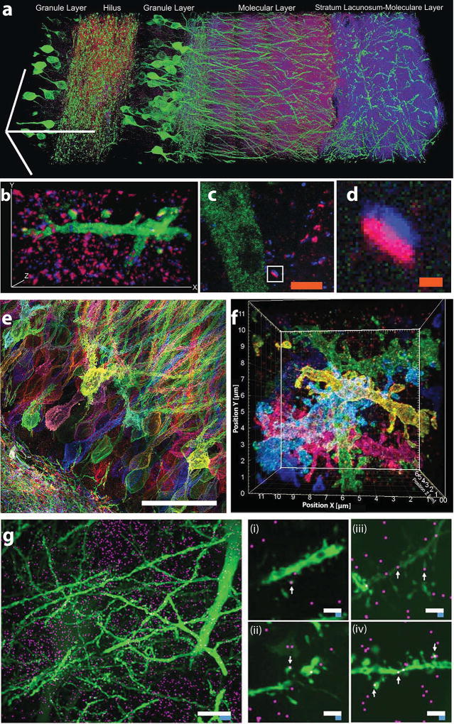
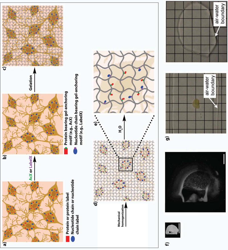
Many neuroscience questions center around understanding how the molecules and wiring in neural circuits mechanistically yield behavioral functions, or go awry in disease states. However, mapping the molecules and wiring of neurons across the large scales of neural circuits has posed a great challenge. We recently developed expansion microscopy (ExM), a process in which we physically magnify biological specimens such as brain circuits. We synthesize throughout preserved brain specimens a dense, even mesh of a swellable polymer such as sodium polyacrylate, anchoring key biomolecules such as proteins and nucleic acids to the polymer. After mechanical homogenization of the specimen-polymer composite, we add water, and the polymer swells, pulling biomolecules apart. Due to the larger separation between molecules, ordinary microscopes can then perform nanoscale resolution imaging. We here review the ExM technology as well as applications to the mapping of synapses, cells, and circuits, including deployment in species such as Drosophila, mouse, non-human primate, and human.


Figures

Figure 1
Figure 1. Expansion microscopy workflow
Biomolecules, or labels highlighting biomolecules of interest, in fixed cells or tissues (a), are functionalized with chemical handles (AcX, green, binds proteins; LabelX, purple, binds nucleic acids such as mRNA) that enable them to be covalently anchored (b) to a swellable polymer mesh (composed of crosslinked sodium polyacrylate) that is evenly and densely synthesized throughout the specimen (c). The sample is mechanically homogenized by treatment with heat, detergent, and/or proteases (d). Adding water initiates polymer swelling (e), which results in biomolecules or labels being pulled apart from each other in an even, isotropic fashion, and thus enabling nanoscale resolution imaging on conventional microscopes (f, adapted from ref. ). Expansion significantly reduces scattering of the sample, since the sample is mostly water (g, adapted from ref. ). In g, a 200 µm thick fixed mouse brain slice is opaque before ExM, but after expansion is completely transparent.
Figure 2
Figure 2. Expansion microscopy of brain circuitry
a) Expanded mouse hippocampus, with YFP-expressing neurons (green) antibody stained for the postsynaptic protein Homerl (magenta) and the presynaptic protein bassoon (blue) (bars, 100 µm). b) A hippocampal neuron from a piece of mouse brain tissue expanded and labeled as in (a), highlighting a single branch bearing multiple synapses (bars, 13.5 µm ×, 7.3 µm y, 2.8 µm z). c) and d) mouse cortex, expanded and labeled as in (a), with a single synapse (box in c) highlighted in d. (scale bars: c, 2.5 µm; d, 250 nm). Panels a–d adapted from ref. . e) Protein retention ExM (proExM, ref. ) and f) iterative ExM (iExM, ref. ) of mouse hippocampus expressing Brainbow (i.e., combinatorially expressed fluorophores for randomly labeling neurons with different colors). g) Expansion microscopy fluorescent in situ hybridization (ExFISH, ref. ) imaging of single RNA molecules (magenta) in mouse hippocampus with simultaneous visualization of protein (green, YFP). Left, Dlg4 mRNA (magenta) visualized simultaneously with YFP (green) (white scale bar, 10 µm; blue scale bar is divided by the expansion factor of 3). Middle (i and ii), dendrites with spine-localized Dlg4 mRNA highlighted with arrows. Right (iii and iv), dendrites with Camk2a mRNA highlighted with arrows (white scale bars, 2µm; blue scale bars are divided by the expansion factor of 3). Panels e–g adapted from the references indicated.

References

    1. Kasthuri N, Hayworth KJ, Berger DR, Schalek RL, Conchello JA, Knowles-Barley S, Lee D, Vázquez-Reina A, Kaynig V, Jones TR, Roberts M, Morgan JL, Tapia JC, Seung HS, Roncal WG, Vogelstein JT, Burns R, Sussman DL, Priebe CE, Pfister H, Lichtman JW. Saturated Reconstruction of a Volume of Neocortex. Cell. 2015;162(3):648–661. doi: 10.1016/j.cell.2015.06.054. - DOI - PubMed
    1. Eichler K, Li F, Litwin-Kumar A, Park Y, Andrade I, Schneider-Mizell CM, Saumweber T, Huser A, Eschbach C, Gerber B, Fetter RD, Truman JW, Priebe CE, Abbott LF, Thum AS, Zlatic M, Cardona A. The complete connectome of a learning and memory centre in an insect brain. Nature. 2017;548(7666):175–182. doi: 10.1038/nature23455. - DOI - PMC - PubMed
    1. He J, Zhou R, Wu Z, Carrasco MA, Kurshan PT, Farley JE, Simon DJ, Wang G, Han B, Hao J, Heller E, Freeman MR, Shen K, Maniatis T, Tessier-Lavigne M, Zhuang X. Prevalent presence of periodic actin–spectrin-based membrane skeleton in a broad range of neuronal cell types and animal species. Proc Natl Acad Sci. 2016;113(21):6029–6034. doi: 10.1073/pnas.1605707113. - DOI - PMC - PubMed
    1. D’Este E, Kamin D, Göttfert F, El-Hady A, Hell SW. STED Nanoscopy Reveals the Ubiquity of Subcortical Cytoskeleton Periodicity in Living Neurons. Cell Rep. 2015;10(8):1246–1251. doi: 10.1016/j.celrep.2015.02.007. - DOI - PubMed
    1. Chen F, Tillberg PW, Boyden ES. Expansion microscopy. Science (80-) 2015;347(6221):543–548. doi: 10.1126/science.1260088. The paper introducing the concept, demonstration, and initial validation of expansion microscopy. - DOI - PMC - PubMed

Publication types