Review
. 2018 Feb;39(1):106-113.
doi: 10.1053/j.sult.2017.09.001. Epub 2017 Sep 5.

Newer Technologies in Breast Cancer Imaging: Dedicated Cone-Beam Breast Computed Tomography

Affiliations
Review

Newer Technologies in Breast Cancer Imaging: Dedicated Cone-Beam Breast Computed Tomography

Avice M O'Connell et al. Semin Ultrasound CT MR. 2018 Feb.

Abstract

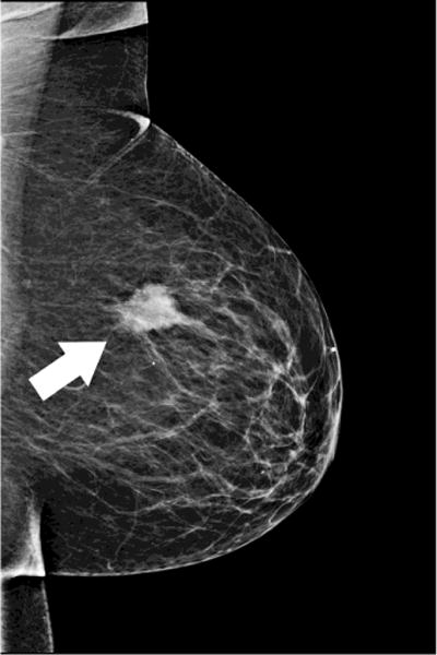
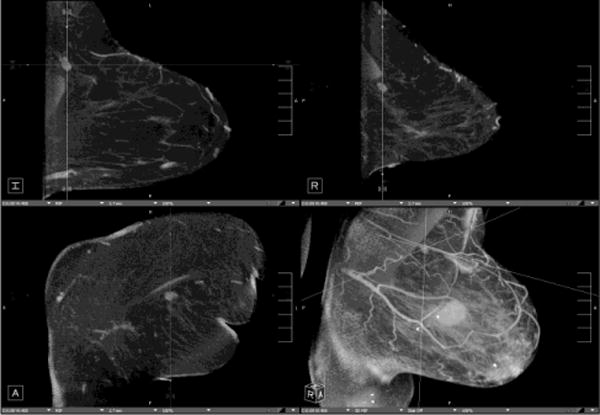
Dedicated breast computed tomography (CT) is the latest in a long history of breast imaging techniques dating back to the 1960s. Breast imaging is performed both for cancer screening as well as for diagnostic evaluation of symptomatic patients. Dedicated breast CT received US Food and Drug Administration approval for diagnostic use in 2015 and is slowly gaining recognition for its value in diagnostic 3-dimensional imaging of the breast, and also for injected contrast-enhanced imaging applications. Conventional mammography has known limitations in sensitivity and specificity, especially in dense breasts. Breast tomosynthesis was US Food and Drug Administration approved in 2011 and is now widely used. Dedicated breast CT is the next technological advance, combining real 3-dimensional imaging with the ease of contrast administration. The lack of painful compression and manipulation of the breasts also makes dedicated breast CT much more acceptable for the patients.


Figures

Fig. 1
Fig. 1. Ductal Carcinoma In-situ
64 year-old woman presents for screening mammogram. Mammographic CC (A) and MLO (B) views demonstrate grouping of pleomorphic calcifications in the left breast at the 12 o’clock position (arrows). Reformatted non-enhanced CBCT images at 0.273 mm standard resolution (C) and 0.122 mm high-resolution (D) clearly depict grouping of calcifications; morphology of calcifications are more clearly depicted in the higher resolution images. Lesion diagnosed as DCIS at time of biopsy.
Fig. 2
Fig. 2. Small Invasive Ductal Carcinoma
55 year-old woman presents for screening mammogram. Mammographic CC (A) and MLO (B) views demonstrate heterogeneously dense breast tissue with ill-defined focal asymmetry in the deep right breast at the 12 o’clock position (white circles). Contrast enhanced CBCT demonstrates more clearly a subcentimeter irregular mass (crosshairs) at the 12 o’clock location (C). Lesion diagnosed as invasive ductal carcinoma at time of biopsy.
Fig. 3
Fig. 3. Assessment of index lesion and extent of disease
Patient presents with palpable mass in the right breast. Mammographic CC (A) and MLO (B) views demonstrate large irregular mass at the 12 o’clock position consistent with BIRADS-5:highly suspicious (white arrows). (c) Contrast enhanced CBCT was performed to evaluate extent of disease, which demonstrates the index lesion in exquisite detail (crosshairs). No satellite lesions or axillary adenopathy is identified.
Fig. 4
Fig. 4. Benign lesion and potential for BI-RADS downgrade
51 year-old woman presents with palpable lump in the left breast and refused conventional work-up using compression mammography because of discomfort. Following initial evaluation with ultrasound and ultrasound-guided biopsy results consistent with benign fibroadenoma, the patient presented for post-clip imaging using CBCT as a more comfortable alternative to compression mammography. Nonenhanced CBCT clearly demonstrates benign features and biopsy clip in place (crosshairs). Notably, this lesion would been initially categorized as BIRADS-3:probably benign by CBCT appearance, with a recommendation for short interval follow-up imaging – clearly a preferable alternative to ultrasound-guided biopsy in a patient concerned about breast discomfort.
Fig. 5
Fig. 5. Evaluation of response to therapy
66 year-old woman presents with palpable lump in the left breast. Mammographic CC (A)and MLO (B) views demonstrate large irregular mass in the upper outer breast (white arrows). Nonenhanced CBCT (C) demonstrates known mass (crosshairs). In addition, an abnormal-appearing lymph node (D, crosshairs) is clearly seen in the axilla, not evident in the standard mammographic views. Repeat nonenhanced CBCT 6 months later following neoadjuvant chemotherapy (E) demonstrates interval enlargement of index lesion (with biopsy clip in place), consistent with lack of therapeutic response.

References

    1. Chang CH, Sibala JL, Fritz SL, Dwyer SJ, 3rd, Templeton AW, Lin F, et al. Computed tomography in detection and diagnosis of breast cancer. Cancer. 1980;46(4 Suppl):939–46. - PubMed
    1. Raptopoulos V, Baum JK, Hochman M, Karellas A, Houlihan MJ, D’Orsi CJ. High resolution CT mammography of surgical biopsy specimens. J Comput Assist Tomogr. 1996;20(2):179–84. - PubMed
    1. Boone JM, Nelson TR, Lindfors KK, Seibert JA. Dedicated breast CT: radiation dose and image quality evaluation. Radiology. 2001;221(3):657–67. doi: 10.1148/radiol.2213010334. Epub 2001/11/24. - DOI - PubMed
    1. Lindfors KK, Boone JM, Nelson TR, Yang K, Kwan AL, Miller DF. Dedicated breast CT: initial clinical experience. Radiology. 2008;246(3):725–33. doi: 10.1148/radiol.2463070410. Epub 2008/01/16. - DOI - PMC - PubMed
    1. O’Connell A, Conover DL, Zhang Y, Seifert P, Logan-Young W, Lin CF, et al. Cone-beam CT for breast imaging: Radiation dose, breast coverage, and image quality. AJR Am J Roentgenol. 2010;195(2):496–509. doi: 10.2214/AJR.08.1017. Epub 2010/07/24. - DOI - PubMed

MeSH terms