Review
. 2018 Feb 5;150(2):189-210.
doi: 10.1085/jgp.201711889. Epub 2018 Jan 9.

The relationship between form and function throughout the history of excitation-contraction coupling

Affiliations
Review

The relationship between form and function throughout the history of excitation-contraction coupling

Clara Franzini-Armstrong. J Gen Physiol. .

Erratum in

Abstract

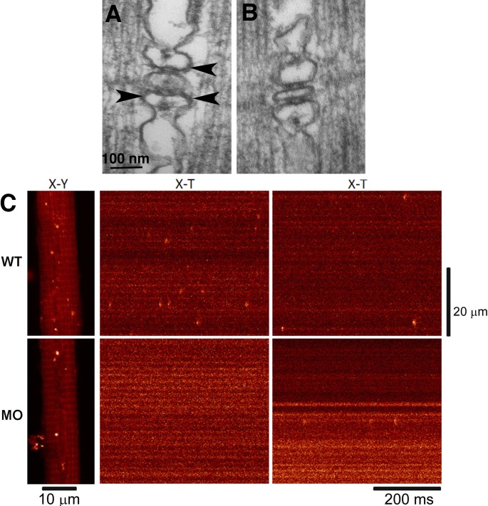
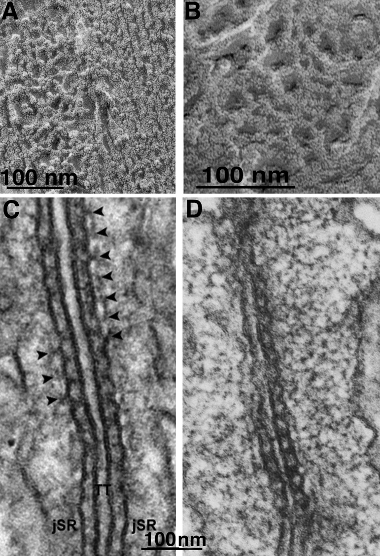
The concept of excitation-contraction coupling is almost as old as Journal of General Physiology It was understood as early as the 1940s that a series of stereotyped events is responsible for the rapid contraction response of muscle fibers to an initial electrical event at the surface. These early developments, now lost in what seems to be the far past for most young investigators, have provided an endless source of experimental approaches. In this Milestone in Physiology, I describe in detail the experiments and concepts that introduced and established the field of excitation-contraction coupling in skeletal muscle. More recent advances are presented in an abbreviated form, as readers are likely to be familiar with recent work in the field.


Figures

Figure 1.
Figure 1.
Local stimulation experiments. (A–D) Images at the edge of a single frog muscle fiber. A small fire-polished pipette establishes a fairly high resistance contact (A and C), and current is passed through the pipette (B and D). Positive spots, responding with a local contraction, are located at the center of the I band, where T tubules invaginate (B), but not opposite the A band (D). Reprinted from Huxley and Taylor (1958). (E and F) Images of a skinned fiber’s edge under oil. Application of calcium through a very small pipette activates contraction both at the I band (E) and the A band (F) level. Reprinted from Costantin and Podolsky (1965).
Figure 2.
Figure 2.
T tubules have different positions in frog and lizard muscles. (A and B) Thin sections of muscle fibers from frog (A) and lizard (B), covering the length of one sarcomere. These are the same type of muscle fibers that were used by A.F. Huxley and collaborators in the local stimulation experiments. The T tubules (arrows) run transversely at the level of the Z line in frog and at the edges of the A band in lizard, corresponding to the location of the “hot spots” through which activation can be initiated.
Figure 3.
Figure 3.
T tubules are continuous, but jSR is not. (A) A slow fiber from fish tail imaged by SEM after extraction of myofibrils. The transverse tubules (T) appear as continuous thin tubes running obliquely from lower left to upper right corners (arrows). Segmented SR cisternae face T tubules forming triads (SR/T/SR). Between triads, the SR forms extensive fenestrated collars (asterisks). Notice the abundance of SR membranes relative to those of T tubules. (B) Frog fiber infiltrated by the Golgi “stain.” The paired dark SR cisternae are the lateral sacs of the triad. The intervening T tubules between them are not visible because the electron dense contrasting agent did not infiltrate them. Note that the SR cisternae are divided into discontinuous segments. A and B are unpublished images from the collection of M. Lavorato (printed here with permission).
Figure 4.
Figure 4.
The SR as a calcium sink. Images from a series of experiments by Leroy Costantin and Richard Podolsky, 1965. (A) Mechanically skinned fiber a la Natori. (B) Sequential images from a brief movie showing a short fiber segment exposed to a droplet of calcium under oil. The segment contracts and then relaxes because of calcium uptake by the SR. (C) Images from a movie illustrating the effect of a drop of calcium solution applied to the edges of a skinned fiber under oil. A strong but very delimited contraction is followed by relaxation. (D) Calcium oxalate deposit in the SR of a skinned muscle fiber exposed to calcium and oxalate. Reprinted from Costantin and Podolsky (1965).
Figure 5.
Figure 5.
Calcium ATPase is a major component of longitudinal SR. All images are from rotary shadowed replicas. (A) Freeze fracture of a fish muscle showing the split SR membrane for an entire sarcomere, from one T tubule to the next (arrows). The cytoplasmic leaflet reveals a uniform distribution of tightly arranged particles, covering the whole free SR and the sides of the triadic SR. Each particle represents a small group of 2–3 ATPase molecules. (B) Cytoplasmic surfaces of freeze-dried SR vesicles from a crude SR fraction of rabbit muscle homogenate. Each dot indicates the position of an individual SR ATPase, at a density of ∼30,000/µm2. (C) Treatment with vanadate (Taylor et al., 1986) induces the formation of two-dimensional “crystals” that impose a tubular shape to the SR vesicles. (D) At higher magnification, the dimeric arrangement of molecules is clear. Such vanadate-treated SR tubes provided the first high-resolution images of the CaATPase.
Figure 6.
Figure 6.
Couplons in e-c coupling. (A) Diagram of local stimulation experiments in crab fibers (reprinted from Peachey, 1965). Depolarization at the level of the Z line did not produce a response, but hot spots were found at the A-I junction level within deep clefts. (B) Phase contrast image of a crab fiber infiltrated with the “Golgi” stain. The orange structures are deep clefts. Two types of invagination arise from the clefts: ribbon like tubules at the Z line levels (double arrow) and tubules with a more complex shape at the edges of the A band (single arrows). (C and D) Electron micrographs of Golgi-stained crab fibers. In C, only tubules at the Z line (double arrow) and T tubules the A-I junction (single arrows) are “stained.” The latter have a convoluted shape. In D the SR is visible as an extensive lacy network and the T tubules are darker. Flat cisternae of T tubules (arrows) are located opposite equivalent cisternae of the SR (arrowhead) to form dyads. Activation of calcium release occurs at these sites. See Franzini-Armstrong et al. (1986).
Figure 7.
Figure 7.
Images of “feet” or RYR1 from skeletal muscle. (A) RYR were first visualized in thin sections of triads in skeletal muscle, where their cytoplasmic domains appeared as evenly spaced rows of feet spanning the junctional gap from SR to T tubules (Franzini-Armstrong, 1970). The content of the triad cisternae is calsequestrin. This image is from a fish muscle. (B) In a grazing view of the junction, feet are seen as small squares engaged in a two-dimensional array. From a tadpole. (C) The heavy SR fraction is composed of vesicles derived from triads (or couplons). They have a dense content (calsequestrin) and show a thin dark ring (extension of the CaATPase) and feet (arrows). This demonstrated that feet are part of the SR membrane (Campbell et al., 1980). (D) Rotary shadowed images of feet on a heavy SR vesicle show ordered arrangement and a 4-subunit structure. Reprinted from Ferguson et al. (1984). (E–I) Images of feet in situ (E and F) and after isolation of the RYR molecule (G–I). The in situ molecules show a central depression on the side facing the cytoplasm. The isolated proteins face the opposite way and show a central raised platform representing the channel domain. Compare Ferguson et al. (1984) and Block et al. (1988).
Figure 8.
Figure 8.
The disposition of DHPR tetrads in T tubules is linked to that of feet. (A–D) The first images of DHPR tetrads were seen in T tubules of a small fish (A; Franzini-Armstrong and Nunzi, 1983) and in the plasmalemma of frog slow fibers (B–D; Franzini-Armstrong, 1984), where it was determined that the distance between tetrads is twice the distance between feet. (E–I) DHPR tetrads in T tubules of 4-d-old zebrafish larvae. E is from a wild type fish, F after the expression of DHPR cDNA. (G and H) From larvae of a zebrafish null for Stac3, but carrying small amounts of maternal DNA. (I) After silencing all DHPR RNA with a morpholino. See Linsley et al. (2017).
Figure 9.
Figure 9.
RYR3 in a parajunctional position are necessary for the production of sparks in zebrafish muscle. (A) Triad in a 72 h postfertilization larva. Two sets of feet (RYR1) connect SR to the central T tubule profile. Additional feet profiles in a parajunctional position (arrowheads) have been proposed to be RYR3 (Felder and Franzini-Armstrong, 2002). (B) One-cell-stage embryos were injected with a morpholino designed to specifically silence RYR3 expression. In triads of larvae at 72 h postfertilization, RYR1 position was normal, but parajunctional feet were essentially missing. (C) The Ca2+ sparks frequency in WT and morpholino-injected (MO) embryos dropped in correspondence to the absence of parajunctional feet. Reprinted from Perni et al. (2015).
Figure 10.
Figure 10.
Triadin and calsequestrin. (A and B) Images of calsequestrin within the SR of a snake muscle, after freeze-fracture, deep etch, and rotary shadowing. The CASQ network is composed of a branching network of elongated elements that form a continuous CASQ polymer (Perni et al., 2013). (C and D) Triads in a mouse (C) and frog (D) muscle. The image in C is from a thicker section. Periodic densities (arrowheads) are located in alternate position relative to feet. The densities are not present in triadin-null muscle fibers, demonstrating that they are composed of triadin polymers (C; reprinted from Boncompagni et al. [2012]). In a thinner section (D), the triadin densities are less clearly visible, but the network nature of the CASQ gel that fills the SR is more obvious. Compare with A and B.

Similar articles

Cited by

References

    1. Adams B.A., Tanabe T., Mikami A., Numa S., and Beam K.G.. 1990. Intramembrane charge movement restored in dysgenic skeletal muscle by injection of dihydropyridine receptor cDNAs. Nature. 346:569–572. 10.1038/346569a0 - DOI - PubMed
    1. Adrian R.H., and Almers W.. 1976. Charge movement in the membrane of striated muscle. J. Physiol. 254:339–360. 10.1113/jphysiol.1976.sp011235 - DOI - PMC - PubMed
    1. Adrian R.H., and Huang C.L.. 1984. Charge movements near the mechanical threshold in skeletal muscle of Rana temporaria. J. Physiol. 349:483–500. 10.1113/jphysiol.1984.sp015169 - DOI - PMC - PubMed
    1. Adrian R.H., Costantin L.L., and Peachey L.D.. 1969. Radial spread of contraction in frog muscle fibres. J. Physiol. 204:231–257. 10.1113/jphysiol.1969.sp008910 - DOI - PMC - PubMed
    1. Almers W. 1980. Potassium concentration changes in the transverse tubules of vertebrate skeletal muscle. Fed. Proc. 39:1527–1532. - PubMed

Substances