. 2018 Jan 10;8(1):8.
doi: 10.1038/s41398-017-0057-7.

Common basis for orofacial clefting and cortical interneuronopathy

Affiliations

Common basis for orofacial clefting and cortical interneuronopathy

Lydia J Ansen-Wilson et al. Transl Psychiatry. .

Abstract

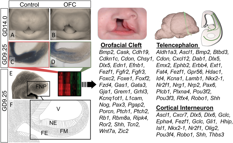
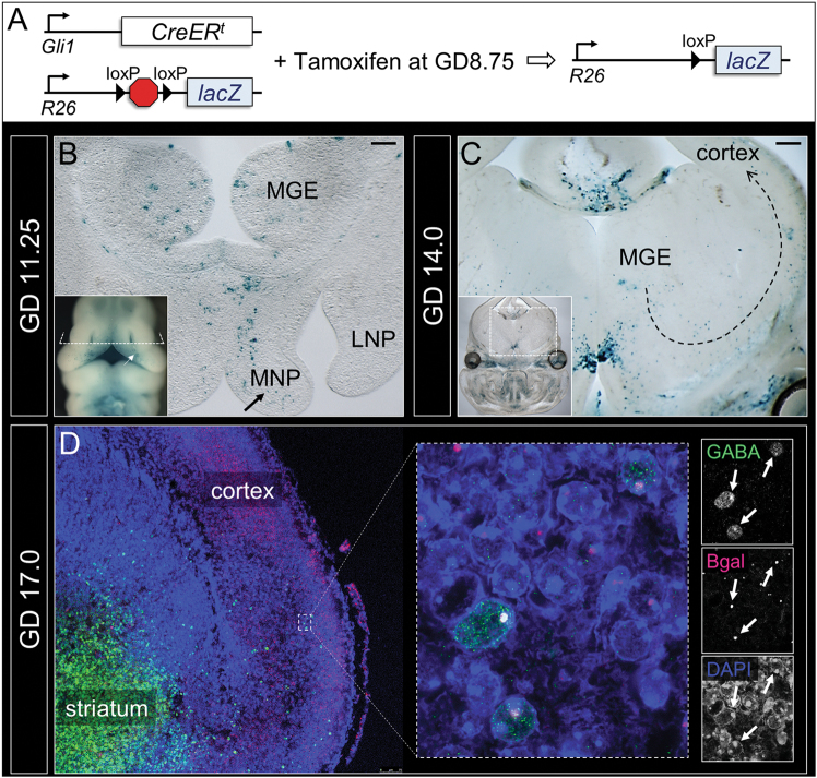
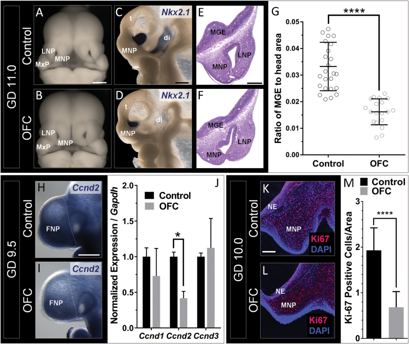
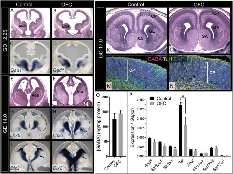
Orofacial clefts (OFCs) of the lip and/or palate are among the most common human birth defects. Current treatment strategies focus on functional and cosmetic repair but even when this care is available, individuals born with OFCs are at high risk for persistent neurobehavioral problems. In addition to learning disabilities and reduced academic achievement, recent evidence associates OFCs with elevated risk for a constellation of psychiatric outcomes including anxiety disorders, autism spectrum disorder, and schizophrenia. The relationship between these outcomes and OFCs is poorly understood and controversial. Recent neuroimaging studies in humans and mice demonstrate subtle morphological brain abnormalities that co-occur with OFCs but specific molecular and cellular mechanisms have not been investigated. Here, we provide the first evidence directly linking OFC pathogenesis to abnormal development of GABAergic cortical interneurons (cINs). Lineage tracing revealed that the structures that form the upper lip and palate develop in molecular synchrony and spatiotemporal proximity to cINs, suggesting these populations may have shared sensitivity to genetic and/or teratogenic insult. Examination of cIN development in a mouse model of nonsyndromic OFCs revealed significant disruptions in cIN proliferation and migration, culminating in misspecification of the somatostatin-expressing subgroup. These findings reveal a unified developmental basis for orofacial clefting and disrupted cIN development, and may explain the significant overlap in neurobehavioral and psychiatric outcomes associated with OFCs and cIN dysfunction. This emerging mechanistic understanding for increased prevalence of adverse neurobehavioral outcomes in OFC patients is the entry-point for developing evidence-based therapies to improve patient outcomes.


Conflict of interest statement

The authors declare that they have no competing financial interests.

Figures

Fig. 1
Fig. 1
Changes in telencephalon and cortical interneuron transcripts during OFC pathogenesis. a,b Representative vehicle-exposed control and cyclopamine-exposed OFC embryos at GD14.0. c,d ISH staining for Gli1 on parasagittal sections through the frontonasal prominence (FNP) of GD9.25 embryos illustrates reduced Shh-signaling activity during initial OFC pathogenesis. e,f Along with a scanning electron micrograph image of a GD9.25 mouse embryo, a schematic of a parasagittal section shows the cell populations comprising the FNP including the neuroectoderm (NE), facial mesenchyme (FM) and facial ectoderm (FE). (Right) Microarray analyses of microdissected FNP tissue from GD9.25 control versus OFC groups revealed differential expression of orofacial cleft, telencephalon, and cortical interneuron-related genes. V ventricle
Fig. 2
Fig. 2
Orofacial and cortical interneuron development occur in spatiotemporal proximity and molecular synchrony. a To identify the lineage of SHH ligand-responding cells, mice with Gli1 promoter-driven and Tamoxifen-inducible Cre recombinase were bred to mice carrying a lacZ reporter with an upstream floxed STOP cassette and administered Tamoxifen at GD8.75. b Whole mount (inset) and sectional staining show reporter-positive cell populations in the medial nasal processes (MNPs) (arrows), and adjacently developing MGEs at GD11.25. c At GD14.0, reporter-positive populations appear along the presumptive tangential migration path of cINs (dotted arrow). d At GD17.0, a population of reporter-positive cells (red) at the deep edge of the cortical plate also express GABA (green). LNP lateral nasal process
Fig. 3
Fig. 3
Disrupted MGE development parallels OFC pathogenesis. Deficiency of the MNPs and MGEs in the OFC group is shown in whole embryos (a,b) and hemisected embryos stained for Nkx2.1 (c,d). e,f The anatomical relationship and paralleled hypoplasia of the MNPs and MGEs is shown by histological staining of transverse sections. g Quantification of MGE area relative to total head area (Fig. S2) was calculated from images depicted in c and d. Individual data points are shown with mean±s.d. of n = 24 embryos from three litters for the control group and n = 19 embryos from three litters for the OFC group; ****P ≤ 0.0001, two-tailed Student’s t-test. Reduced expression of Ccnd2 in the OFC group is shown by ISH staining (h,i) and RT-PCR analysis of microdissected FNP tissue of GD9.5 embryos (j). Values represent mean±s.e.m. of n = 3 pooled litters per group; *P ≤ 0.05, two-tailed Student’s t-test. Staining on frontal sections through the neuroectoderm (NE) of GD10.0 embryos (k,l) demonstrated a reduction in the number of Ki67-positive cells per total area in the OFC group. Values represent the mean±s.e.m.; ****P ≤ 0.0001, two-tailed Student’s t-test. LNP lateral nasal process, MNP medial nasal process, t telencephalon, di diencephalon
Fig. 4
Fig. 4
OFC pathogenesis co-occurs with disrupted cIN migration and specification. At GD12.25, the MGEs in the OFC group are dysmorphic (a,b) but retain expression of the GABAergic precursor marker Gad1 (c,d). By GD14.0, differences in MGE morphology in the OFC group are less apparent (e,f) but the domains of expression of Lhx6 and Dlx2 are abnormally expanded (arrows; gj). At GD17.0, no major differences are observed between groups in cortical plate histology (k,l) or in the spatial distribution of GABA-positive cells (m,n). o GABA levels measured in cortical homogenate relative to total protein. Values represent the mean±s.e.m. of n = 5 fetuses from two litters for the control group and n = 7 fetuses with overt upper lip clefts from two litters for the OFC group; *P ≤ 0.05, two-tailed Student’s t-test. p RT-PCR analysis of cortical mRNA levels of GABAergic (Gad1, Slc32a1, Slc6a1, Sst, and Abat) and glutamatergic (Slc17a7, Slc17a6, and Slc17a8) markers from the cortex at GD17 illustrates a specific reduction of Sst in the OFC group. Values represent the mean ± s.e.m. of n = 5 fetuses from two litters for the control group and n = 7 fetuses with overt upper lip clefts from two litters for the OFC group; *P ≤ 0.05, two-tailed Student’s t-test). SA septal area, GE ganglionic eminence, CP cortical plate

References

    1. Robin NH, et al. The multidisciplinary evaluation and management of cleft lip and palate. South. Med. J. 2006;99:1111–1120. doi: 10.1097/01.smj.0000209093.78617.3a. - DOI - PubMed
    1. Waitzman N., Scheffler R. M. & Romano P. S. The Cost of Birth Defects: Estimates of the Value of Prevention, xiv (University Press of America: Lanham, MD), 1996, 262pp.
    1. Endriga MC, Kapp-Simon KA. Psychological issues in craniofacial care: state of the art. Cleft Palate Craniofac. J. 1999;36:3–11. doi: 10.1597/1545-1569(1999)036<0001:PIICCS>2.3.CO;2. - DOI - PubMed
    1. Collett BR, Speltz ML. A developmental approach to mental health for children and adolescents with orofacial clefts. Orthod. Craniofac. Res. 2007;10:138–148. doi: 10.1111/j.1601-6343.2007.00394.x. - DOI - PubMed
    1. Richman LC. Cognitive patterns and learning disabilities in cleft palate children with verbal deficits. J. Speech Language Hear. Res. 1980;23:447–456. doi: 10.1044/jshr.2302.447. - DOI - PubMed

Publication types

Supplementary concepts