. 2018 Jan 9;8(1):185.
doi: 10.1038/s41598-017-18502-8.

Silica nanoparticle stability in biological media revisited

Affiliations

Silica nanoparticle stability in biological media revisited

Seon-Ah Yang et al. Sci Rep. .

Abstract

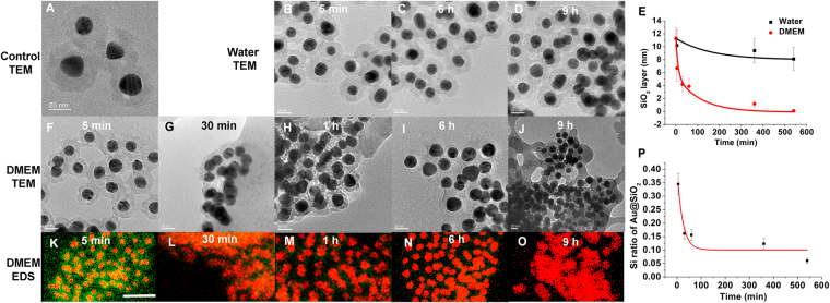
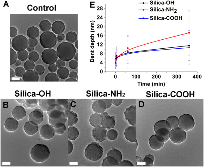
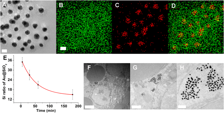
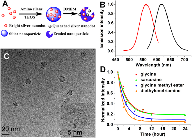
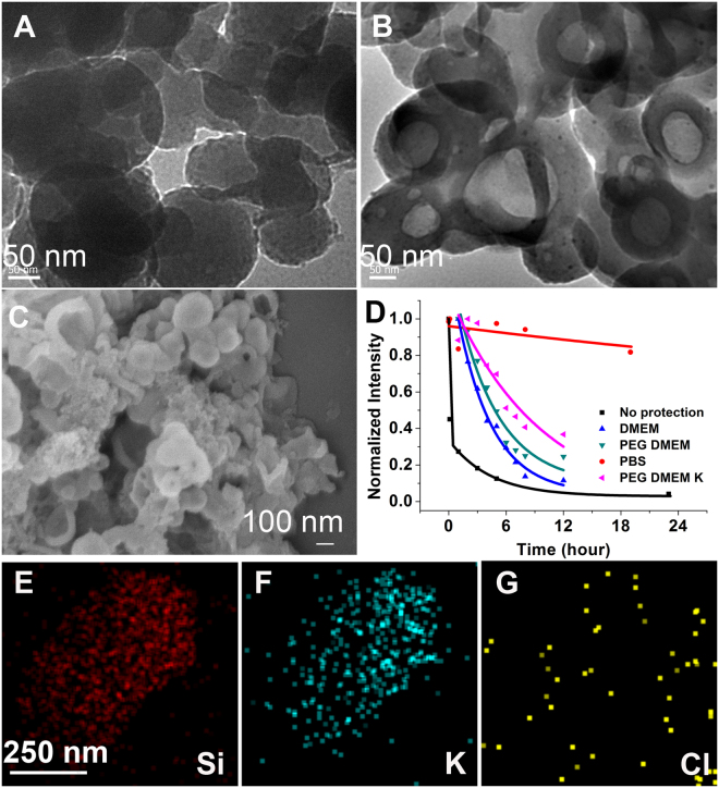
The stability of silica nanostructure in the core-silica shell nanomaterials is critical to understanding the activity of these nanomaterials since the exposure of core materials due to the poor stability of silica may cause misinterpretation of experiments, but unfortunately reports on the stability of silica have been inconsistent. Here, we show that luminescent silver nanodots (AgNDs) can be used to monitor the stability of silica nanostructures. Though relatively stable in water and phosphate buffered saline, silica nanoparticles are eroded by biological media, leading to the exposure of AgNDs from AgND@SiO2 nanoparticles and the quenching of nanodot luminescence. Our results reveal that a synergistic effect of organic compounds, particularly the amino groups, accelerates the erosion. Our work indicates that silica nanostructures are vulnerable to cellular medium and it may be possible to tune the release of drug molecules from silica-based drug delivery vehicles through controlled erosion.


Conflict of interest statement

The authors declare that they have no competing interests.

Figures

Figure 1
Figure 1
Schematic showing that silver nanodots-encapsulated in silica reveals the erosion of silica.
Figure 2
Figure 2
In vitro stability of silica shells. (AD) TEM images of Au@SiO2 (A, control) and in water at 5 min, 6 h and 9 h, respectively (BD). Scale bars, 20 nm. (E) Plots of the thickness of silica shells versus time in water (black) and DMEM (red), measured in TEM images. The error bars indicate standard error. The p value in each statistical analysis was less than 0.001. (FJ) TEM images of Au@SiO2 in DMEM at 5 min, 30 min, 1 h, 6 h and 9 h, respectively. Scale bars, 20 nm. (KO) EDS images of Au@SiO2 in DMEM at 5 min, 30 min, 1 h, 6 h and 9 h, respectively. The images were merged from signals of gold in pseudocolor red and that of silicon in pseudocolor green. Scale bars, 100 nm. (P) Plots of the EDS intensity of silicon versus time in DMEM, measured from EDS images.
Figure 3
Figure 3
Degradation of mesoporous silica nanoparticles in DMEM. (A) Representative TEM image of mesoporous silica nanoparticles. (BD) TEM images of original (B), amino group-modified (C) and carboxylic group-modified (D) MSNs in DMEM at 9 h. Scale bars, 200 nm. (E) Plots of the dent depth of MSNs versus time in DMEM. The error bars indicate standard error. The p value in each statistical analysis was less than 0.001.
Figure 4
Figure 4
Ex vivo stability of silica in sheep blood. (AD) TEM (A) and EDS (BD) images of Au@SiO2, incubated in blood for 5 min. Scale bars, 20 nm. (E) Plots of the EDS intensity of silicon versus time, measured from EDS images. (FH) TEM images of Au@SiO2 in sheep blood at various magnification. Scale bars, 2 μm, 0.5 μm and 100 nm for (F,G and H), respectively.
Figure 5
Figure 5
Detection of silica degradation with luminescent silver nanodots. (A) Schematic showing the encapsulation of silver nanodots in silica nanoparticles and their optical detection of silica degradation. (B) Emission (black) and excitation (red) spectra of the 615-emitter. (C) TEM image of silver nanodot-encapsulating silica nanoparticles (AgND@SiO2). Scale bar, 20 nm. Inset showing a high resolution image of the above nanoparticles. Scale bar, 5 nm. (D) Normalized luminescence intensity decay of the above AgND@SiO2 in the presence of glycine (red), sarcosine (green), glycine methyl ester (blue) and diethylenetriamine (orange).
Figure 6
Figure 6
Improved silica stability. (A + B) TEM images of larger AgND@SiO2 after 5 min (A) and 9 h (B) incubation in DMEM. Scale bars, 50 nm. (C) SEM image of the sample in (B). Scale bar, 100 nm. (D) Comparison of luminescence intensity decay of silver nanodots under various conditions. (EG) EDS images of the above AgND@SiO2 in PBS showing the signal of silicon (E), potassium (F) and chlorine (G). Scale bars, 250 nm.

References

    1. Kao J, Thorkelsson K, Bai P, Rancatore BJ, Xu T. Toward functional nanocomposites: taking the best of nanoparticles, polymers, and small molecules. Chem. Soc. Rev. 2013;42:2654–2678. doi: 10.1039/C2CS35375J. - DOI - PubMed
    1. Davis ME, Chen Z, Shin DM. Nanoparticle therapeutics: an emerging treatment modality for cancer. Nat Rev Drug Discov. 2008;7:771–782. doi: 10.1038/nrd2614. - DOI - PubMed
    1. Caruso F, Caruso RA, Mohwald H. Nanoengineering of inorganic and hybrid hollow spheres by colloidal templating. Science. 1998;282:1111–1114. doi: 10.1126/science.282.5391.1111. - DOI - PubMed
    1. Kim J, et al. Generalized fabrication of multifunctional nanoparticle assemblies on silica spheres. Angew. Chem.-Int. Edit. 2006;45:4789–4793. doi: 10.1002/anie.200504107. - DOI - PubMed
    1. Law WC, et al. Optically and magnetically doped organically modified silica nanoparticles as efficient magnetically guided biomarkers for two-photon imaging of live cancer cells. J. Phys. Chem. C. 2008;112:7972–7977. doi: 10.1021/jp712090y. - DOI

Publication types