. 2018 Jan 9;8(1):153.
doi: 10.1038/s41598-017-18530-4.

Loss of the Mitochondrial Fatty Acid β-Oxidation Protein Medium-Chain Acyl-Coenzyme A Dehydrogenase Disrupts Oxidative Phosphorylation Protein Complex Stability and Function

Affiliations

Loss of the Mitochondrial Fatty Acid β-Oxidation Protein Medium-Chain Acyl-Coenzyme A Dehydrogenase Disrupts Oxidative Phosphorylation Protein Complex Stability and Function

Sze Chern Lim et al. Sci Rep. .

Abstract

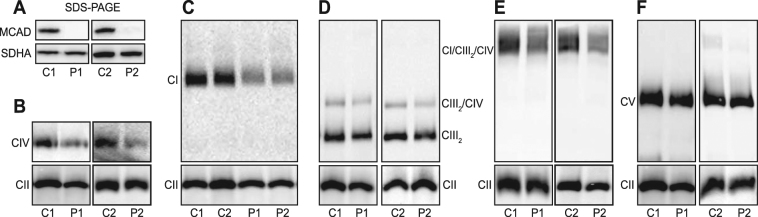
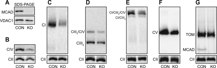
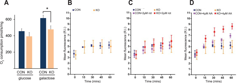
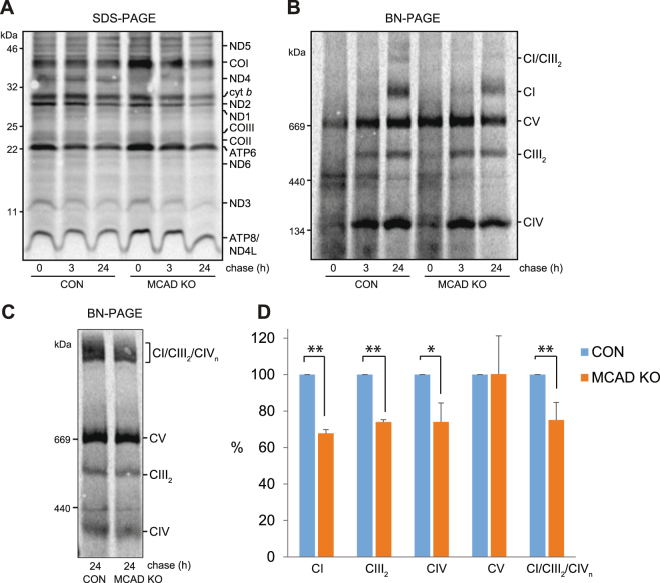
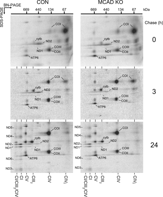
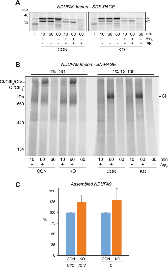
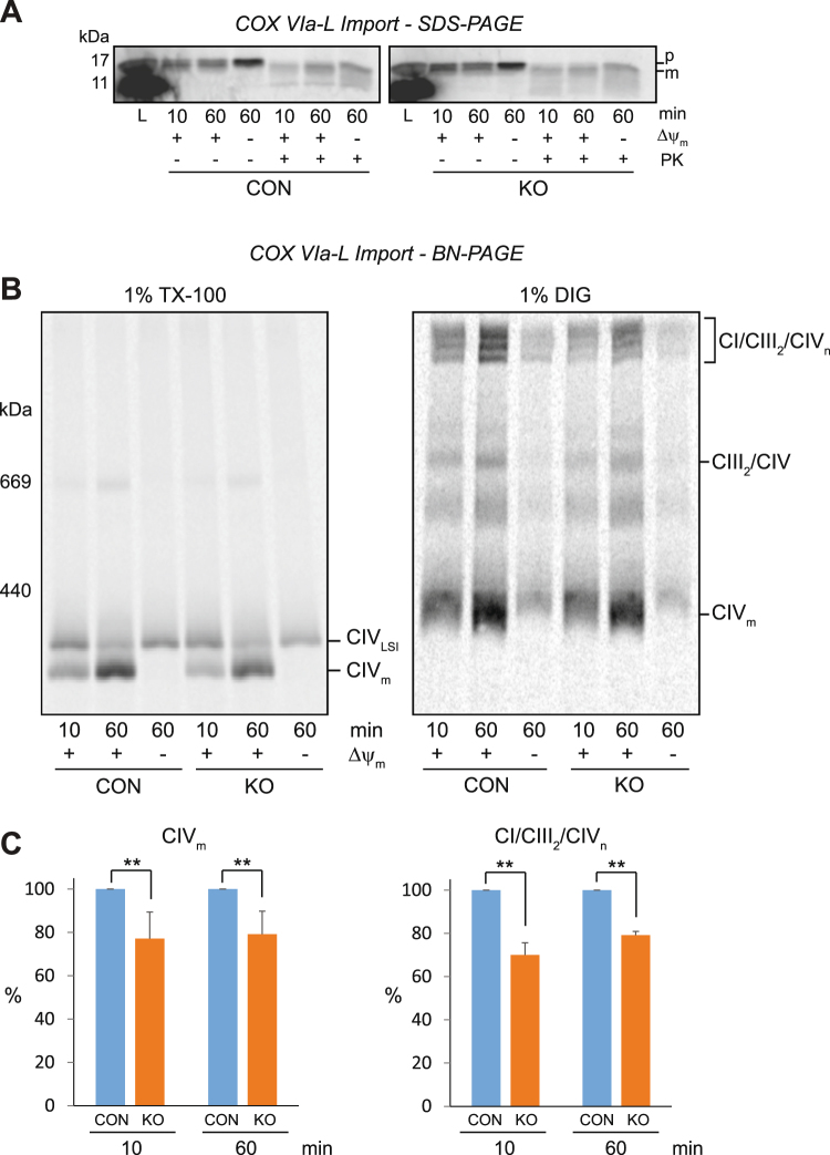
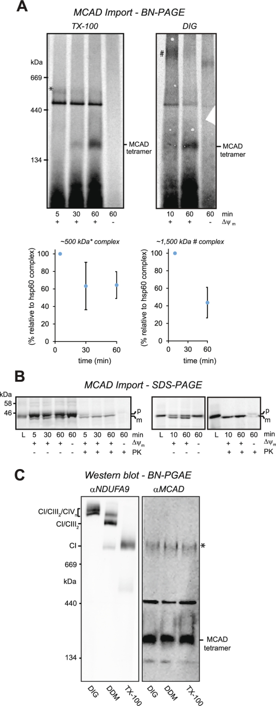
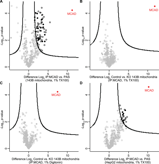
Medium-chain acyl-Coenzyme A dehydrogenase (MCAD) is involved in the initial step of mitochondrial fatty acid β-oxidation (FAO). Loss of function results in MCAD deficiency, a disorder that usually presents in childhood with hypoketotic hypoglycemia, vomiting and lethargy. While the disruption of mitochondrial fatty acid metabolism is the primary metabolic defect, secondary defects in mitochondrial oxidative phosphorylation (OXPHOS) may also contribute to disease pathogenesis. Therefore, we examined OXPHOS activity and stability in MCAD-deficient patient fibroblasts that have no detectable MCAD protein. We found a deficit in mitochondrial oxygen consumption, with reduced steady-state levels of OXPHOS complexes I, III and IV, as well as the OXPHOS supercomplex. To examine the mechanisms involved, we generated an MCAD knockout (KO) using human 143B osteosarcoma cells. These cells also exhibited defects in OXPHOS complex function and steady-state levels, as well as disrupted biogenesis of newly-translated OXPHOS subunits. Overall, our findings suggest that the loss of MCAD is associated with a reduction in steady-state OXPHOS complex levels, resulting in secondary defects in OXPHOS function which may contribute to the pathology of MCAD deficiency.


Conflict of interest statement

The authors declare that they have no competing interests.

Figures

Figure 1
Figure 1
MCAD-deficient patient fibroblasts have reduced steady-state levels of OXPHOS complexes and supercomplexes. Mitochondria were isolated from control (C1 and C2) or MCAD-deficient patient (P1 and P2) fibroblasts for SDS-PAGE and BN-PAGE Western blot analysis. (A) No mature MCAD protein is detectable in P1 or P2 patient mitochondria by SDS-PAGE (the OXPHOS complex II subunit SDHA is shown as a loading control). BN-PAGE revealed that the steady-state levels of mature OXPHOS complex IV (detected with an anti-COI antibody) (B), complex I (anti-NDUFA9 antibody) (C), the complex III dimer (CIII2) and the CIII2/CIV supercomplex (anti-UQCRC1 antibody) (D) and the CI/CIII2/CIV supercomplex (anti-NDUFA9 antibody) (E) were all reduced in P1 and P2 patient mitochondria compared to C1 and C2 controls respectively. (F) Steady-state levels of OXPHOS complex V (anti-ATP5A antibody) were no different in P1 and P2 patient mitochondria compared to controls. Mitochondria were solubilised in TX-100 (B–D,F) or in digitonin (E). Quantitation is relative to the steady-state levels of OXPHOS complex II (anti-SDHA antibody) (n = 3).
Figure 2
Figure 2
143B MCAD knockout (KO) cells also exhibit reduced steady-state levels of OXPHOS complexes and supercomplexes. The ACADM gene was edited in 143B cells using lentiviral-based CRISPR/Cas9. Mitochondria were isolated from 143B control cells (CON) or a 143B MCAD knockout clone (KO) for SDS-PAGE and BN-PAGE Western blot analysis. (A) No mature MCAD protein is detectable in KO mitochondria by SDS-PAGE (the mitochondrial protein VDAC1 is shown as a loading control). BN-PAGE revealed that the steady-state levels of mature OXPHOS complex IV (detected with an anti-COI antibody) (B), complex I (anti-NDUFA9 antibody) (C), the complex III dimer (CIII2) and the CIII2/CIV supercomplex (anti-UQCRC1 antibody) (D) and the CI/CIII2/CIV supercomplex (anti-NDUFA9 antibody) (E) were all significantly reduced in KO mitochondria compared to the control (CON). Steady-state levels of OXPHOS complex V (anti-ATP5A antibody) (F) and the Translocase of the Outer Mitochondrial Membrane (anti-TOMM40 antibody) (G) were not different in KO mitochondria compared to the control (CON). (G) The homotetrameric MCAD complex is detectable in CON, but not MCAD KO mitochondria. Mitochondria were solubilised in TX-100 (B–D) or in digitonin (E–G). Quantitation is relative to the steady-state levels of OXPHOS complex II (anti-SDHA antibody) (n = 3).
Figure 3
Figure 3
143B MCAD knockout (KO) cells exhibit defects in mitochondrial respiration and are more sensitive to OXPHOS inhibitor-induced oxidative stress. (A) Mitochondrial oxygen consumption in MCAD knockout (KO) cells was not significantly different to control (CON) cells in the presence of glucose. However, oxygen consumption was significantly lower in MCAD KO cells in the presence of galactose. Data is mean ± s.d., n = 3. *p < 0.05. Mitochondrial reactive oxygen species generation was assessed in live cells using the superoxide probe MitoSOXTM. There was no difference in mitochondrial superoxide generation between CON and MCAD KO cells over a 60 min time course in either untreated conditions (B) or with the addition of the OXPHOS complex I inhibitor rotenone (C). (D) Mitochondrial superoxide generation was significantly greater in MCAD KO cells compared to CON cells after 30 min (p = 0.001), 45 min (p = 0.0005) and 60 min (p = 0.0034) in the presence of the OXPHOS complex III inhibitor antimycin A. Data is mean ± s.d., n = 5.
Figure 4
Figure 4
Incorporation of newly-translated mtDNA-encoded subunits into the OXPHOS complexes and the OXPHOS supercomplex is disrupted in MCAD knockout cells. MtDNA-encoded-proteins were radiolabeled in the presence of cycloheximide and chased for 0, 3 or 24 h. (A) SDS-PAGE showing similar amounts of newly-translated complex I (ND1, ND2, ND3, ND4L, ND5 and ND6), complex III (cyt b), complex IV (COI, COII and COIII) and complex V (ATP6 and ATP8) subunits in both 143B control (CON) and 143B MCAD knockout (KO) mitochondria. (B) BN-PAGE analysis of mitochondria solubilised in TX-100 shows that the amount of newly-translated mtDNA-encoded proteins incorporated into complex I (CI), the complex III dimer (CIII2) and complex IV (CIV) after 24 h chase is less in MCAD KO mitochondria than CON mitochondria. Levels of complex V were not different (C) BN-PAGE following solubilisation in digitonin. Reduced levels of newly-translated subunits in the CI/CIII2/CIV supercomplex are evident in MCAD KO mitochondria compared to control (CON). (D) Quantitation of OXPHOS complex and supercomplex levels. Data is mean ± s.d., n = 3. *p < 0.05, **p < 0.01.
Figure 5
Figure 5
The amount of newly-translated mtDNA-encoded subunits incorporated into the OXPHOS supercomplex is reduced in MCAD knockout mitochondria. MtDNA-encoded proteins were radiolabeled in both 143B control (CON) and 143B MCAD knockout (KO) cells in the presence of cycloheximide and chased for 0, 3 or 24 h. Mitochondria were then isolated for two dimensional (2D) native-PAGE analysis. OXPHOS complex subunits are identified according to,. Following 24 h chase, the incorporation of subunits ND1, ND2, ND3, ND4, ND5 and ND6 (complex I), Cyt b (complex III) and COI, COII and COIII (complex IV) into the CI/CIII2/CIV supercomplex is reduced in MCAD KO mitochondria compared to the control (CON) (p = 0.001).
Figure 6
Figure 6
The nuclear-encoded subunit NDUFA9 is imported and assembled efficiently into OXPHOS complex I in 143B MCAD knockout mitochondria. NDUFA9 was radiolabeled by in vitro transcription/translation, followed by incubation for 10 and 60 min with isolated 143B control (CON) or 143B MCAD knockout (KO) mitochondria. (A) SDS-PAGE showing NDUFA9 in its precursor (p) form and as a proteinase K (PK) resistant mature (m) form. NDUFA9 is imported efficiently into both CON and MCAD KO mitochondria in a mitochondrial membrane potential (Δψm) dependent manner. (B) BN-PAGE showing the assembly of NDUFA9 into the CI/CIII2 and CI/CIII2/CIV supercomplexes (following solubilisation in digitonin, left) or mature complex I (CI, following solubilisation in TX-100, right). (C) Quantitation of NDUFA9 assembly after 60 min of import. There was no difference in the amount of NDUFA9 assembled into the CI/CIII2/CIV supercomplex (p = 0.07) or complex I (p = 0.14).
Figure 7
Figure 7
The assembly of the nuclear-encoded subunit COX VIa-L into mature complex IV and the OXPHOS supercomplexes is reduced in 143B MCAD knockout mitochondria. COX VIa-L was radiolabeled by in vitro transcription/translation, followed by incubation for 10 and 60 min with isolated 143B control (CON) or 143B MCAD knockout (KO) mitochondria. (A) SDS-PAGE showing COX VIa-L in its precursor (p) form and as a proteinase K (PK) resistant mature (m) form. COX VIa-L is imported efficiently into both CON and MCAD KO mitochondria. (B) BN-PAGE showing the assembly of COX VIa-L into the late-stage intermediate (CIVLSI) and mature complex IV (CIVm) [following solubilisation in 1% (v/v) TX-100, left] or the CI/CIII2/CIVn supercomplexes [following solubilisation in 1% (w/v) digitonin, right]. The amount of newly-translated COX VIa-L assembled into CIVm and the CI/CIII2/CIVn supercomplexes was significantly less in MCAD KO mitochondria compared to CON mitochondria after import times of both 10 and 60 min. (C) Quantitation of assembled COX VIa-L into CIVm (n = 4) and the CI/CIII2/CIVn supercomplexes (n = 3). Data is mean ± s.d. **p < 0.01.
Figure 8
Figure 8
High molecular weight complexes containing MCAD are detectable by BN-PAGE. (A) MCAD was radiolabeled by in vitro transcription/translation, followed by incubation for 5, 10, 30 and 60 min with isolated 143B control mitochondria. Mitochondria were solubilised in 1% (v/v) TX-100 or 1% (w/v) digitonin, followed by BN-PAGE analysis. MCAD can be detected in complexes of ~175 kDa (MCAD homotetramer) and ~450 kDa (mitochondrial Hsp60 complex), as well as unknown complexes of ~500 kDa (marked *) and ~1,500 kDa (marked #). The intensities of both the ~500 kDa (*) and ~1,500 kDa (#) MCAD-containing complexes (relative to the ~450 kDa mitochondrial Hsp60 complex) was reduced over the 60 min import time course (p = 0.016 and 0.005 respectively). (B) SDS-PAGE showing MCAD in its precursor (p) form and as a proteinase K (PK) resistant mature (m) form. MCAD is imported in a mitochondrial membrane potential (Δψm) dependent manner, with the levels of mature MCAD protein (m) similar at each time point for both the TX-100 and DIG experiments. (C) BN-PAGE and Western blot analysis of HepG2 cell mitochondria solubilised in 1% (w/v) digitonin (DIG), 0.2% (w/v) dodecyl maltoside (DDM) or 1% (v/v) TX-100. Anti-NDUFA9 antibodies detect monomeric complex I and the CI/CIII2/CIVn and CI/CIII2 OXPHOS supercomplexes, while anti-MCAD antibodies detect MCAD in complexes of ~130 kDa, ~175 kDa (homotetramer), ~450 kDa (mitochondrial Hsp60 complex) and ~1,000 kDa (*).
Figure 9
Figure 9
Identification of MCAD interacting proteins. (A) Co-immunoprecipitation (Co-IP) and mass-spectrometry analysis of 143B cell mitochondria. Crude mitochondria were solubilised in 1% (v/v) Triton X-100 and proteins incubated with Protein A-sepharose coupled MCAD antibodies or protein A-sepharose alone. Eluates were analyzed by label-free quantitative mass-spectrometry (LFQ). Log2 LFQ intensities were submitted to a modified two-sided two-sample t-test with significance determined through permutation-based false discovery rate (FDR) statistics. Closed circles, mitochondrial protein; empty circles, non-mitochondrial protein; black, proteins with <1% FDR. (B) As for (A) using either control 143B cell mitochondria or mitochondria from 143B MCAD knockout (KO) cells solubilised in 1% (v/v) Triton X-100, bound to Protein A-sepharose coupled MCAD antibodies in both cases. (C) As for (B) using either control 143B mitochondria or mitochondria from 143B MCAD knockout (KO) cells solubilised in 1% (w/v) digitonin, bound to Protein A-sepharose coupled MCAD antibodies in both cases. (D) As for (A) using HepG2 control cell mitochondria and 1% (w/v) digitonin, bound to Protein A-sepharose coupled MCAD antibodies or protein A-sepharose alone.

References

    1. Matsubara Y, et al. Molecular cloning of cDNAs encoding rat and human medium-chain acyl-CoA dehydrogenase and assignment of the gene to human chromosome 1. Proc. Natl. Acad. Sci. USA. 1986;83:6543–6547. doi: 10.1073/pnas.83.17.6543. - DOI - PMC - PubMed
    1. Saijo T, Welch WJ, Tanaka K. Intramitochondrial folding and assembly of medium-chain acyl-CoA dehydrogenase (MCAD). Demonstration of impaired transfer of K304E-variant MCAD from its complex with hsp60 to the native tetramer. J. Biol. Chem. 1994;269:4401–4408. - PubMed
    1. Andresen BS, et al. Medium-chain acyl-CoA dehydrogenase (MCAD) mutations identified by MS/MS-based prospective screening of newborns differ from those observed in patients with clinical symptoms: identification and characterization of a new, prevalent mutation that results in mild MCAD deficiency. Am. J. Hum. Genet. 2001;68:1408–1418. doi: 10.1086/320602. - DOI - PMC - PubMed
    1. Grosse SD, Khoury MJ, Greene CL, Crider KS, Pollitt RJ. The epidemiology of medium chain acyl-CoA dehydrogenase deficiency: an update. Genet. Med. 2006;8:205–212. doi: 10.1097/01.gim.0000204472.25153.8d. - DOI - PubMed
    1. Leal J, Ades AE, Wordsworth S, Dezateux C. Regional differences in the frequency of the c.985A > G ACADM mutation: findings from a meta-regression of genotyping and screening studies. Clin. Genet. 2014;85:253–259. doi: 10.1111/cge.12157. - DOI - PubMed

Publication types

MeSH terms

Supplementary concepts