. 2018 Jan 9;8(1):201.
doi: 10.1038/s41598-017-18590-6.

Shape dependent physical mutilation and lethal effects of silver nanoparticles on bacteria

Affiliations

Shape dependent physical mutilation and lethal effects of silver nanoparticles on bacteria

Debashish Acharya et al. Sci Rep. .

Abstract

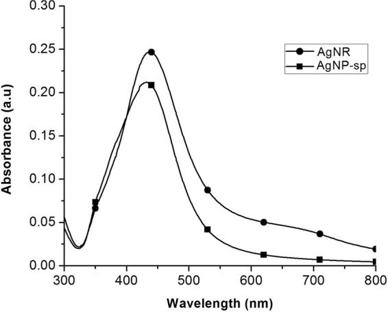
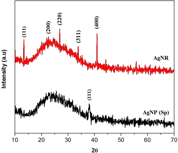
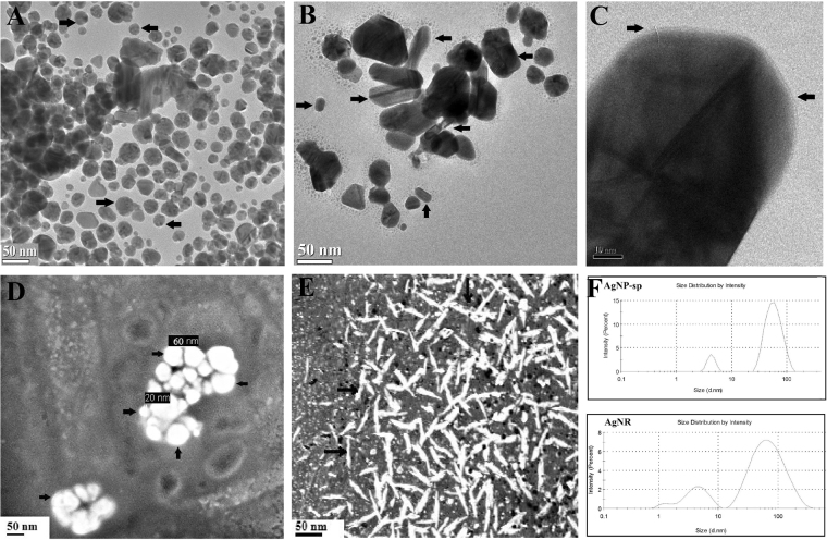
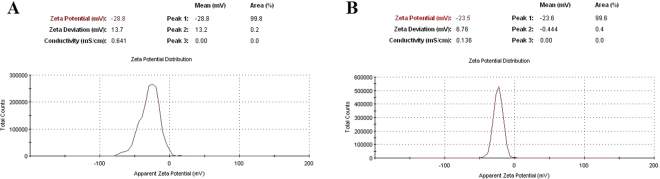
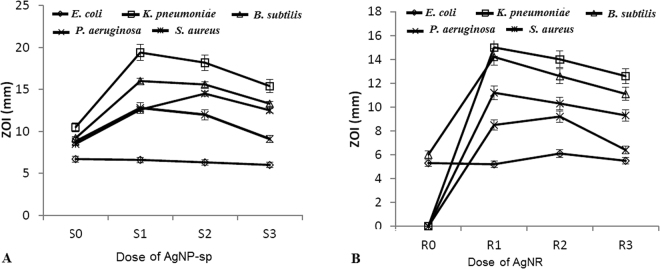
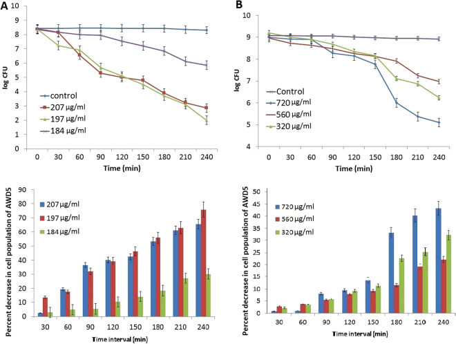
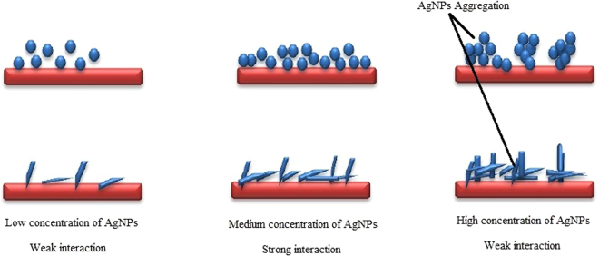
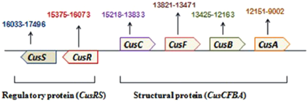
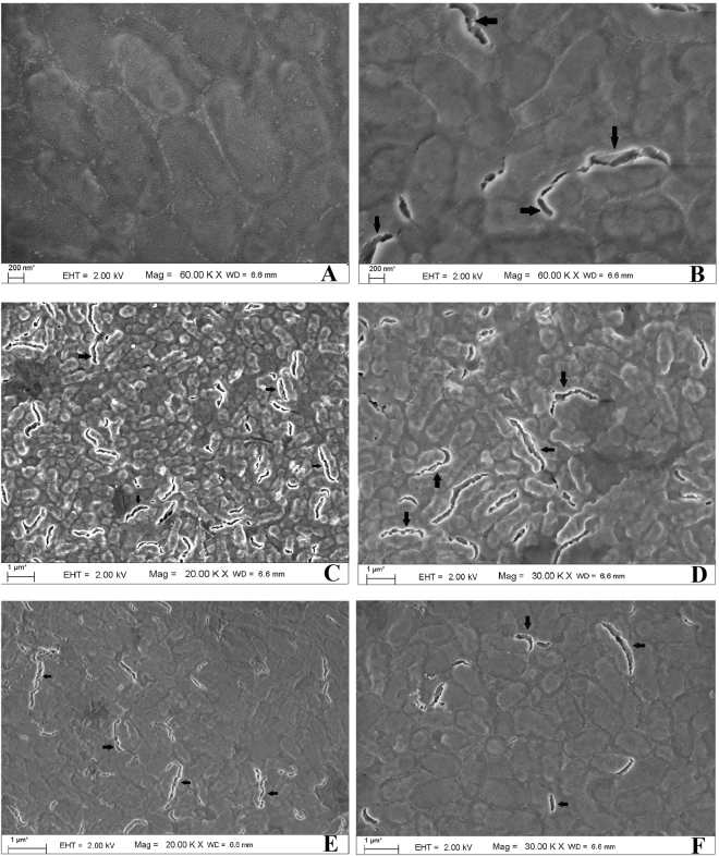
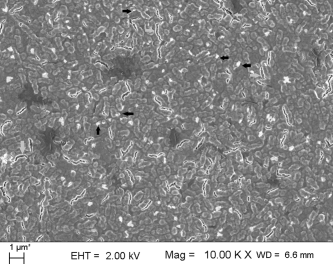
In this report, spherical silver nanoparticle (AgNP-sp) and rod-shaped silver nanoparticle (AgNR) were prepared by chemical reduction method and their antibacterial activity against various Gram-positive and Gram-negative bacteria had been evaluated for their efficiency. Minimal inhibitory concentration (MIC) tests were conducted to study the antibacterial properties, and substantiated with killing kinetics of silver nanoparticles (AgNPs). The study revealed that both AgNP-sp and AgNRs are good antibacterial candidates. Bacterial sensitivity to nanoparticles (NPs) was found to vary depending on microbial species. Disc diffusion studies revealed the greater effectiveness of AgNP-sp and AgNR against Klebsiella pneumoniae AWD5 at the doses of 249 and 392 µg. The dose dependent activities of prepared NPs were also observed on the batch studies of disc diffusion and MIC with various strains. The optical and morphological structures of NPs were analyzed by UV-visible, XRD, FE-SEM and TEM. Further, FESEM of bacterial culture treated with AgNPs confirmed antibacterial activity of NPs by showing rupture of bacterial cell wall. Also, the genome of test organism was found to have CusCFBA and CusRS operons. The killing kinetics confirmed that the death rate of K. pneumoniae was higher against AgNP-sp as compared to AgNR.


Conflict of interest statement

The authors declare that they have no competing interests.

Figures

Figure 1
Figure 1
UV-Vis absorption spectra of AgNP-sp and AgNR in aqueous solution.
Figure 2
Figure 2
XRD spectra of AgNR and AgNP-sp.
Figure 3
Figure 3
TEM images of spherical AgNPs (A), rod AgNPs (B), and HR-TEM images of nanorod showing sharp edges (C). FE-SEM images of spherical (D) and rod shaped AgNPs (E), The size distribution profile of AgNP-sp and AgNR by the DLS techniques (F).
Figure 4
Figure 4
Surface zeta potential analysis of (A) AgNP-sp and (B) AgNR.
Figure 5
Figure 5
Antibacterial activity of AgNPs against different bacteria (E. coli ATCC 25922, S. aureus ATCC 25923, B. subtilis, P. aeruginosa, K. pneumoniae AWD5) at different concentrations of (A) AgNP-sp and (B) AgNR. 255, 249, 242 and 230 µg assigned as S0, S1, S2 and S3 for AgNP-sp and 399, 392, 380, 364 µg for AgNR were assigned as R0, R1, R2 and R3 respectively.
Figure 6
Figure 6
Killing kinetics of K. pneumoniae AWD5 exposed to (A) AgNP-sp at concentrations of 184–207 µg/ml and (B) AgNR at a concentration of 320–720 µg/ml. The percent decrease in cell population at different time intervals (0–4 h) is also given below the respective figure. Error bar represents the standard error of mean for three independent assessments.
Figure 7
Figure 7
Schematic representation of the release of AgNP-sp and AgNR at lower, medium and high concentrations.
Figure 8
Figure 8
Arrangements of genes in the genome of K. pneumoniae AWD5 for CusCFBA and CusRS operons.
Figure 9
Figure 9
FE-SEM images of nanoparticle-untreated K. pneumoniae AWD5 (A) with intact cell surfaces, in contrast to AgNP-sp treated K. pneumoniae AWD5 (B). The cells of K. pneumoniae AWD5 were disrupted due to interaction with AgNP-sp (C,D) and AgNR (E,F). Arrows indicate disrupted regions on cell surface.
Figure 10
Figure 10
FE-SEM image showing the nanoparticles (AgNP-sp) (black arrow), overlaid with cells of K. pneumoniae AWD5 with damaged cell surfaces and disrupted cells (grey arrows) due to interaction with NPs.

References

    1. Lazar V. Quorum sensing in biofilms-how to destroy the bacterial citadels or their cohesion/power? Anaerobe. 2011;17:280–285. doi: 10.1016/j.anaerobe.2011.03.023. - DOI - PubMed
    1. Donlan RM, Costerton JW. Biofilms: survival mechanisms of clinically relevant microorganisms. Costerton, Clin. Microbiol. Rev. 2002;15:167–193. doi: 10.1128/CMR.15.2.167-193.2002. - DOI - PMC - PubMed
    1. Morones JR, et al. The bactericidal effect of silver nanoparticles. Nanotechnology. 2005;16:2346–2353. doi: 10.1088/0957-4484/16/10/059. - DOI - PubMed
    1. Periasamy S, et al. How staphylococcus aureus biofilms develop their characteristics structure. Proc. Natl. Acad. Sci. USA. 2012;109:1281–1286. doi: 10.1073/pnas.1115006109. - DOI - PMC - PubMed
    1. Rolim JP, et al. The antimicrobial activity of photodynamic therapy against Streptococcus mutans using different photosensitizers. Photochem. Photobiol. B. 2012;106:40–46. doi: 10.1016/j.jphotobiol.2011.10.001. - DOI - PubMed

Publication types

MeSH terms