. 2017:2017:3619879.
doi: 10.1155/2017/3619879. Epub 2017 Nov 26.

Tabetri™ (Tabebuia avellanedae Ethanol Extract) Ameliorates Osteoarthritis Symptoms Induced by Monoiodoacetate through Its Anti-Inflammatory and Chondroprotective Activities

Affiliations

Tabetri™ (Tabebuia avellanedae Ethanol Extract) Ameliorates Osteoarthritis Symptoms Induced by Monoiodoacetate through Its Anti-Inflammatory and Chondroprotective Activities

Jae Gwang Park et al. Mediators Inflamm. 2017.

Abstract

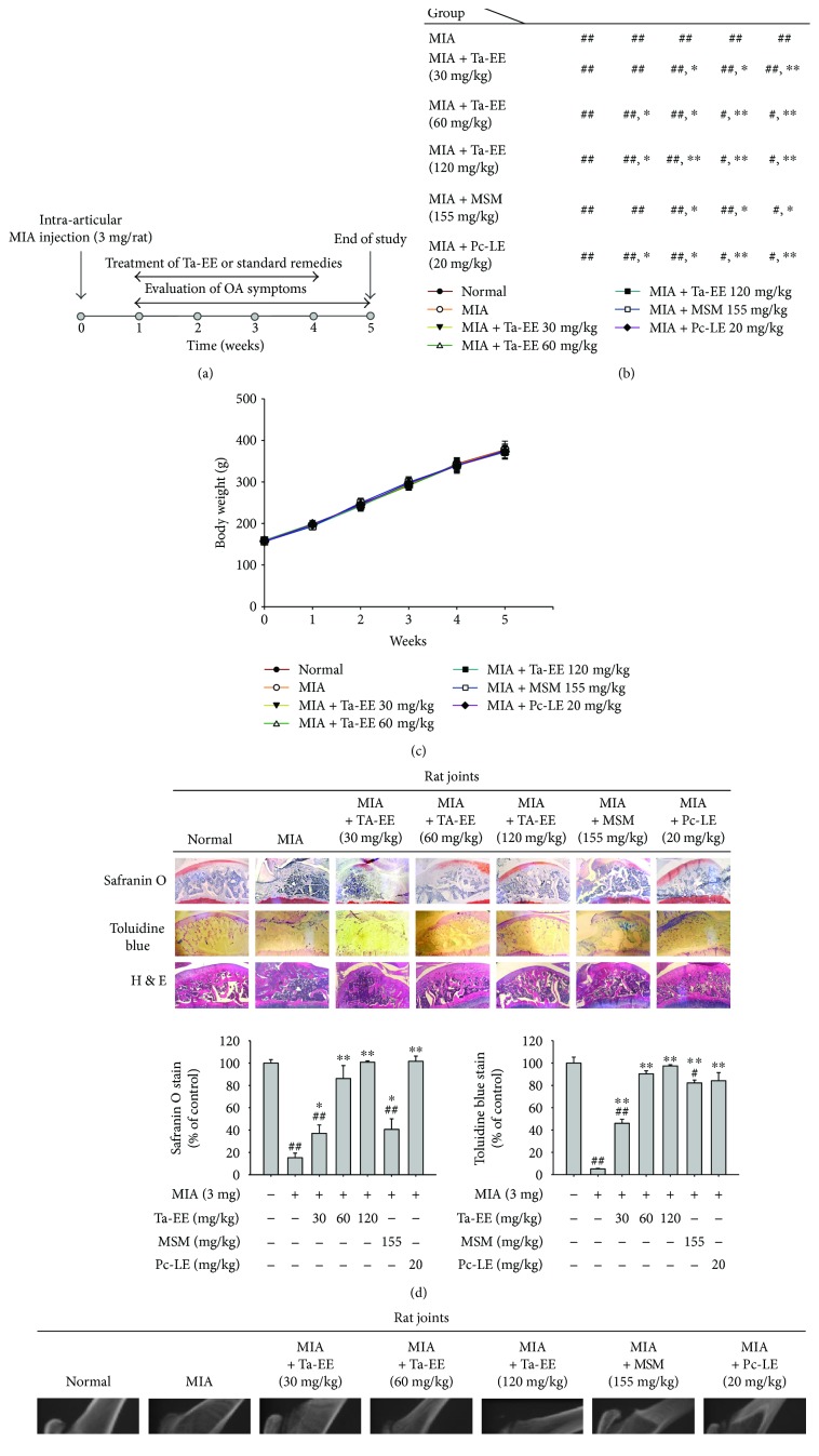
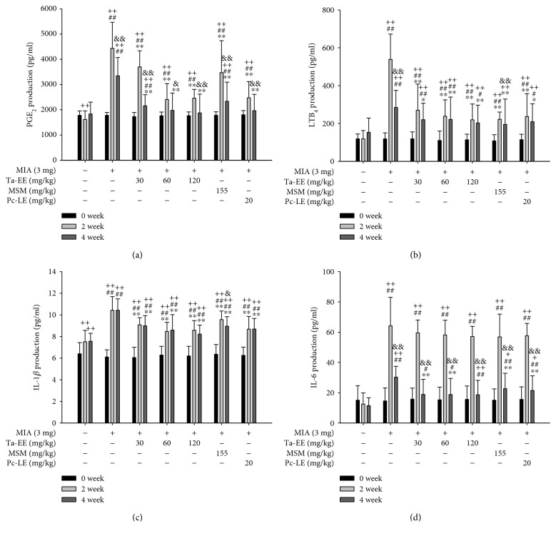
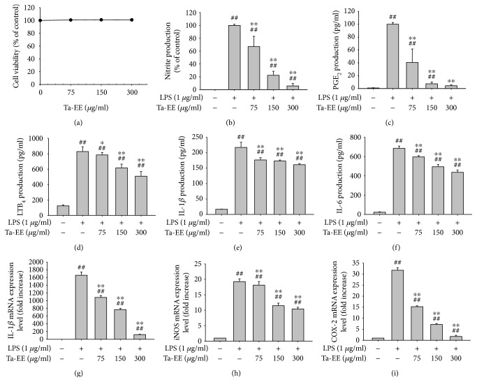
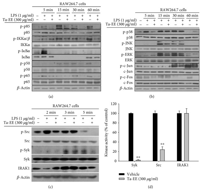
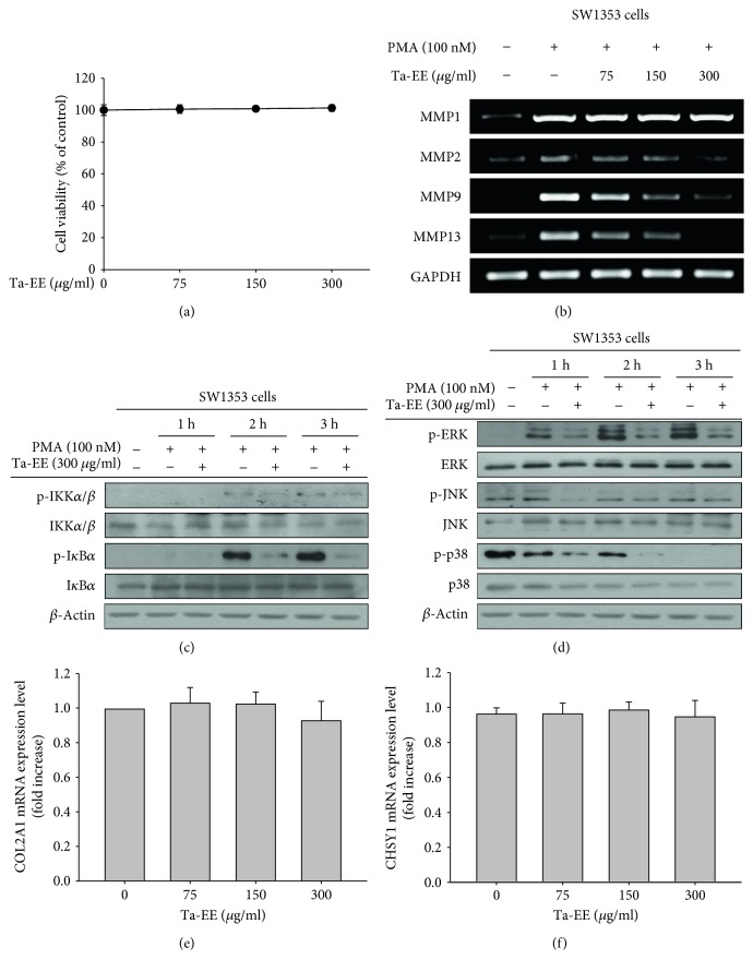
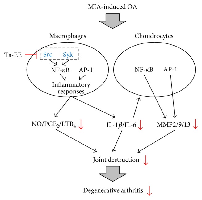
Although osteoarthritis (OA), a degenerative joint disease characterized by the degradation of joint articular cartilage and subchondral bones, is generally regarded as a degenerative rather than inflammatory disease, recent studies have indicated the involvement of inflammation in OA pathogenesis. Tabebuia avellanedae has long been used to treat various diseases; however, its role in inflammatory response and the underlying molecular mechanisms remain poorly understood. In this study, the pharmacological effects of Tabetri (Tabebuia avellanedae ethanol extract (Ta-EE)) on OA pathogenesis induced by monoiodoacetate (MIA) and the underlying mechanisms were investigated using experiments with a rat model and in vitro cellular models. In the animal model, Ta-EE significantly ameliorated OA symptoms and reduced the serum levels of inflammatory mediators and proinflammatory cytokines without any toxicity. The anti-inflammatory activity of Ta-EE was further confirmed in a macrophage-like cell line (RAW264.7). Ta-EE dramatically suppressed the production and mRNA expressions of inflammatory mediators and proinflammatory cytokines in lipopolysaccharide-stimulated RAW264.7 cells without any cytotoxicity. Finally, the chondroprotective effect of Ta-EE was examined in a chondrosarcoma cell line (SW1353). Ta-EE markedly suppressed the mRNA expression of matrix metalloproteinase genes. The anti-inflammatory and chondroprotective activities of Ta-EE were attributed to the targeting of the nuclear factor-kappa B (NF-κB) and activator protein-1 (AP-1) signaling pathways in macrophages and chondrocytes.


Figures

Figure 1
Figure 1
Ta-EE ameliorated OA symptoms in MIA-induced OA rats. (a) Experimental schedule of OA induction, treatment, and evaluation. Sprague-Dawley rats (10 rats/group) were intra-articularly injected with MIA (3 mg/rat). One week later, the rats were subjected to different treatments once a day for four weeks. The study finished at week five. (b) MIA-induced OA rats were orally administered with either Ta-EE (30–120 mg/kg), MSM (155 mg/kg), or Pc-LE (20 mg/kg) once a day for four weeks, and paw withdrawal threshold was measured once a week for five weeks. The significance of each group with respect to the MIA group each week is summarized in the box. (c) Body weights of the rats were measured once a week for five weeks. ((d); left panel) MIA-induced OA rats were orally administered with Ta-EE (30–120 mg/kg), MSM (155 mg/kg), or Pc-LE (20 mg/kg) once a day for four weeks, and the tibia bones were stained with hematoxylin and eosin, safranin O, or toluidine blue at week five. The areas stained by ((d); middle panel) safranin O and ((d); right panel) toluidine blue were measured and plotted. (e) The femur and tibia bones of the MIA-induced OA rats administered with Ta-EE (30–120 mg/kg), MSM (155 mg/kg), or Pc-LE (20 mg/kg) were analyzed by X-ray radiography. P < 0.05 and ∗∗P < 0.005 versus a control group; #P < 0.05 and ##P < 0.005 versus a normal group.
Figure 2
Figure 2
Ta-EE reduced the serum levels of inflammatory mediators and proinflammatory cytokines in MIA-induced OA rats. MIA-induced OA rats were orally administered with either Ta-EE (30–120 mg/kg), MSM (155 mg/kg), or Pc-LE (20 mg/kg) once a day, and the levels of (a) PGE2, (b) LTB4, (c) IL-1β, and (d) IL-6 in the sera of the rats in each group were measured at weeks zero, two, and four. P < 0.05, ∗∗P < 0.005 versus a control group; #P < 0.05 and ##P < 0.005 versus a normal group; +P < 0.05 and ++P < 0.005 week 0 versus week 2 or 4; &P < 0.05 and &&P < 0.005 week 2 versus week 4.
Figure 3
Figure 3
Ta-EE suppressed the production and mRNA expressions of inflammatory mediators and proinflammatory cytokines in LPS-stimulated RAW264.7 cells. (a) RAW264.7 cells were treated with Ta-EE (0–300 μg/ml) for 24 h, and cell viability was measured by MTT assay. (b) RAW264.7 cells pretreated with Ta-EE (0–300 μg/ml) for 30 min were treated with LPS (1 μg/ml) for 24 h, and nitrite production levels in the cell culture media were measured using Griess reagents. RAW264.7 cells pretreated with Ta-EE (0–300 μg/ml) for 30 min were treated with LPS (1 μg/ml) for 24 h, and the production levels of (c) PGE2, (d) LTB4, (e) IL-1β, and (f) IL-6 in the cell culture media were measured by ELISA. RAW264.7 cells pretreated with Ta-EE (0–300 μg/ml) for 30 min were treated with LPS (1 μg/ml) for 6 h, and the mRNA expression levels of (g) IL-1β, (h) IL-6, and (i) COX-2 in the cells were measured by quantitative real-time PCR. P < 0.05 and ∗∗P < 0.005 versus a control group; #P < 0.05 and ##P < 0.005 versus a normal group.
Figure 4
Figure 4
Ta-EE suppressed the activation of the NF-κB and AP-1 signaling pathways in LPS-stimulated RAW264.7 cells. RAW264.7 cells pretreated with Ta-EE (300 μg/ml) for 30 min were treated with LPS (1 μg/ml) for the indicated time, and the total and phosphorylated protein levels of (a) p85, IKKα/β, IκBα, p50, and p65; (b) p38, JNK, ERK, c-Jun, and c-Fos; and (c) Src, Syk, and IRAK1 in the total cell lysates were determined by Western blot analysis. β-Actin was used as an internal control. (d) Effects of Ta-EE on the kinase activities of Src, Syk, and IRAK1 were determined by in vitro kinase assay using purified Src, Syk, and IRAK1 as described in Materials and Methods. P < 0.05 and ∗∗P < 0.005 versus a control group.
Figure 5
Figure 5
Ta-EE exerted a chondroprotective effect by downregulating MMP gene expression rather than upregulating the gene expressions of COL2A1 and CHSY1 in SW1353 cells. (a) SW1353 cells were treated with Ta-EE (0–300 μg/ml) for 24 h, and cell viability was measured by MTT assay. (b) SW1353 cells pretreated with Ta-EE (0–300 μg/ml) for 30 min were treated with PMA (100 nM) for 6 h, and the mRNA expression levels of MMP1, MMP2, MMP9, and MMP13 in the cells were measured by semiquantitative PCR. SW1353 cells pretreated with Ta-EE (300 μg/ml) for 30 min were treated with PMA (100 nM) for the indicated time, and the total and phosphorylated protein levels of (c) IKKα/β and IκBα and (d) ERK, JNK, and p38 in the total cell lysates were determined by Western blotting. β-Actin was used as an internal control. (e) SW1353 cells were treated with Ta-EE (0–300 μg/ml) for 6 h, and the mRNA expression levels of COL2A1 and CHSY1 in the cells were measured by quantitative real-time PCR.
Figure 6
Figure 6
Proposed model showing the inhibitory pathways associated Ta-EE-mediated anti-inflammatory and chondroprotective activities in OA pathogenesis.

References

    1. Glyn-Jones S., Palmer A. J., Agricola R., et al. Osteoarthritis. The Lancet. 2015;386(9991):376–387. doi: 10.1016/S0140-6736(14)60802-3. - DOI - PubMed
    1. Wang Y., Teichtahl A. J., Cicuttini F. M. Osteoarthritis year in review 2015: imaging. Osteoarthritis and Cartilage. 2016;24(1):49–57. doi: 10.1016/j.joca.2015.07.027. - DOI - PubMed
    1. Abusarah J., Benabdoune H., Shi Q., et al. Elucidating the role of protandim and 6-gingerol in protection against osteoarthritis. Journal of Cellular Biochemistry. 2017;118(5):1003–1013. doi: 10.1002/jcb.25659. - DOI - PubMed
    1. Goldring S. R., Goldring M. B. Clinical aspects, pathology and pathophysiology of osteoarthritis. Journal of Musculoskeletal & Neuronal Interactions. 2006;6(4):376–378. - PubMed
    1. Shane Anderson A., Loeser R. F. Why is osteoarthritis an age-related disease? Best Practice & Research Clinical Rheumatology. 2010;24(1):15–26. doi: 10.1016/j.berh.2009.08.006. - DOI - PMC - PubMed

MeSH terms

LinkOut - more resources