. 2017 Dec 20:13:63-76.
doi: 10.2147/IJN.S150918. eCollection 2018.

Lipopolysaccharide-induced inflammation in monocytes/macrophages is blocked by liposomal delivery of Gi-protein inhibitor

Affiliations

Lipopolysaccharide-induced inflammation in monocytes/macrophages is blocked by liposomal delivery of Gi-protein inhibitor

Monica Madalina Tucureanu et al. Int J Nanomedicine. .

Abstract

Background: Lipopolysaccharide (LPS) is widely recognized as a potent activator of monocytes/macrophages, and its effects include an altered production of key mediators, such as inflammatory cytokines and chemokines. The involvement of Gi protein in mediating LPS effects has been demonstrated in murine macrophages and various cell types of human origin.

Purpose: The aim of the present work was to evaluate the potential of a Gi-protein inhibitor encapsulated in liposomes in reducing the inflammatory effects induced by LPS in monocytes/macrophages.

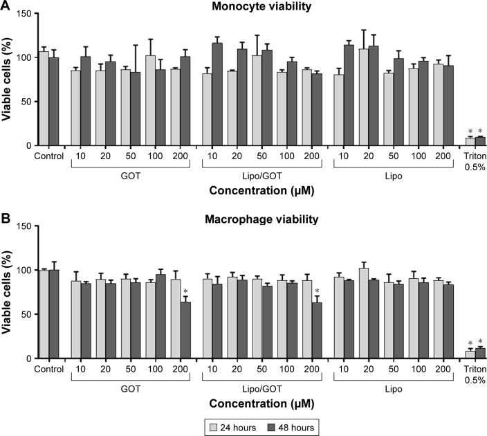
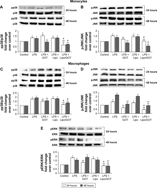
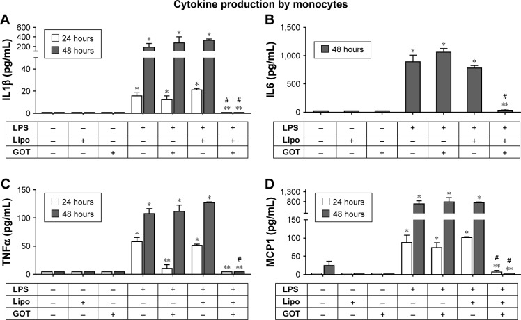
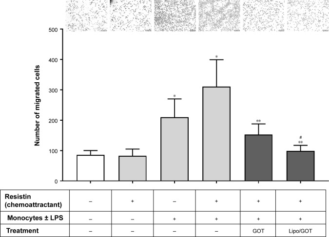
Materials and methods: Guanosine 5'-O-(2-thiodiphosphate) (GOT), a guanosine diphosphate analog that completely inhibits G-protein activation by guanosine triphosphate and its analogs, was encapsulated into liposomes and tested for anti-inflammatory effects in LPS-activated THP1 monocytes or THP1-derived macrophages. The viability of monocytes/macrophages after incubation with different concentrations of free GOT or liposome-encapsulated GOT was assessed by MTT assay. MAPK activation and production of IL1β, TNFα, IL6, and MCP1 were assessed in LPS-activated monocytes/macrophages in the presence or absence of free or encapsulated GOT. In addition, the effect of free or liposome-encapsulated GOT on LPS-stimulated monocyte adhesion to activated endothelium and on monocyte chemotaxis was evaluated.

Results: We report here that GOT-loaded liposomes inhibited activation of MAPK and blocked the production of the cytokines IL1β, TNFα, IL6, and MCP1 induced by LPS in monocytes and macrophages. Moreover, GOT encapsulated in liposomes reduced monocyte adhesion and chemotaxis. All demonstrated events were in contrast with free GOT, which showed reduced or no effect on monocyte/macrophage activation with LPS.

Conclusion: This study demonstrates the potential of liposomal GOT in blocking LPS proinflammatory effects in monocytes/macrophages.

Keywords: MAPK activation; chemotaxis; cytokine; guanosine 5′-O-(2-thiodiphosphate) (GOT); monocyte adhesion.


Conflict of interest statement

Disclosure The authors report no conflicts of interest in this work.

Figures

Figure 1
Figure 1
Concentration-dependent effect of GOT on monocyte and macrophage viability. Notes: THP1 monocytes (A) or THP1 cells differentiated to macrophages (B) were treated with different concentrations of free GOT, Lipo/GOT, or Lipo for 24 and 48 hours. Cell viability was measured by MTT assay and compared with the viability of untreated cells, considered 100%. A solution of 0.5% Triton X-100 was used as a positive control. Values shown are mean ± SE. *P<0.05. Abbreviations: GOT, guanosine 5′-O-(2-thiodiphosphate); Lipo, hydrated lipidic film with phosphate buffered saline; Lipo/GOT, GOT-encapsulated liposomes.
Figure 2
Figure 2
The effect of Lipo/GOT on MAPK phosphorylation in monocytes and macrophages. Notes: THP1 monocytes or THP1 cells differentiated to macrophages were activated with 100 ng/mL LPS for 24 (white bars) or 48 (gray bars) hours and treated with GOT or Lipo/GOT for 2 hours prior to LPS activation. p38 (A) and JNK (B) activation in monocytes, and p38 (C), JNK (D), and ERK (E) activation in macrophages were determined by Western blot. The phosphorylation of ERK in monocytes was also assessed, but showed no activation. In these experiments, cells unstimulated with LPS served as control (considered 1) and the phosphorylated proteins were correlated with total protein expression. Cells treated with Lipo in the presence of LPS (LPS + Lipo) were used as control. Every subsection of the figure shows in the upper panel a representative image of the Western blot analysis and in the lower panel the statistical results. Results given as mean ± SE (n=3). *P<0.05 (over control); **P<0.05 over LPS activation; #P<0.05 over LPS + GOT. Abbreviations: GOT, guanosine 5′-O-(2-thiodiphosphate); Lipo/GOT, GOT-encapsulated liposomes; LPS, lipopolysaccharide.
Figure 3
Figure 3
Effect of Lipo/GOT on production of IL1β, IL6, TNFα, and MCP1 by monocytes. Notes: THP1 cells were activated with 100 ng/mL LPS for 24 (white bars) or 48 (gray bars) and treated with GOT or Lipo/GOT for 2 hours prior to LPS activation. IL1β (A), IL6 (B), TNFα (C), and MCP1 (D) secretion was determined by enzyme-linked immunosorbent assay. Unstimulated cells served as controls. Cells treated with Lipo or free GOT in the presence or absence of LPS were used as experimental controls. Concentrations of IL1β, IL6, TNFα, and MCP1 given as mean ± SE (n=4). *P<0.05 over control; **P<0.05 over LPS activation; #P<0.05 over LPS + GOT. Abbreviations: GOT, guanosine 5′-O-(2-thiodiphosphate); Lipo, hydrated lipidic film with phosphate buffered saline; Lipo/GOT, GOT-encapsulated liposomes; LPS, lipopolysaccharide.
Figure 4
Figure 4
Effect of Lipo/GOT on the production of IL1β, IL6, TNFα, and MCP1 by macrophages. Notes: THP1 cells differentiated with phorbol 12-myristate 13-acetate for 3 days were activated with 100 ng/mL LPS for 24 (white bars) or 48 (gray bars) hours and treated with GOT or Lipo/GOT for 2 hours prior to LPS activation. IL1β (A), IL6 (B), TNFα (C), and MCP1 (D) secretion was determined by enzyme-linked immunosorbent assay. Unstimulated cells served as controls. Cells treated with Lipo or free GOT in the presence or absence of LPS were used as experimental controls. Concentrations of IL1β, IL6, TNFα, and MCP1 given as mean ± SE (n=4). *P<0.05 over control; **P<0.05 over LPS activation; #P<0.05 over LPS + GOT. Abbreviations: GOT, guanosine 5′-O-(2-thiodiphosphate); Lipo, hydrated lipidic film with phosphate buffered saline; Lipo/GOT, GOT-encapsulated liposomes; LPS, lipopolysaccharide.
Figure 5
Figure 5
Effect of Lipo/GOT on monocyte adhesion to endothelium. Notes: HECs were stimulated with LPS for 24 hours (black bars). Monocytes were preincubated with GOT or Lipo/GOT prior to activation for 24 hours with LPS. Unstimulated cells were used as controls. The number of adhered monocytes was quantified after 1 hour’s incubation of monocytes together with HECs, and results are given as mean ± SE (n=6). *P<0.05 over unstimulated HECs and monocytes (white bar); **P<0.05 over LPS-activated monocytes; #P<0.05 over LPS-activated monocytes treated with GOT; &P<0.05 for LPS-activated HECs vs unstimulated HECs. Scale bars are 50 μm. Abbreviations: GOT, guanosine 5′-O-(2-thiodiphosphate); HECs, human endothelial cells; Lipo/GOT, GOT-encapsulated liposomes; LPS, lipopolysaccharide.
Figure 6
Figure 6
Effect of Lipo/GOT on monocyte migration. Notes: THP1 monocytes were preincubated with GOT or Lipo/GOT prior to activation for 24 hours with LPS. After activation, monocytes were added to a chemotaxis chamber, where resistin was found in the lower compartment. The number of migrated monocytes toward resistin was quantified after 18 hours. Unstimulated monocytes were used as controls. Results are given as mean ± SE (n=6). *P<0.05 over control monocytes; **P<0.05 over LPS-activated monocytes; #P<0.05 over LPS-activated monocytes treated with GOT. Scale bars are 50 μm. Abbreviations: GOT, guanosine 5′-O-(2-thiodiphosphate); Lipo/GOT, GOT-encapsulated liposomes; LPS, lipopolysaccharide.

References

    1. Calin M, Manduteanu I. Emerging nanocarriers-based approaches to diagnose and reduce vascular inflammation in atherosclerosis. Curr Med Chem. 2017;24:550–567. - PubMed
    1. Ngkelo A, Meja K, Yeadon M, Adcock I, Kirkham PA. LPS induced inflammatory responses in human peripheral blood mononuclear cells is mediated through NOX4 and Giα dependent PI-3 kinase signalling. J Inflamm (Lond) 2012;9:1. - PMC - PubMed
    1. Takashiba S, Van Dyke TE, Amar S, Murayama Y, Soskolne AW, Shapira L. Differentiation of monocytes to macrophages primes cells for lipopolysaccharide stimulation via accumulation of cytoplasmic nuclear factor kB. Infect Immun. 1999;67:5573–5578. - PMC - PubMed
    1. Borzęcka K, Płóciennikowska A, Björkelund H, Sobota A, Kwiatkowska K. CD14 mediates binding of high doses of LPS but is dispensable for TNF-α production. Mediators Inflamm. 2013;2013:824919. - PMC - PubMed
    1. Solomon KR, Kurt-Jones EA, Saladino RA, et al. Heterotrimeric G proteins physically associated with the lipopolysaccharide receptor CD14 modulate both in vivo and in vitro responses to lipopolysaccharide. J Clin Invest. 1998;102:2019–2027. - PMC - PubMed

MeSH terms